

 PDF
PDF ePub
ePub Citation
 Print
Print


Introduction
Tendons of extensor musculature of the forearm can be compartmentalized into six synovial tendon sheaths which pass deep to the extensor retinaculum. The fourth tendon sheath, in most cases, envelop tendons of extensor digitorum (ED) and extensor indicis proprius (EIP) [1]. In literature, numerous reports describe variations of the extensor muscles as well as flexor muscles [23456]. Particularly, supernumerary muscles might be present in this fourth compartment. Initially coined extensor brevis digiti indicis vel medii by Albinus in 1734, this variant muscle is now commonly accepted as the extensor digitorum brevis manus (EDBM) [7]. Also, the extensor medii proprius (EMP) might be found as a muscle analogous to the EIP which inserts into second digit, but the EMP inserts into the third digit instead [8910].
Herein the authors report a case of bilateral yet asymmetric variant extensor muscles on the dorsum of the hands, and discuss their embryological backgrounds and clinical significance.
Go to : 
Case Report
During a routine dissection of the upper limb in anatomy class of 2017 at Eulji University School of Medicine, couple of anomalous muscles were found after removing the skin and connective tissue on the dorsum of each hand. This male cadaver was born in 1938 and deceased in 2016 with no medical history in both upper limbs. Out of 22 cadavers dissected in the university from 2015 to 2017, it was the only case with a variation in the forearm extensor musculature. The photos were taken using a digital camera (SM-G930S, Samsung Inc., Seoul, Korea) and schematic diagrams were illustrated using Adobe Photoshop (Adobe System Inc., San Jose, CA, USA).
The variant muscle on the dorsum of the right hand was EDBM (Fig. 1A, B). It originated from the distal end of the radius and radiocarpal ligaments, and inserted into the metacarpophalangeal joint of the third digit. The EDBM had a single belly with 55 mm in length and 20 mm in width. Another anomalous muscle, EMP is located on the same region of the left hand (Fig. 2A, B). It originated from the distal third of the ulna near the EIP and the interosseous membrane. The muscle belly of the EMP was fusiform shape with 35 mm in length, and its thin tendon was inserted into the dorsal aponeurosis of the third digit. Both muscle tendons were inserted into palmar- and ulnar-side of the insertion of the ED tendon of the third digit, respectively.
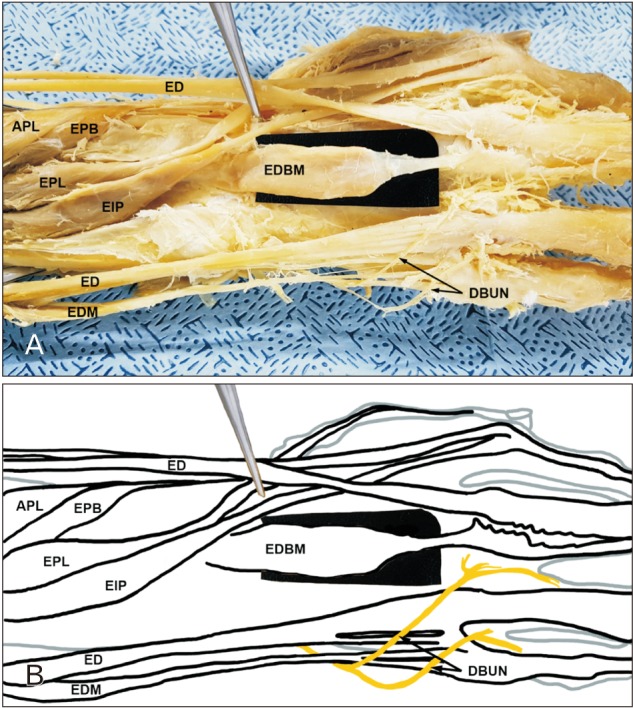 | Fig. 1(A, B) Digital photograph and a sketch show the origin and insertion of the extensor digitorum minimi (EDBM) on the dorsum of the right hand. APL, abductor pollicis longus; DBUN, dorsal branch of the ulnar nerve; ED, extensor digitorum; EDM, extensor digitorum minimi; EIP, extensor indicis proprius; EPB, extensor pollicis brevis; EPL, extensor pollicis longus.
|
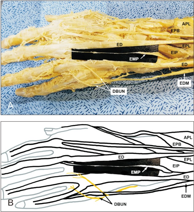 | Fig. 2(A, B) Digital photograph and a sketch show the origin and insertion of the extensor medii proprius (EMP) on the dorsum of the left hand. APL, abductor pollicis longus; DBUN, dorsal branch of the ulnar nerve; ED, extensor digitorum; EDM, extensor digitorum minimi; EIP, extensor indicis proprius; EPB, extensor pollicis brevis; EPL, extensor pollicis longus.
|
Go to :
Discussion
The EDBM and EMP are rare muscles found on the dorsum of the hand. A meta-analytic review demonstrated that EDBM has 4% of crude cadaveric prevalence, which is the number of cadavers who have either one or two EDBM compared to the number of cadavers in available, and has 2.5% of true cadaveric prevalence, which is the number of hands appeared EDBM compared to the number of hands in available. Only 26.3% of these cases showed bilateral presence of EDBM [3]. Though slightly variable among case, the origin of EDBM is known to be near the distal end of the radius, carpal ligaments and carpal joint capsule. Its most common insertion is the extensor expansion of the second digit with 70% probability, remaining 30% of cases had inserted into the third digit [2]. Although less frequently reported than the EDBM, EMP is also an uncommon muscle with a frequency of 0.8% to 10.4% [8]. Because the EMP has a small muscle belly and slender tendon, it very rarely causes clinical symptoms and is seen only during cadaver dissections [10].
In the present case, bilateral extensor variations were found in one of 22 cadavers (4.5%) and both muscles were inserted into the dorsal aponeurosis of the third digit with a presence of the EIP. It is distinctive feature of the present case, because majority of anomalous extensors were inserted into the second digit, and were liable to be lack of EIP [23]. According to the classification of Ogura et al. [2], the variation of the right hand was type III. In addition, according to the novel classification of the EIP variation from Georgiev et al. [10], the variation of the right hand was type III.b.2, whereas the variation of the left hand was type III.c.3.
As far as the author's knowledge, there is only one case report of bilateral variation including the EDBM and EMP from Cigali et al. [9], but it is different from the present case in that EDBM and EMP were observed on the same hand and their tendons are merged. Ogura et al. [2] demonstrated that ten out of 20 hands in the absence of the EIP had an EDBM, suggesting that the EDBM may compensate for the lack of an EIP. However, absence of the EIP was not seen in the case described here. This discrepancy is may be due to the racial difference or population size.
In the human embryo, the precursor extensor muscle of the upper and lower limb migrates and differentiates into three parts: radial, superficial and deep. These parts lead to the indivisualization of about 19 muscles in the upper limb and 14 muscles in the lower limb [111]. The deep part of the forearm extensor precursor, which is innervated by the posterior interosseous nerve, gives rise to the abductor pollicis longus and the extensor pollicis brevis on the radial side, while the extensor pollicis longus and the EIP on the ulnar side [11]. Lines of evidences support that anatomical variations including the EDBM and EMP are associated with the EIP, which have been developed from the relatively unstable part of the forearm extensor precursors [5810].
These muscles are usually identified through clinical symptoms as chronic pain or swelling on the hand after repeated exercise, and can be misdiagnosed as dorsal wrist ganglion, exostosis, tendon sheath cyst, rheumatoid tenosynovitis or benign tumor mass [4]. However, most EDBM are asymptomatic as in the case here, and their presence is incidentally recognized during hand surgery that was operated to treat other injuries or illnesses. Therefore, prior knowledge of EDBM and EMP may be important in that it helps clinicians to make an accurate diagnosis from other diseases, and to plan reconstructive tendon transfer surgeries in response to functional defect of forearm extensor muscles [510].
In the present study, two anomalous extensors, EDBM and EMP, were found on both hands asymmetrically in the same cadaver. To the best of our knowledge, this could be the first report on multi-muscular variation of the dorsum of each hand with regard to the EDBM and the EMP.
Go to :
 XML Download