

 PDF
PDF ePub
ePub Citation
 Print
Print


Introduction
Thymus is a content of superior and anterior mediastinum. It intervenes between sternum in front and the pericardium, arch of aorta with its three branches, brachiocephalic veins and trachea behind. It is prominent in children and it extends from the lower pole of lateral lobes of thyroid gland up to the fourth costal cartilage [1]. At birth it weighs 10–35 g. It grows till puberty where it gains a maximum weight of 20–50 g, thereafter in elderly it undergoes progressive involution to 5–15 g [2]. It appears to be a single encapsulated organ but actually it is bilobed [3]. The capsule sends fibrous extensions that divides each lobe into numerous lobules, which has an outer cortex enclosing central medulla. Diverse types of cells are present in thymus but thymic epithelial cells and immature T lymphocytes predominate. Progenitor cells from bone marrow migrate to the thymus where they are educated to distinguish between self and non self antigen and then they are exported to the periphery. In adulthood, the organ atrophies and production of T cells also declines [2]. Embryologically epithelium of thymus is a derivative of pair of third pharyngeal pouch. The epithelium become thymic corpuscles and they also form a network of epithelial reticular cells which is believed to be a source of thymic hormones that helps in differentiation of T lymphocytes [3]. Here we present a large thymus in an elderly cadaver with persistent active tissue in it.
Case Report
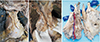
During routine undergraduate dissection class of thorax in department of Anatomy, an embalmed formalin fixed elderly male cadaver of around 65 years was dissected. Skin, pectoralis major, and pectoralis minor were reflected. When the thoracic cage was removed by cutting the costochondral junction a large mass (thymus) was found in the mediastinum (Fig. 1A). The position, extent and relation of the thymus was noted (Fig. 1B) and it was carefully dissected out (Fig. 1C) and weighed using analytic balance. The length, breadth and thickness of both lobes of the thymus was measured using digital caliper.
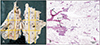
For microscopic anatomy, each lobe of thymus was then divided into nine quadrants and small piece of tissue were taken from each quadrant and were preserved in 10% formalin solution. These tissues were processed, embedded in paraffin and sections were taken using rotatory microtome and the slides were prepared and stained with hematoxylin and eosin stain (H&E) to see whether there is any persistence of active thymic tissue. Tissue sections from each quadrants of both lobes were examined under light microscope. In the right lobe, persistent thymic tissue was found in the eighth quadrant (Fig. 2).
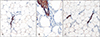
Further, immunohistochemistry (IHC) was done to look for cytokeratin expression (to confirm the epithelial origin of the tissue). Paraffin sections were deparaffinised using xylene and ethanol. Sections were rehydrated. Antigen retrieval was done in microwave oven using a citrate buffer. Endogenous peroxidase activity was blocked by adding H2O2. Slides were incubated with Pan Cytokeratin primary antibody (Prediluted, Dako, Hamburg, Germany) for 2 hours. Bound antibodies were detected with the EnVision detection system (Dako) following the protocol provided with the kit. For this methods, 3,3′-diaminobenzidine was used as chromogen and mild counterstaining was performed with haematoxylin.
Observation of the gross specimen
A diffuse symmetrical enlarged thymus was found in the anterior mediastinum that was extending from the lower part of thyroid gland to fifth costal cartilage. It was present in the anterior mediastinum between the two lungs and in front of pericardium and behind the sternum (Fig. 1A). It was pyramidal shaped with concave margin, yellowish brown in colour, bilobed separated only in the lower part (Fig. 1B). No other anterior mediatinal mass were detected. Weight of the specimen was 89 g and the length, breadth and thickness of right and left lobe was 147.52 mm, 69.74 mm, 7.44 mm and 124.17 mm, 60.3 mm, 10.45 mm, respectively.
Observation of H&E stained sections
Under microscope, H&E stained sections from the left lobe and sections from most of the quadrants of right lobe of the thymus showed fatty infiltration and blood vessels. In all sections, lobules were not demarcated and interlobular septas not seen, but H&E stained section from eighth quadrant (Fig. 2A, B) of the right lobe of thymus showed presence of thymic tissue and lymphocytes beside fatty infiltration and presence of blood vessel.
Observation of IHC
When further section obtained from the eighth quadrant (which showed presence of thymic tissue) was stained with cytokeratin antibody, it showed expression of cytokeratin in epithelial cells of the thymus (Fig. 3).
Discussion
Thymus acts as a central organ of lymphatic system and provides uncommitted immunologically competent lymphocytes to the circulating pool and to the peripheral lymphoid tissue. It is small at the time of birth and its size progressively increases up to age of puberty and then it undergoes involution and is converted into fibro-fatty mass. In the mid-adult life, it weighs about 10 g [1]. Literatures on thymus are available but most of them are based on ultrasonography, radiology, and histology. Very few literatures are available regarding its IHC. In our present case we have studied the gross, histology and IHC of a large thymic mass to see the active tissue in an elderly cadaver. Susimitha and Anitha [4] has reported presence of flat large bilobed thymus in adult male cadaver. Nayak et al. [5] has reported a 25 g irregular shaped thymus in 70 years old cadaver in which the two lobes were separated only in the upper part. H&E stained sections under low magnification showed distinct cortex and medulla in which cortex were densely packed with T lymphocyte and medulla consisted of few lymphocytes [5]. Araki et al. [6] has investigated computed tomography appearance of normal thymus in adult in which 74% of cases showed complete fatty replacement of thymus, 18% showed fatty attenuation, 7% showed half fatty and half soft tissue attenuation. Shrinkage of thymus have been reported by Nasseri and Eftekhari [7] when the thymus is subjected to stress followed by its recovery in which it regains its size or even it becomes larger. This phenomenon of rebound hyperplasia is commonly found in children but can be seen in adults. Rebound hyperplasia is also observed in patients who have undergone chemotherapy. Therefore, a complete knowledge of this persistent thymus is essential to distinguish thymic tissue from pathologic masses [7]. Cardiothoracic surgeons should have a detailed knowledge of these variation to avoid unnecessary surgical intervention. Follicular hyperplasia occurs in a number of chronic inflammatory and immunological states, e.g., myasthenia gravis. In Graves disease, scleroderma, rheumatoid arthritis, and other autoimmune disorder thymic changes are found. Therefore, the size of the thymus varies widely and whether it is a true hyperplasia or is merely a variant of normal has to be determined. Thymic hyperplasia is radiologically mistaken for thymoma [2]. Therefore, a detailed knowledge of large thymic mass, histological and IHC features will help clinicians to differentiate any mediastinal mass. Our present case resembles with the case of Susimitha and Anitha [4] and Nayak et al. [5] up to some extent. We got a huge thymic mass weighing 89.02 g separated in the lower part. In our case the cortico-medullary pattern was not maintained and interlobular septa was not seen. Instead we got a very small area of active persistent thymic tissue in a large thymic mass by IHC. Our method of identification (macroscopic as well as microscopic and immunostaining techniques) of the thymic tissue was more robust than the methods used by earlier authors.
Though the thymus undergoes involution in elderly and becomes a content of superior mediastinum, it persists in some cases and even a normal thymus can present as a huge anterior mediastinal mass. This knowledge will be helpful to the radiologists and cardiothoracic surgeons in making differential diagnosis of anterior mediastinal mass and to avoid any unnecessary surgical incision in the thorax.
 XML Download