

 PDF
PDF ePub
ePub Citation
 Print
Print


Introduction
The median nerve is a branch of brachial plexus that is formed by the union of lateral and medial roots of corresponding cords of brachial plexus, conveying the fibers of C5, C6, C7, C8, and T1 spinal nerves. In the arm, the median nerve lies deep to biceps brachii muscle but superficial to brachialis muscle. In the upper part of the arm, the median nerve lies lateral to the brachial artery. However, in the middle of the front of the arm, the nerve crosses the artery from lateral to medial side superficially. Thereafter, the nerve accompanies the medial side of the artery and appears in the cubital fossa as a most medial content of cubital fossa. In its further course, the median nerve leaves the fossa by passing through the pronator teres [1].
Nerve entrapment syndrome is one of the major clinical complications of the median nerve, which affects the movement and sensation in the hand. Three common syndromes associated with the entrapment are carpal tunnel syndrome (CTS), pronator teres syndrome (PTS), and anterior interosseous nerve syndrome (AINS). CTS is manifested because of compression of the median nerve at the wrist and is characterized by numbness, tingling, or burning sensations in the thumb and radial half of the palm. PTS results from the median nerve compression in the forearm and can cause pain and/or numbness in the distribution of the distal median nerve. The AINS results from injury to the anterior interosseous nerve in the forearm and is characterized by partial or complete loss of motor function of the long flexor muscle of the thumb, the deep flexor muscles of the index and middle fingers, and the pronator quadratus muscle.
Case Report
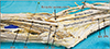
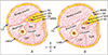
During routine human cadaveric dissection of upper limb for the medical undergraduate students, we noticed a variant course and branching pattern of the median nerve in the right arm. The musculocutaneous nerve (MCN) was absent and the muscles of front of the arm namely, coracobrachialis, biceps brachii and brachialis were supplied by the median nerve. In the lower part of arm, median nerve was deeply placed, beneath an abnormally persistent musculo-fascial tunnel. The tunnel was formed by the medial intermuscular septum supplemented by few fibers of brachialis muscle. The brachial artery also accompanied the median nerve laterally throughout its course beneath the tunnel. At the cubital fossa, the brachial artery emerged out of the tunnel and terminated by dividing into radial and ulnar artery. However, the median nerve continued its deep course, as a result it was not found as a content of the cubital fossa. Later it appeared in the front of the forearm underneath the pronator teres muscle (Fig. 1). A schematic diagram of the cross section at the lower part of the arm, shows the variation of the median nerve and brachial artery with respect to its relationship with the medial intermuscular septum (Fig. 2). These variations were found unilaterally in an elderly male human cadaver aged approximately 65 years.
Discussion
The compression of the median nerve can occur at various sites along its course [2], causing specific and variable signs and symptoms or nerve entrapment syndromes. One of the uncommon sites of compression of the median nerve is presented in this report. In this case, the median nerve together with the brachial artery were entrapped by an abnormally formed musculo-fascial tunnel in the lower one-third of the front of the arm. The tunnel was formed by the medial intermuscular septum which was reinforced by few muscle fibers of brachialis.
Scientific literatures state that, most proximal site of median nerve compression occurs at the vicinity of the lower end of humerus by the presence of ligament of Struthers, causing PTS. The ligament of Struthers courses from the supracondylar process of the humerus to the medial humeral epicondyle [3]. Struthers ligament is not a constant ligament and often encases median nerve together with the brachial artery and is believed to be rarely producing nerve entrapment symptoms. This is probably due to existence of adequate space for the passage of neurovascular structures. Topographically, the present variation is comparable with this, but no typical ligamentous structure was identified. In the present case, the entrapment was due to an unyielding nature of the musculo-fascial tunnel. In such cases, the possibilities of median nerve and brachial artery compression with the complications of PTS cannot be ruled out.
Symptoms of PTS can also be manifested because of the imprisonment of median nerve between the humeral and ulnar heads of pronator teres muscle in the forearm [4] or also by the abnormal presence of third head of biceps brachii [5].
Melanie et al. [6], reported a rare sub-brachialis course of the median nerve in the arm, wherein, the entire course of the median nerve was deep to the brachialis muscle. But, near the cubital fossa, it returned to its normal course and appeared as the most medial content of the cubital fossa [6]. However, in the present case, the deep coursed median nerve did not appear in the cubital fossa and passed underneath the pronator teres muscle.
Absence of the median nerve in the cubital fossa is an exceptional variation. There may be variations in the arrangement of contents in the fossa, as reported in a case where the median nerve was in between brachial artery and biceps tendon [7]. A very rare case of absence of median nerve in the cubital fossa was reported by Shetty et al. [8]. Here in their case, the median nerve was enclosed within the fleshy fibers of pronator teres. In the present case, deep course of median nerve under the abnormal tunnel prevented its appearance in the cubital fossa. This variation has all possibilities of entrapment neuropathy since the median nerve and brachial artery were found compressed under the musculo-fascial structure, which had an unyielding nature. Hence, we assume that, the clinical indications possibly presented in such entrapment might be similar to that of pronator syndrome. Additionally, the brachial artery might also be compressed resulting in compression-related symptoms. In advance stages, compression could lead to endothelial damage and thrombotic occlusion of brachial artery [9].
Absence of MCN may not lead to impairment of actions of muscles of anterior compartment of arm, its morphological variation has been considered as relevant while performing neurotization of brachial plexus lesions and several other surgical and reconstructive procedures [1011].
Median nerve entrapment syndrome affects the movement and sensation in the hand. Impingement of median nerve and brachial artery under an abnormally formed musculo-fascial tunnel could also be one of the rare causes of neurovascular compression syndrome. It should be considered in the differential diagnosis of upper limb ischemia, especially in young patients with weak or absent pulses. Knowledge regarding such unusual variations is very important during the assessment of median nerve entrapment syndromes.
 XML Download