

 PDF
PDF ePub
ePub Citation
 Print
Print


Introduction
The buccal nerve (BN) is important for dentists and oral and maxillofacial surgeons during clinical procedures such as regional anesthesia. The BN is a terminal branch of the mandibular division of the trigeminal nerve (CN V). The usual course of the BN is medial to the ramus of the mandible onto the cheek while passing anterolaterally to the tendon of the temporalis. The BN supplies the skin lateral to the lips, the buccal alveolar mucosa and the buccal gingivae of the second and third molars [1]. The BN is also known to sometimes join with buccal branches of the facial nerve.
There are variations in the medical literature describing the specifics of BN including the additional anatomical structures that it innervates, its course, and its origin from the mandibular division of the CN V. In dissections of multiple human cadaveric heads, Takezawa et al. [2] detailed the BN course and distribution concluding that “broader distribution of the BN was found than described previously.” Here, we report an unusual case of a BN that to our knowledge has been previously unreported.
Case Report
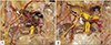
A routine anatomical dissection of the left infratemporal fossa in an embalmed Caucasian male cadaver aged 89-year-old at death revealed a left sided BN with two roots. The BN originated from the anterior division of the mandibular nerve (Fig. 1). The BN originated as one trunk from the main trunk of the mandibular nerve then divided into two branches. The anterior branch (1.0 mm in diameter) of the BN was found sending branches into the superior head of the lateral pterygoid muscle. The posterior branch (0.9 mm in diameter) of the BN continued between the superior and inferior heads of the lateral pterygoid muscle, after which it rejoined (1.1 mm in diameter) the anterior root to continue along its course. The BN terminated on the skin above the surface of the buccinator muscle. The two roots of the BN were superficial to the second part of the maxillary artery and posterior to the buccal artery. There were no additional anatomical structures that passed through two roots of the BN. No other anatomical anomalies were noted in the specimen relating to the regional nerves. No gross findings of previous surgical intervention to the dissected region were identified.
Discussion
While several variations of the BN can occur, the variant reported here, to our knowledge, has not been reported. According to Kamijo [3], 4% of the Japanese population has two roots of origin of the BN. From our understanding of the BN, such a variation would not alter the sensory supply to the region. The two roots of the BN is proximally located and after its origin from the mandibular nerve. Anatomical complications to consider in this BN variant are compression of the two branches between the superior and inferior heads of the lateral pterygoid muscles. This could compromise sensory innervation of the BN in the region.
Takezawa et al. [2] discussed four distinct variations of the BN. These variations are categorized by the region they supply which are termed: posterior distribution, anterior distribution, superior distribution, and inferior distribution. Takezawa et al. [2] also discussed the implications of nerve block to the BN variations and their effectiveness.
The retromolar foramen, a variation of the mandibular canal, has recently been focused on by oral surgeons because this anatomical variant could cause sensory disturbance of the buccal gingiva of the lower second and third molars when cut [45]. Some of the nerve fibers, which are supposed to be included in the BN, occasionally run with the inferior alveolar nerve and derive from the retromolar foramen. Thus, the BN could travel with different nerves. In this case, two roots of the BN join and form one main trunk and did not seem to travel with other nerves. However, we believe that the BN is variable and could affect the outcome of clinical procedures such as a BN block.
In conclusion, the existence of this unusual BN variant should be appreciated by both anatomists and clinicians.
 XML Download