

 PDF
PDF ePub
ePub Citation
 Print
Print


Abstract
Purpose
To assess the diagnostic performance in detecting rotator cuff tears at 3T of non-arthrographic shoulder magnetic resonance imaging (MRI) using 3D isotropic turbo spin-echo (TSE-SPACE) sequence as compared with 2D sequences.
Materials and Methods
Seventy-four patients who were arthroscopically confirmed to have underwent non-arthrographic shoulder MRI with 2D sequences and TSE-SPACE were included. Three independent readers retrospectively scored supraspinatus and infraspinatus tendon (SST-IST) and subscapularis tendon (SCT) tears on 2D sequences and TSE-SPACE.
Results
The mean sensitivity, specificity, and accuracy of the three readers were 95%, 100%, and 95% on TSE-SPACE and 99%, 93%, and 98% on 2D sequences for detecting SST-IST tears, respectively, whereas those were 87%, 49%, and 68% on TSE-SPACE and 88%, 66%, and 77% on 2D sequences for detecting SCT tears, respectively. There was no statistical difference between the two sequences, except for in the specificity of one reader for detecting SCT tears. The mean AUCs of the three readers on TSE-SPACE and 2D sequences were 0.96 and 0.98 for detecting SST-IST tears, respectively, which were not significantly different, while those were 0.71 and 0.82 for detecting SCT tears, respectively, which were significantly different (P < 0.05).
Go to : 
References
1. de Jesus JO, Parker L, Frangos AJ, Nazarian LN. Accuracy of MRI, MR arthrography, and ultrasound in the diagnosis of rotator cuff tears: a metaanalysis. AJR Am J Roentgenol. 2009; 192:1701–1707.

2. Shapiro L, Staroswiecki E, Gold G. Magnetic resonance imaging of the knee: optimizing 3 Tesla imaging. Semin Roentgenol. 2010; 45:238–249.
3. Gold GE, Suh B, Sawyer-Glover A, Beaulieu C. Musculoskeletal MRI at 3.0 T: initial clinical experience. AJR Am J Roentgenol. 2004; 183:1479–1486.
4. Steinbach LS, Palmer WE, Schweitzer ME. Special focus session. MR arthrography. Radiographics. 2002; 22:1223–1246.
5. Jung JY, Jee WH, Park MY, Lee SY, Kim YS. Supraspinatus tendon tears at 3.0 T shoulder MR arthrography: diagnosis with 3D isotropic turbo spin-echo SPACE sequence versus 2D conventional sequences. Skeletal Radiol. 2012; 41:1401–1410.
6. Jung JY, Jee WH, Park MY, Lee SY, Kim YS. SLAP tears: diagnosis using 3-T shoulder MR arthrography with the 3D isotropic turbo spin-echo space sequence versus conventional 2D sequences. Eur Radiol. 2013; 23:487–495.
7. Jung JY, Yoon YC, Choi SH, Kwon JW, Yoo J, Choe BK. Three-dimensional isotropic shoulder MR arthrography: comparison with two-dimensional MR arthrography for the diagnosis of labral lesions at 3.0 T. Radiology. 2009; 250:498–505.
8. Magee T. Can isotropic fast gradient echo imaging be substituted for conventional T1 weighted sequences in shoulder MR arthrography at 3 Tesla? J Magn Reson Imaging. 2007; 26:118–122.
9. Lee JH, Yoon YC, Jee S, Kwon JW, Cha JG, Yoo JC. Comparison of three-dimensional isotropic and two-dimensional conventional indirect MR arthrography for the diagnosis of rotator cuff tears. Korean J Radiol. 2014; 15:771–780.
10. Park SY, Lee IS, Park SK, Cheon SJ, Ahn JM, Song JW. Comparison of three-dimensional isotropic and conventional MR arthrography with respect to the diagnosis of rotator cuff and labral lesions: focus on isotropic fat-suppressed proton density and VIBE sequences. Clin Radiol. 2014; 69:e173–182.
11. Choo HJ, Lee SJ, Kim OH, Seo SS, Kim JH. Comparison of three-dimensional isotropic T1-weighted fast spin-echo MR arthrography with two-dimensional MR arthrography of the shoulder. Radiology. 2012; 262:921–931.
12. Kijowski R, Davis KW, Blankenbaker DG, Woods MA, Del Rio AM, De Smet AA. Evaluation of the menisci of the knee joint using three-dimensional isotropic resolution fast spin-echo imaging: diagnostic performance in 250 patients with surgical correlation. Skeletal Radiol. 2012; 41:169–178.
13. Kijowski R, Davis KW, Woods MA, et al. Knee joint: comprehensive assessment with 3D isotropic resolution fast spin-echo MR imaging–diagnostic performance compared with that of conventional MR imaging at 3.0 T. Radiology. 2009; 252:486–495.
14. Notohamiprodjo M, Horng A, Pietschmann MF, et al. MRI of the knee at 3T: first clinical results with an isotropic PDfs-weighted 3D-TSE-sequence. Invest Radiol. 2009; 44:585–597.
15. Notohamiprodjo M, Kuschel B, Horng A, et al. 3D-MRI of the ankle with optimized 3D-SPACE. Invest Radiol. 2012; 47:231–239.
16. Ristow O, Steinbach L, Sabo G, et al. Isotropic 3D fast spin-echo imaging versus standard 2D imaging at 3.0 T of the knee–image quality and diagnostic performance. Eur Radiol. 2009; 19:1263–1272.
17. Yao L, Pitts JT, Thomasson D. Isotropic 3D fast spin-echo with proton-density-like contrast: a comprehensive approach to musculoskeletal MRI. AJR Am J Roentgenol. 2007; 188:W199–201.
18. Altman DG. Practical statistics for medical research. London: Chapman & Hall. 1991. 404–409.
19. Oh DK, Yoon YC, Kwon JW, et al. Comparison of indirect isotropic MR arthrography and conventional MR arthrography of labral lesions and rotator cuff tears: a prospective study. AJR Am J Roentgenol. 2009; 192:473–479.
20. Park HJ, Lee SY, Kim MS, et al. Evaluation of shoulder pathology: three-dimensional enhanced T1 high-resolution isotropic volume excitation MR vs two-dimensional fast spin echo T2 fat saturation MR. Br J Radiol. 2015; 88:20140147.
21. Stevens KJ, Busse RF, Han E, et al. Ankle: isotropic MR imaging with 3D-FSE-cube–initial experience in healthy volunteers. Radiology. 2008; 249:1026–1033.
22. Van Dyck P, Gielen JL, Vanhoenacker FM, et al. Diagnostic performance of 3D SPACE for comprehensive knee joint assessment at 3 T. Insights Imaging. 2012; 3:603–610.
23. Koo SS, Burkhart SS. Subscapularis tendon tears: identifying mid to distal footprint disruptions. Arthroscopy. 2010; 26:1130–1134.
24. David TS, Bravo H, Scobercea R. Arthroscopic visualization of subscapularis tendon lesions. Orthopedics. 2009; 32.
25. Magee T, Williams D. 3.0-T MRI of the supraspinatus tendon. AJR Am J Roentgenol. 2006; 187:881–886.
26. Jung JY, Yoon YC, Yi SK, Yoo J, Choe BK. Comparison study of indirect MR arthrography and direct MR arthrography of the shoulder. Skeletal Radiol. 2009; 38:659–667.
27. Pfirrmann CW, Zanetti M, Weishaupt D, Gerber C, Hodler J. Subscapularis tendon tears: detection and grading at MR arthrography. Radiology. 1999; 213:709–714.
28. Gyftopoulos S, O'Donnell J, Shah NP, Goss J, Babb J, Recht MP. Correlation of MRI with arthroscopy for the evaluation of the subscapularis tendon: a musculoskeletal division's experience. Skeletal Radiol. 2013; 42:1269–1275.
29. Jung JY, Jee WH, Chun CW, Kim YS. Diagnostic performance of MR arthrography with anterior trans-subscapularis versus posterior injection approach for subscapularis tendon tears at 3.0T. Eur Radiol. 2017; 27:1303–1311.
Go to :
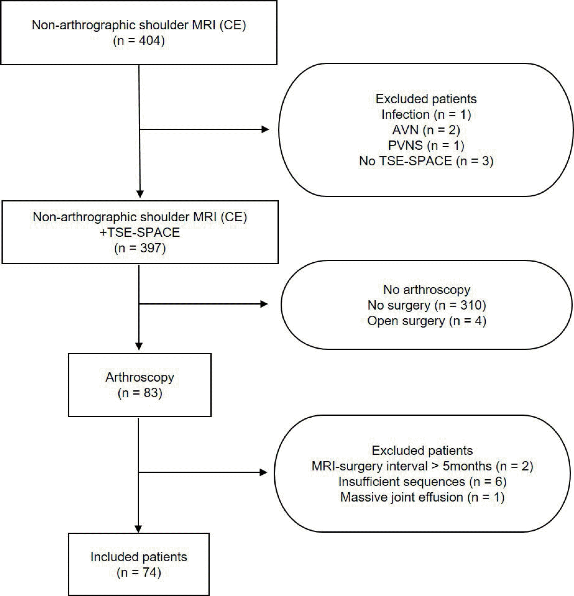 | Fig. 1.Flow diagram of the study. AVN = avascular necrosis; CE = contrast-enhancement; GCT = giant cell tumor; PVNS = pigmented villonodular synovitis; TSE-SPACE = three-dimensional isotropic fat-suppressed turbo spin-echo SPACE intermediate-weighted image |
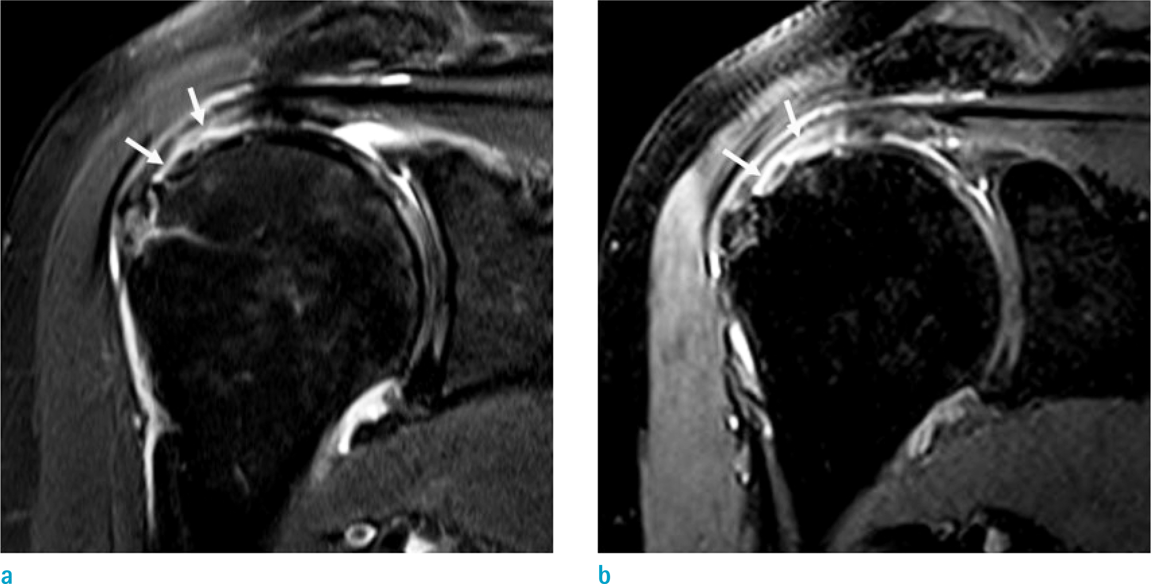 | Fig. 2.Non-arthrographic MR images show arthroscopically-proven partial-thickness articular-sided tear of the SST-IST in a 68-year-old woman. (a) Oblique coronal fat-suppressed T2-weighted image and (b) 3D isotropic fat-suppressed intermediate-weighted TSE-SPACE image show a partial-thickness articular-sided tear (arrows) of the SST-IST. All three readers correctly interpreted this case on both 2D sequences and TSE-SPACE. |
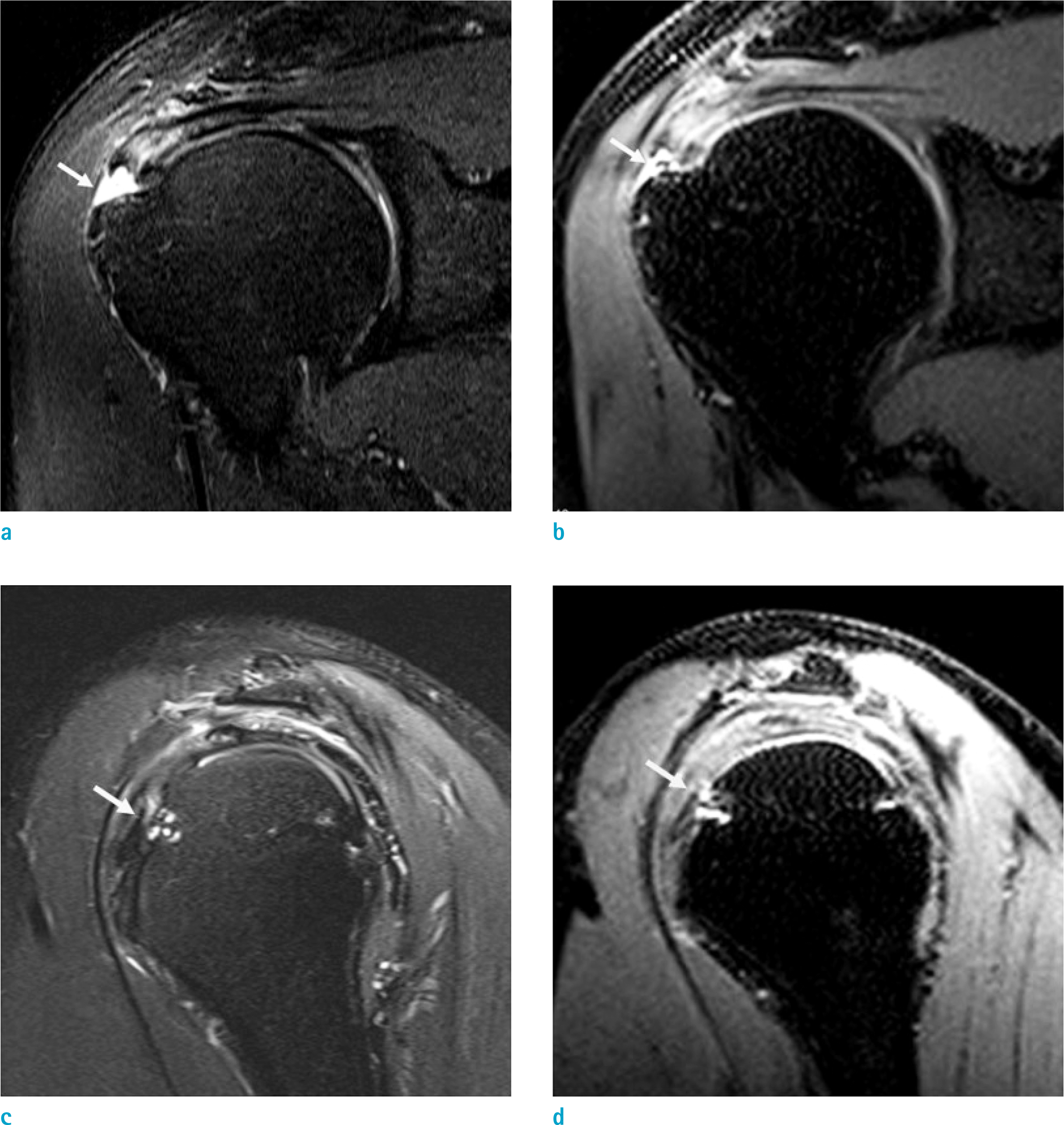 | Fig. 3.Non-arthrographic MR images show arthroscopically-proven partial-thickness bursal-sided tear of SST-IST and intact SCT in a 55-year-old man. (a) Oblique coronal fat-suppressed T2-weighted image and (b) 3D isotropic fat-suppressed intermediate-weighted TSE-SPACE image show a partial-thickness bursal-sided tear (arrows) of the SST-IST. (c) Oblique sagittal fat-suppressed T2-weighted image and (d) 3D isotropic fat-suppressed intermediate-weighted TSE-SPACE image with an oblique sagittal reformatted image show a subtle high signal intensity focus at the articular surface of SCT, which involves the superior portion of the tendon (arrows). A subcortical cyst is noted in the lesser tuberosity of proximal humerus. All three readers correctly interpreted the SST-IST on both 2D sequences and TSE-SPACE. However, they misinterpreted the SCT on both 2D sequences and TSE-SPACE. |
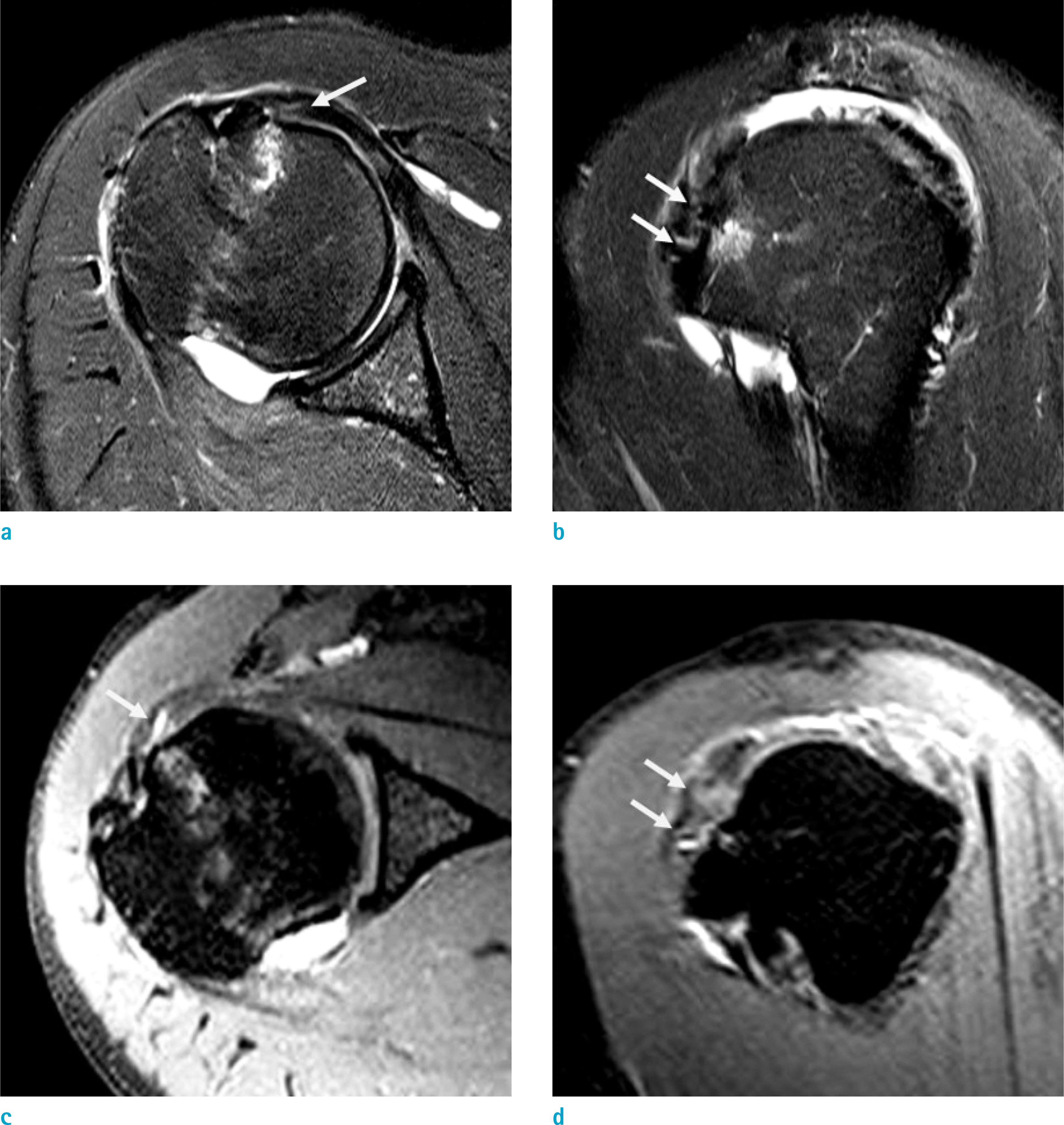 | Fig. 4.Non-arthrographic MR images show arthroscopically-proven grade 2 partial-thickness articular-sided tear of the SCT in a 56-year-old woman. (a, b) Axial and oblique sagittal fat-suppressed T2-weighted images and (c, d) 3D isotropic fat-suppressed intermediate-weighted TSE-SPACE images with an axial and an oblique sagittal reformation show a high signal intensity area at the articular surface of SCT, which involves more than one third of the craniocaudal diameter of the tendon (arrows). A subcortical cyst is present in the lesser tuberosity of proximal humerus. All three readers correctly interpreted this case on both 2D sequences and TSE-SPACE. |
Table 1.
MR Imaging Parameters
Table 2.
Diagnostic Performance of Conventional 2D Sequences and 3D TSE-SPACE in Evaluating Rotator Cuff Tears
Data in parentheses are numbers of lesions. Data in square brackets are 95% confidence interval. 2D = two-dimensional; 3D TSE-SPACE = three-dimensional isotropic fat-suppressed turbo spin-echo SPACE intermediate-weighted image; FT = full-thickness tear; NA = non-applicable; PT = partial-thickness tear; SCT = subscapularis tendon; SST-IST = supraspinatus and infraspinatus tendon
Table 3.
Area Under Receiver Operating Characteristics Curve (AUC) of Three Readers in Evaluating Rotator Cuff Tears
Table 4.
Interobserver Agreements of Three Readers in Evaluating Rotator Cuff Tears
 XML Download