

 PDF
PDF ePub
ePub Citation
 Print
Print


Introduction
Scrub typhus, or tsutsugamushi disease is an acute febrile illness caused by bacteria from the Rickettsiaceae family, named Orientia tsutsugamushi. This illness is characterized by a fever, rash, and lymphadenopathy. The clinical illness varies from mild and self-limiting to fatal. We describe a rare case of a pulmonary artery thrombosis (PAT) associated with scrub typhus.
This study was approved by the Institutional Review Board of the Yeungnam University Medical Center of Korea with a waiver of informed consent (Subject number: 2018-12-038).
Case report
A 67-year-old man was admitted with a fever and skin rash, which developed 2 days prior to admission. He had a previous history of diabetes and hypertension. He has been living in a rural area, and regularly works on his farm. On the physical examination, multiple erythematous macules on his neck, trunk, and both proximal extremities were observed and two pea-sized eschars were found on his left axilla and right upper abdomen (Fig. 1). There were no palpable masses suggesting lymphadenopathy around the eschars. The laboratory findings revealed a white blood cell count of 4,500/mm3, platelet count of 137,000 /mm3, and elevated liver enzyme (aspartate aminotransferase/alanine aminotransaminase 857/614 [IU/L]) and C-reactive protein (>8 mg/dL; normal range <0.5 mg/dL) levels. A serologic test for O. tsutsugamushi using an indirect immunofluorescence assay antibody was positive (quantitative value 1:80). Antibodies for the Hantaan virus and leptospira were negative. An electrocardiogram (ECG) revealed sinus rhythm (75 beats/min) with non-specific flattening of the T waves in the inferior leads. His clinical symptoms and laboratory findings gradually improved with oral doxycycline (200 mg/day), and he was discharged on the 5th hospital day. Oral doxycycline was additionally administered for 2 more days after discharge (7 days, totally).
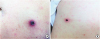 | Figure 1Two pea-sized black-crusted shallow ulcers (eschars) were observed with multiple erythematous macules on the left axilla (A) and right upper abdomen (B).
|
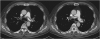
One week after discharge, he was referred with a complaint of dyspnea for 3 days. His D-dimer level was elevated at 20.636 ug/ml FEU, and his troponin I and N-terminal pro brain natriuretic levels were normal. An arterial blood gas analysis on room air revealed the following: pH 7.45, partial pressure of carbon dioxide (PaCO2) 34 mmHg, partial pressure of oxygen (PaO2) 84 mmHg, and oxygen saturation 97.5%. A serologic antibody test for O. tsutsugamushi was markedly elevated at >1:10,240. His fibrinogen and protein C antigen levels had mildly decreased to 193 mg/dL (normal range, 200–400 mg/dL) and 69% (normal range, 72–160%), respectively. His antiphospholipid antibodies (immunoglobulin G [IgG] and IgM) were negative. An ECG revealed sinus rhythm (79 beats/min) without any definite change compared to the previous ECG. Chest computed tomography revealed multifocal contrast filling defects along both pulmonary arteries (Fig. 2). There was no evidence of deep vein thrombosis (DVT) on duplex ultrasonography of the lower extremities. On transthoracic echocardiography, the right ventricular systolic pressure was mildly elevated at 44 mmHg without any significant right ventricular dysfunction. The symptoms improved with anticoagulation using unfractionated heparin for 7 days and he was discharged with an oral non-vitamin K anticoagulant (rivaroxaban) on the 9th hospital day. A followed up serologic antibody titer on the day of discharge revealed the O. tsutsugamushi antigen level had decreased to 1:5,120.
Discussion
It is known that the main pathologic findings in scrub typhus are systemic vasculitis and perivasculitis, which are caused by a proliferation of O. tsutsugamushi in endothelial cells of the microvascular system [1]. A previous report suggested the concept that acute infections are associated with a transient increase in the risk of vascular events [2]. However, the mechanism by which an acute infection or inflammation affect the risk of vascular events is uncertain.
The so called ‘rickettsial vasculitis’ can affect the skin, lungs, liver, kidneys, central nervous system, and skeletal and cardiac muscles. Injury to endothelial cells in the lungs and brain results in the most severe manifestations including non-cardiogenic pulmonary edema, interstitial pneumonia, acute respiratory distress syndrome, meningoencephalitis, seizures, and comas [3]. The manifestations of the cardiovascular system are mainly due to endothelial cell damage, vasculitis, inflammatory cell infiltration, new endothelial cell proliferation, thrombosis, and ischemia [4]. Myocardial infarctions [5] as well as DVTs [6], retinal vein occlusions [7], and central venous sinus thromboses [8] have been reported. Although the exact pathogenesis of the PAT in our patient with scrub typhus was unclear, it is possible that vasculitis or a vascular occlusion may have been involved via endothelial dysfunction.
Considering the involvement of the pulmonary vasculature without any evidence of an embolic source, the scrub typhus may have affected the procoagulability via inflammation. Specific infections can result in thrombo-hemorrhagic syndromes associated with bacterial as well as non-bacterial pathogens [9]. Recently, a study demonstrated the coagulation changes in scrub typhus, which are characterized by an inflammation-induced coagulopathy and coagulation activation [10]. A previous study reported a hypofibrinogenetic coagulopathy associated with thrombophlebitis in scrub typhus [11]. Further, disseminated intravascular coagulation caused by O. tsutsugamushi has also been reported [12]. The fibrinogen level was mildly decreased in the current patient, and an infection-induced activation of coagulation could have been associated.
We described a rare case of PAT associated with scrub typhus. The vasculitis with endothelial dysfunction caused by the scrub typhus could be considered as the main pathologic mechanism. Further studies are needed to clarify this issue in terms of the epidemiological and pathological aspects in the future.
 XML Download