

 PDF
PDF ePub
ePub Citation
 Print
Print


Introduction
Gastrointestinal (GI) diseases are common in patients with human immunodeficiency virus (HIV) infection and have been attributed to opportunistic diseases that resulted from advanced immunosuppression in the era before combination antiretroviral therapy (cART) was available [1]. With the advent of potent cART, the spectrum of GI diseases has changed in these patients, requiring a broader range of diagnostic considerations. As cART utilization has increased the life expectancy of patients with HIV infection, malignancies, particularly non-acquired immune deficiency syndrome (AIDS)-defining cancers, have become a growing cause of death in this population [23]. Cancer screening is now considered an important component of health maintenance in HIV clinical practice.
Several studies have reported cancer epidemiology and screening interventions in patients with HIV infection [45678]. However, gastric cancer data have been too limited to allow specific screening recommendations. Recent studies showed that the incidence of gastric cancer in patients with HIV infection was 1.8-1.9 times higher than that in the general population [910]. Although there have been studies of upper GI endoscopic findings in symptomatic patients with HIV infection [1111213], few studies have investigated endoscopic findings for gastric cancer in those receiving cART.
In the general population of Korea, gastric cancer is the second most common cancer after thyroid cancer [14], and Helicobacter pylori infection is found in approximately 60% of adults [15]. The National Cancer Screening Program (NCSP) of the National Health Insurance Service (NHIS) in Korea provides upper GI series or endoscopy for gastric cancer screening every 2 years for individuals aged ≥40 years [16]. Previous studies on Korean patients with HIV infection have reported that gastric cancer accounted for 3.1% of 32 cancers and 6.3% of 48 cancers [1718]. However, there are limited data on upper GI endoscopic findings and gastric cancer incidence in Korean patients with HIV infection. In this study, we retrospectively analyzed upper GI endoscopic findings in patients with HIV infection and investigated their role as gastric cancer screening.
Materials and Methods
1. Study population and data collection
We retrospectively reviewed the medical records of patients with HIV infection who underwent upper GI endoscopy at Pusan National University Hospital (Busan, Korea) between January 2004 and December 2018. This hospital is a 1,450-bed university-affiliated teaching hospital and provides HIV care for patients with HIV infection in the southeastern region of Korea. Patients were excluded from analysis if they had undergone gastric resection for gastric cancer treatment before presentation to the study hospital. Ethnic minorities and patients with unsuccessful endoscopy were also excluded.
We collected data on patient characteristics, including age, sex, timing of diagnosis, HIV status, CD4 cell count, viral load, use of antiretroviral therapy, presence of GI symptoms, purpose of endoscopy, and endoscopic and pathologic findings. Each endoscopic examination was classified as either diagnostic or screening endoscopy. Screening endoscopy included endoscopies performed as part of the NCSP of the NHIS or the health check program (HCP) of the health promotion center (HPC) of the study hospital. Endoscopic examinations performed owing to the presence of GI symptoms were classified as diagnostic endoscopy. Endoscopic findings were reviewed by 2 gastroenterologists, and all diagnoses were made on the basis of minimal standard terminologies for GI endoscopy [14]. Atrophic gastritis was assessed endoscopically using the Kimura-Takemoto classification [15]. Intestinal metaplasia was diagnosed either endoscopically by visualizing whitish plaque-like elevations in the gastric antrum and/or body or pathologically. Endoscopic biopsy specimens were graded using the Sydney system. H. pylori infection was assessed using the rapid urease test or biopsy results.
To examine the influence of the duration of cART on the endoscopic findings, we stratified the patients who received cART for at least 6 months and whose time to follow-up loss was less than 10% of the total follow-up duration into 4 groups according to the duration of therapy: 1) ≤6 months of cART, 2) 6 months to 5 years after cART initiation, 3) 5 to 10 years after cART initiation, and 4) >10 years after cART initiation. Similar findings with no significant change in multiple endoscopies within each period were not duplicated.
To determine the role of upper GI endoscopy as gastric cancer screening, we analyzed the incidence of gastric cancer in patients with HIV infection who underwent upper GI endoscopy for screening purpose. The incidence was computed as events per 1,000 person-years (PYs). The observation periods were measured from the date of the first visit to the study hospital to either the last date on which upper GI endoscopy was performed or the date of endoscopic gastric cancer diagnosis, whichever was earlier.
2. Statistical analysis
Statistical analyses were performed using SPSS for window version 25.0 (SPSS Inc., Chicago, IL, USA). All continuous variables were summarized as median and interquartile range (IQR). Categorical variables were described using frequencies and percentiles. Categorical variables were compared using Pearson's χ2 test or Fisher's exact test, whereas noncategorical variables were tested using the t-test or one-way ANOVA. All results were considered significant by P <0.05.
Results
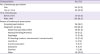
During the study period, 1,139 patients with HIV infection visited the study hospital. Three hundred thirty-four upper GI endoscopies were performed in 210 patients (18.4%). Of these, 4 patients were excluded from the analysis owing to gastric resection before enrollment (1.9%); further, 1 ethnic minority patient (0.5%) and 4 patients with unsuccessful endoscopy (1.9%) were also excluded. The remaining 310 endoscopies in 201 patients were included in the analysis; the baseline characteristics and endoscopic findings are presented in Tables 1 and 2, respectively. The median age at the time of endoscopy was 52.5 years (IQR, 44–60 years). The median number of endoscopies per patient was 1 (IQR, 1–2). The average number of endoscopies per patient was 1.5; 32.8% of the patients underwent more than 2 endoscopies. The median follow-up was 4.6 years (IQR, 0.6–8.8 years).
Table 1
Clinical characteristics of the 310 endoscopies in the 201 patients with human immunodeficiency virus
![]()
Table 2
Clinical characteristics and endoscopic findings according to purpose of endoscopies in the patients with human immunodeficiency virus infection
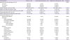
aComparison between screening endoscopy and diagnostic endoscopy
IQR, interquartile range; HIV, human immunodeficiency virus; cART, combination antiretroviral therapy; CMV, cytomegalovirus; HSV, herpes simplex virus.
![]()
Of the total of 310 endoscopies in 201 patients, 192 (61.9%) endoscopies in 142 patients were performed for diagnostic purposes. Their most commonly reported GI symptom was abdominal pain (14.6%), followed by nausea/vomiting/anorexia (11.5%) and dysphagia (10.9%); 30 (15.7%) endoscopies were performed to evaluate GI bleeding, including active bleeding and anemia. Atrophic gastritis (34.9%) was the most common endoscopic finding, followed by reflux esophagitis (25.5%), erosive gastritis (18.8%), and intestinal metaplasia (16.7%) (Table 2). Gastric cancer was found in 1 patient (0.5%). The patient was presented with early gastric cancer (EGC, type IIa+IIc, differentiated-type adenocarcinoma with atrophic gastritis and intestinal metaplasia on histologic examination) and diagnosed with HIV in the preoperative assessment. He was surgically cured (Table 3).
Table 3
Characteristics of human immunodeficiency virus-infected patients who were diagnosed with gastric cancer
HIV, human immunodeficiency virus; yrs, years; cART, combination antiretroviral therapy; EGC, early gastric cancer; ESD, endoscopic mucosal dissection.
![]()
One hundred eighteen (38.1%) endoscopies in 81 patients were performed for cancer screening purposes; 103 (87.3%) for the NCSP of the NHIS; and 15 (12.7%) for the HCP of the study hospital's HPC (Table 1). Their observation period was 728.1 years. Atrophic gastritis (55.9%) was the most common endoscopic finding, followed by erosive gastritis (25.4%), intestinal metaplasia (23.7%) and reflux esophagitis (17.8%) (Table 2). Gastric cancer was found in 3 patients (2.5%) and their median observation period until cancer detection was 7.3 years (Table 3). Their median number of endoscopies performed until cancer detection was 5 (range, 1–7). All 3 patients were diagnosed as EGC (type IIc and differentiated-type adenocarcinoma with atrophic gastritis and intestinal metaplasia on histologic examination) and underwent endoscopic submucosal dissection (ESD). Two patients received curative resection; however, 1 patient underwent additional distal gastrectomy with lymph node dissection because of confirmed submucosal invasion on pathologic examination for ESD (Table 3). On follow-up endoscopy, low-grade dysplasia was found in 1 patient who underwent curative ESD, which was treated with additional ESD. The incidence of gastric cancer was 4.1 per 1,000 PYs in 81 HIV-infected patients who undertaken upper GI endoscopy for gastric cancer screening purpose.
The patients who underwent screening endoscopy were older (56 vs. 50 years, P <0.001), immunologically more stable (CD4 cell count, 590.5 vs. 338.5, P <0.001), and more commonly diagnosed with atrophic gastritis (55.9% vs. 34.9%, P <0.001) than the patients who underwent diagnostic endoscopy (Table 2).
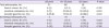
H. pylori tests were performed on 86 endoscopies in 73 patients. Of these, 30 (41.1%) patients had confirmed H. pylori infection. The prevalence of atrophic gastritis increased with age, from 25.8% in the patients younger than 39 years to 59.5% in the patients aged 40-60 years and to 66.1% in the patients older than 60 years (P = 0.001) (Table 4). The prevalence of intestinal metaplasia also increased with age, from 26.2% in the patients aged 40-60 years to 33.9% in the patients older than 60 years (P = 0.001) (Table 4).
Table 4
Prevalence of gastric cancer and its precursor lesions according to age
![]()
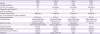
The clinical characteristics and endoscopic findings of the patients with good adherence to cART, according to the duration of cART, are summarized in Table 5. Opportunistic infections were common in the early cART period, and their prevalence gradually decreased thereafter. The prevalence of atrophic gastritis significantly increased with longer durations of cART (P <0.001). Although that of intestinal metaplasia tended to increase over time, it was not statistically significant.
Table 5
Clinical characteristics and endoscopic findings according to combination antiretroviral therapy duration in the patients with good adherence
M, month; Y, year; IQR, interquartile range; HIV, human immunodeficiency virus; cART, combination antiretroviral therapy; CMV, cytomegalovirus.
![]()
Discussion
The life expectancy of patients with HIV infection has dramatically increased since more potent cART has become available. As the lifespan of these patients is extended, an increasing number are at a risk of developing non-AIDS-defining cancers that typically occur at older ages [161718]. Reports on the epidemiology of gastric cancer in patients with HIV infection are very limited. There are also significant differences in the incidence of gastric cancer by region. Herein, we analyzed the endoscopic findings in patients with HIV infection and investigated the prevalence and incidence of gastric cancer on screening endoscopy in Korea, which has a high incidence of gastric cancer. Our study is the first report on the endoscopic findings and their role as gastric cancer screening in Korean patients with HIV infection.
In a recent study conducted in Japan, a country with a high incidence of gastric cancer, 11 gastric cancers developed in 1,001 patients with HIV infection during a median follow-up of 9 years. This incidence was 1.8 times higher than that in an age- and sex-matched general population [9]. Although cancer screening is currently considered an important component of health maintenance in patients with HIV infection, it is still unclear whether screening specifically for gastric cancer is also helpful in these patients, even in a country with a high incidence of gastric cancer. In Korea, gastric cancer was the third leading cause of cancer death in 2015, with an age-standardized incidence of 33.8 per 100,000 PYs (male sex; 49.3 per 100,000 PYs) [19]. Population-based screening for gastric cancer has been in place since 1999 in Korea. Upper GI series or endoscopy is recommended every 2 years for individuals aged 40-75 years [20]. In a study conducted at 40 hospitals in Korea, the prevalence of gastric cancer was 0.25% among 25,536 individuals who underwent gastric cancer screening [21]. In our study, 3 patients were diagnosed with gastric cancer through screening endoscopy; the prevalence of gastric cancer was 3.7%, it seemed that the prevalence of gastric cancer was somewhat higher than that of the general population. All 3 patients were diagnosed with EGC, which was cured with ESD or surgery. This suggests that gastric cancer screening might be helpful in detecting gastric cancer at an early curable stage in patients with HIV infection, at least in a country with a high incidence of gastric cancer.
We also found that as the duration of cART increased, the prevalence of atrophic gastritis increased over time, from 44.8% in the patients who received cART for ≤6 months to 70.9% in those who received cART for >5 years. The prevalence of intestinal metaplasia also showed an increasing trend with increasing cART duration. Atrophic gastritis and intestinal metaplasia are considered precursor lesions of gastric cancer [22232425]. Gastric cancer was found to develop 10.9 times more frequently in the presence of intestinal metaplasia in Korea [26], and several studies have reported atrophic gastritis as a risk factor for gastric tumorigenesis in Korea [2223]. On screening endoscopy of the general population in Korea, the prevalence of atrophic gastritis was reported to be 27.1% and 40.7%, and that of intestinal metaplasia was 7.1% and 12.5% in 2 separate studies [2728]. In 25,536 subjects who underwent health check-ups in Korea, the prevalence of atrophic gastritis increased with age, from 14.9% in the subjects aged <40 years to 28.9% in those aged 40-59 years and to 43.5% in those aged >60 years. The prevalence of intestinal metaplasia also increased with age, from 7.9% in those aged <60 years to 12.3% in those aged >60 years [27]. In our study, the prevalence of atrophic gastritis and intestinal metaplasia showed a similar tendency, increasing from 66.0% and 23.6% in those aged 40-59 years to 73.3% and 43.3% in those aged >60 years, respectively. However, the overall prevalence of atrophic gastritis and intestinal metaplasia was somewhat higher than that of the general population. These results support the idea that gastric cancer screening might be helpful for patients with HIV infection in Korea.
H. pylori infection is an important risk factor for gastric cancer, and its prevalence is very high in Korea [29]. Some studies reported the prevalence of H. pylori infection in patients with HIV infection and GI symptoms [3031]. However, there is no report of its prevalence in patients with HIV infection in Korea. In this study, only 1 patient with gastric cancer had H. pylori infection. The false negative results of the rapid urease test are related to the presence of severe atrophic gastritis and proton pump inhibitor or antibiotic use [32]. Further evaluation of H. pylori infection in patients with HIV infection is needed in areas with a high prevalence of gastric cancer and H. pylori infection.
In this study, the patients who received cancer screening endoscopy were older and more virologically and immunologically stable. In Korea, upper GI endoscopy is provided free of charge by the government for public health promotion. There may have been some patients who received the national cancer screening even though GI symptoms were present. However, regardless of GI symptoms, the prevalence of gastric ulcer and reflux esophagitis was higher than that in subjects who underwent health check-ups (gastric ulcer: 3.3% and reflux esophagitis: 7.9%) [21]. Therefore, even if symptoms are not present, screening endoscopy is important for the diagnosis of GI diseases in patients with HIV infection in the cART era.
This study has some limitations. First, this study was a single-center cohort study, and we could not enroll all patients with HIV infection who underwent endoscopy. Some patients underwent endoscopy in other centers, which is difficult to identify. In addition, many patients do not receive endoscopy because of confidentiality concerns regarding their disease owing to associated stigma. There is a possibility that the prevalence rate is underestimated. On the other hand, the prevalence rate can be overestimated because people with HIV infection received regularly check-up by doctors, so some patients may have had easier access to endoscopy and may have been diagnosed early with gastric cancer. Further studies on cancer screening rates of patients with HIV infection are needed. Second, study subjects were relatively small, we did not perform age- and sex-matched analysis in the assessment of gastric cancer incidence. So, the incidence of gastric cancer in patients with HIV infection was not comparable with that of the general population. Third, routine biopsy and H. pylori tests were not performed on all patients. The rapid urease test was performed only for indications allowed by insurance standards, and we did not include the urea breath test or serum H. pylori antibody test. This limited our ability to evaluate H. pylori infection. Fourth, the longer the cART duration, the older the patients; however, we could not adjust for age in comparing the prevalence of GI diseases according to the duration of cART. Finally, we have some limitations due to retrospective study. We were unable to assess the extent of atrophic gastritis. All cases of intestinal metaplasia and atrophic gastritis were not confirmed on biopsy. Thus, their prevalence may be overestimated or underestimated. Further, we were unable to investigate other risk factors of gastric cancer, such as alcohol use, smoking, and family history of gastric cancer.
In conclusion, we found the prevalence of gastric cancer in screening endoscopies was 3.7%. All gastric cancers confirmed on cancer screening endoscopy were in the early stage. Our study revealed that precursor lesions of gastric cancer, including intestinal metaplasia and atrophic gastritis, as well as other GI diseases were common even in the patients with HIV infection without GI symptoms. However, many patients do not receive periodic endoscopy. Therefore, regular gastric cancer screening might be useful in the early detection of gastric cancer and management of GI diseases in patients with HIV infection even without symptoms.
 XML Download