

 PDF
PDF ePub
ePub Citation
 Print
Print


INTRODUCTION
Positional downbeat nystagmus (pDBN) is a type of central positional nystagmus resulting from disruption of the brainstem or cerebellar vestibular network.12345 pDBN of central origin is often associated with brainstem or cerebellar signs such as gait unsteadiness with falls, motor weakness, clumsiness of extremities, diplopia, and slurred speech.1 However, diagnosing isolated pDBN in the absence of other neurological signs remains challenging.56 It can be ascribed to benign paroxysmal positional vertigo (BPPV) involving an anterior canal (AC).5678910 In AC BPPV, the otoconial debris moves away from the cupula during straight head-hanging (SHH) or Dix-Hallpike (DH) maneuver, giving rise to an ampullofugal deflection of the cupula and excitation of the AC. This situation produces pDBN with a torsional nystagmus in which the top poles of the eyes beat toward the involved ear. Since the AC is closer to the sagittal plane than the posterior canal (PC), the torsional component may be much smaller.
There have been recent reports of a variant of PC BPPV that presents with torsional downbeat nystagmus during SHH and DH maneuver.11121314 If the debris is located in the nonampullary arm of the PC near the common crus, it can induce an ampullopetal deflection of the cupula during positional tests, leading to pDBN via inhibition of the PC. Some patients can be diagnosed with a typical form of PC BPPV before or after the development of pDBN.15 Transient pDBN can also be observed after PC BPPV is treated.16 However, the overall incidence and clinical importance of pDBN in PC BPPV have not been systematically explored previously.
The aim of this study was to determine the incidence and characteristics of pDBN after applying a canalith repositioning procedure (CRP) for PC BPPV. The determined characteristics of pDBN included its latency, duration, maximum slow-phase velocity (SPV), and natural course.
METHODS
Subjects
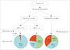
We consecutively recruited 77 patients with a diagnosis of PC BPPV in Pusan National University Yangsan Hospital from January to December 2016. PC BPPV was diagnosed according to the following criteria: 1) a history of vertigo provoked by changes in head position, 2) torsional upbeat nystagmus (with the upper pole of the eyes beating toward the affected ear) in positional tests (SHH and DH maneuver), and 3) vertigo associated with the elicited nystagmus. We excluded patients with multiple canal involvement or atypical positional nystagmus.
Study design
We treated all patients with the Epley maneuver according to the treatment guideline.7 The treatment response was assessed 1 hour after performing a single maneuver, and responders were defined as resolution of positional vertigo and positional torsional upbeat nystagmus (pT-UBN). We evaluated the presence of pDBN in the responder group in follow-up tests performed 1 hour later. Using three-dimensional video-oculography (SLMED, Seoul, Korea), we analyzed the latency, maximum SPV, duration, and presence of a torsional component of pDBN. The nonresponders who failed on initial maneuver, were instructed to perform the Brandt-Daroff exercise as described previously.7
All patients were scheduled to return 1 week after the first visit to receive follow-up positional tests. We determined whether pDBN had disappeared in the responders with pDBN. We reapplied the Epley maneuver if the responders showed pT-UBN. In the nonresponders, we assessed the resolution of pT-UBN or the new development of pDBN. The patients with persistent pDBN were evaluated every month until the pDBN disappeared.
This study was approved by the Institutional Review Board of Pusan National University Yangsan Hospital (IRB No. 05-2018-016). All of the experiments followed the tenets of the Declaration of Helsinki, and informed consents were obtained after the nature and possible consequences of this study had been explained to the participants.
RESULTS
Of 77 patients with unilateral PC BPPV, 50 were female, resulting in a female-to-male ratio of 1.9:1. They were aged 65.9±11.7 years (mean±SD), with an age range from 38 to 90 years. Five patients exhibited pDBN at the initial visit, but developed a typical form of PC BPPV with pT-UBN within 1 week. These patients were also included after they received the Epley maneuver. At initial visit, three had pDBN with a torsional component: counterclockwise (CCW) from the patient's perspective in two who developed right-side PC BPPV at the next visit, and clockwise (CW) in one with left-side PC BPPV.
The Epley maneuver produced an immediate response (within 1 hour) in 57 (74%) of the 77 patients, with resolution of positional vertigo and pT-UBN (Fig. 1). Four of them exhibited the transition of PC BPPV into geotropic horizontal-canal BPPV, which was treated by the Gufoni maneuver. The remaining 20 did not respond to the initial maneuver, and so they were instructed to perform the Brandt-Daroff exercise.
Oculographic analysis of pDBN
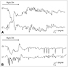
Twenty-two (39%) of the 57 patients in the responder group showed pDBN during the follow-up tests performed 1 hour later (Figs. 1 and 2). A torsional component was observed in six patients (27%) (Fig. 2): CCW in four with a prior right-side PC BPPV and CW in two with a prior left-side PC BPPV.
The evoked positions were SHH and bilateral DH maneuver in six patients, SHH and unilateral DH maneuver in nine (nine ipsilateral and two contralateral), unilateral or bilateral DH maneuver in six (three ipsilateral, two contralateral, and one bilateral), and SHH in one.
The latency and duration of pDBN were 3.2±2.0 and 12.0±10.0 s, respectively. The maximum SPV of pDBN was 5.1±2.5 degrees, and ranged from 2.0 to 12.2 degrees. The nystagmus did not reverse during the sitting position from SHH and DH maneuver in any patients.
Despite having pDBN, none of the patients experienced intense positional vertigo.
Follow-up results
At the 1-week follow-up in the responder group (Fig. 1), the pT-UBN was again observed in five patients with pDBN, but in only one without pDBN. The resolution rate of pT-UBN was higher in the responders without pDBN (30/31, 97%) than in those with pDBN (17/22, 77%). pDBN disappeared in six patients, but the remaining 11 still had pDBN.
The follow-up evaluation was completed in seven of the 11 patients who still had pDBN at the 1-week follow-up. pDBN disappeared within 6 months in five patients (three after 1 month, one after 5 months, and one after 6 months), and the remaining two patients experienced relapse of PC BPPV on the opposite side during follow-up. One patient with pDBN resolution at the 1-week follow-up experienced relapse in the same canal at 2 months.
The 1-week follow-up was completed by 16 of the 20 nonresponders (Fig. 1). Six still had pT-UBN, but the remaining ten showed resolution of positional vertigo and pT-UBN. Six of them did not have nystagmus during positional tests, while four had pDBN without concomitant positional vertigo. The evoked positions were SHH and bilateral DH maneuver in two, SHH and contralateral DH maneuver in one, and ipsilateral DH maneuver in one. Three patients had no torsional component, but one with prior right-side PC BPPV had CCW torsional nystagmus with pDBN. pDBN disappeared in all of the patients within 2 months.
DISCUSSION
Our study shows that transient pDBN can be observed in 40% of patients with PC BPPV after the immediate resolution of positional vertigo and pT-UBN. Although the pDBN was not sufficiently intense to induce positional vertigo, it could last for 6 months. The patients with pDBN were much more likely to develop a typical form of PC BPPV again at the 1-week follow-up.
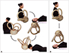
pDBN of peripheral origin has traditionally been linked to BPPV involving the AC.5678910 Therefore, the pDBN in our patients may have been due to translocation of otoconia into the AC from the PC during the Epley maneuver. However, this hypothesis is not supported by the presence of a torsional component of pDBN in some of the patients. If the Epley maneuver resulted in otoconia moving into the ipsilateral AC from the PC, the torsional component would be directed to the upper pole of the eye to the left ear (CCW) for the left AC or to the right ear (CW) for the right AC. In our patients, however, the torsional component of pDBN was directed CCW and CW for prior right- and left-side PC BPPV, respectively, which was not in accordance with the direction evoked by excitation of the ipsilateral AC. Furthermore, some patients who initially showed CCW or CW torsional pDBN consequently developed right- or left-side PC BPPV, respectively. Therefore, pDBN in our patients may have originated from the initially involved PC. The Epley maneuver results in the otoconia within the PC dropping out into the utricle, leading to resolution of pT-UBN. However, some otoconial debris can remain in the nonampullary arm of the PC near the common crus (Fig. 3A). In such cases, when the patient lies down into head-hanging position, the residual debris in the PC could move toward the ampulla producing an ampullopetal flow of endolymph (Fig. 3B).11121314 This would generate an inhibitory discharge of the posterior ampullary nerve and evoke pDBN. The absence of a torsional component in most cases of pDBN may be due to the anatomy of the semicircular canals. Since the nonampullary arm of the PC is closer to the sagittal plane, the vertical component of the nystagmus is greater than the torsional one.17 Alternatively, any debris present in both the AC and PC on one side after the Epley maneuver could move ampullopetally in the AC and ampullofugally in the PC during positional tests, resulting in the absence of torsional components.15
pDBN originating from the PC has already been reported previously. Vannucchi et al.11 described patients who initially presented with vertical torsional downbeat nystagmus suggestive of AC BPPV.12 However, at the next follow-up these patients exhibited a typical form of PC BPPV on the side opposite to the initially diagnosed AC, which is similar to five patients in our study. Those authors hypothesized that the debris could be localized to the distal portion of the PC, leading to pDBN similar to that for the contralateral AC, and named this variant apogeotropic PC BPPV. Cambi et al.15 assessed the natural course of pDBN of presumed peripheral origin. They found that 40% of patients were diagnosed with a typical form of PC BPPV before or after a diagnosis of pDBN, and proposed that pDBN and PC BPPV are related. Our study provides further evidence that pDBN is related to an atypical form of PC BPPV.
Despite having pDBN, none of the patients experienced intense positional vertigo. This is inconsistent with previous reports of patients with pDBN due to apogeotropic PC BPPV experiencing even worse symptoms.1112 However, previous cases showed positional vertigo with pDBN on their first visit before developing a typical form of PC BPPV, whereas our patients had pDBN after the immediate resolution of typical PC BPPV. Our patients might have already adapted to the symptoms due to them experiencing repeated positional vertigo. Furthermore, the amplitude of pDBN was quite small and the nystagmus did not last long. These factors may have contributed to the absence of intense positional vertigo in our patients. Some felt a mild and continuous dizziness, but these symptoms can remain in any BPPV patients following a successful CRP.18
In our study, 23% of the patients with pDBN had developed a typical form of PC BPPV again at a 1-week follow-up. This suggests that the presence of pDBN after a CRP may be a sign of incomplete or partial resolution of PC BPPV. Considering that tiny debris in the distal portion of the PC might induce pDBN, the accumulated debris can move toward the ampullary end of the PC and produce an ampullofugal flow of endolymph. To prevent tiny debris from accumulating within the canal, it may be better to recommend regular physical activity to patients showing pDBN. Alternatively, repeated application of the Epley maneuver may be helpful to remove any residual debris in the canal.
Our study was subject to several limitations, such as the sample being rather small and some patients not completing follow-up evaluations. These features may have reduced the statistical power of the analysis and contributed to an inability to obtain significant findings.
In conclusion, transient pDBN can be observed in patients with PC BPPV after the immediate resolution of positional vertigo and pT-UBN. This can also be present before a diagnosis of typical PC BPPV. pDBN may be ascribed to otoconial debris in the distal portion of the PC, which can move toward the ampulla producing an ampullopetal flow of endolymph during positioning. Although pDBN disappears spontaneously in most cases, it may indicate the incomplete or partial resolution of PC BPPV.
 XML Download