Journal List > J Clin Neurol > v.15(2) > 1119553

Kim, Illigens, McCormick, Wang, and Gibbons: Alpha-Synuclein in Skin Nerve Fibers as a Biomarker for Alpha-Synucleinopathies

Abstract

The common pathological features of synucleinopathies are abnormal aggregates of the synaptic protein alpha-synuclein (αSN) in the cytoplasm of neurons or glia. These abnormal aggregates appear several years before the onset of clinical manifestations, and so the early detection of αSN in body fluids or peripheral tissues (e.g., cerebrospinal fluid, colonic mucosa, salivary glands, and skin) is considered a potential tool for identifying synucleinopathies. Performing a skin biopsy is a practical option because it is a relatively noninvasive, safe, and reliable method to measure αSN deposition in the peripheral nervous system. Moreover, there is growing research interest in the use of cutaneous synuclein deposition as a biomarker for synucleinopathies. The aim of this study was to interpret the current data on cutaneous αSN deposition and present the current perspectives and future prospects.

INTRODUCTION

Alpha-synucleinopathies are a group of chronic, progressive neurodegenerative disorders that include idiopathic Parkinson's disease (IPD), dementia with Lewy bodies (DLB), multiple-system atrophy (MSA), and pure autonomic failure (PAF).1 Similar to other neurodegenerative diseases, the prevalence and incidence of synucleinopathies increase with age. The key pathological findings of α-synucleinopathies are fibrillary aggregates of the protein alpha-synuclein (αSN) in the cytoplasm of neurons, neuritis, and glia, which have usually been found in neurons in IPD, DLB, and PAF, but in glia in MSA, thereby interfering with the axonal transport of synaptic proteins and promoting mitochondrial deficit and oxidative stress.2 While synucleinopathies share the same pathological protein, they exhibit diverse clinical features.
Synucleinopathies are primarily diagnosed based on their respective clinical diagnostic criteria. The overlapping presentations and transitional phenotypes make an accurate diagnosis challenging. Some studies assessing the clinical diagnosis in early stages of the development of synucleinopathy found that the diagnosis accuracy was far lower than expected.345678 One clinicopathological study identified that the accuracy was 53% for a clinical diagnosis of PD in subjects with early PD responsive to medication (disease duration <5 years), and only 26% for PD in untreated or not-clearly-responsive subjects.4 In another clinicopathological study of 100 cases, 76 subjects had neuropathologically confirmed PD, but many cases had a neuropathological diagnosis of progressive supranuclear palsy, MSA, or Alzheimer's disease despite the clinical diagnosis of PD.5 The sensitivity of clinical diagnoses of MSA and DLB is very low, and more than half of patients with MSA or DLB remain either undiagnosed or misdiagnosed.678 The clinical diagnosis of synucleinopathies alone, especially during the early stages, is not particularly accurate, which indicates the need to identify a reliable biomarker.
Early detection in the prodromal stages is important for a prompt and effective intervention to treat and eventually slow the disease progression (as disease-modifying therapies are identified), ultimately leading to improving the patient's quality of life. Accordingly, the need for a more-accurate diagnostic tool for the early or prodromal stages is expanding. There is increasing evidence that the pathological process starts years before the onset of clinical manifestations. In PD, it is known that motor signs first appear when >50% of substantia nigra dopamine neurons are lost.9
αSN deposition can be detected in the early stage of disease in the nerve fibers in colonic submucosa and in neurons of the submucosal Meissner's plexus.1101112 In addition, there is accumulating evidence of multiple organs being involved in synucleinopathies, since αSN pathology is detected in various peripheral tissues including the skin, salivary glands, sympathetic ganglia, vagus nerve, gastrointestinal tract, genitourinary tract, and heart.131415161718 Among the peripheral tissues that have been studied, the skin is one of the most-easily accessible organs, and is suitable for both single and repeated sampling. Although it is well established that αSN deposition can be seen in samples obtained from the gastrointestinal tract in PD, their acquisition procedure involves an invasive colonoscopy or endoscopy, requiring sedation and prior preparation. In addition, skin biopsies are already used for several indications, such as for determining the intraepidermal nerve fiber densities to detect and monitor small-nerve-fiber neuropathy.19 Moreover, performing a skin biopsy is safe and minimally invasive. Thus, the identification and quantification of αSN deposition in cutaneous biopsy samples could become a valuable tool for α-synucleinopathies, as already suggested by several investigators.202122232425262728293031
However, previous studies of cutaneous αSN deposition have produced inconsistent results. Systematic reviews found wide ranges for the sensitivity and specificity in detecting αSN pathology using skin biopsies, of 0–100% and 80–100%, respectively. 1732 These marked differences in study results could be due to several factors such as the study design and other methodological differences. If the quantification of cutaneous αSN deposition using skin biopsy provides a reliable measure across studies, cutaneous αSN deposition could be a useful biomarker for α-synucleinopathies.
The purpose of this study was to evaluate and interpret the current data on cutaneous αSN deposition, to clarify its role as a potential biomarker in synucleinopathies, and to determine problems to be addressed before establishing αSN deposition as a biomarker and surrogate outcome.

THE ROLE AND DISTRIBUTION OF αSN

αSN is one of the key molecules in the pathogenesis of synucleinopathies. It is one of the normal proteins in soluble cytosolic brain fractions and is mainly localized at presynaptic terminals.33 Its detailed physiological functions are still unclear, but recent studies suggest that it plays a key role in synaptic functions in cooperation with cysteine-string protein-α, which contains a typical domain for HSP40-type molecular cochaperones.34 Physiologically, in its normal (or native) form, αSN modulates the stability of the neuronal membrane and influences presynaptic signaling and membrane trafficking via vesicular transport.32 Environmental influences such as neurotoxins, low pH, and high temperature, as well as genetic mutations may result in misfolding of αSN that leads to its polymerizing into fibrils and accumulating throughout the nervous system, as represented by pathological hallmark inclusions such as Lewy bodies, Lewy neurites, and glial cytoplasmic inclusions. 3235 There are increasing reports of αSN within Lewy bodies undergoing several posttranslational modifications, including phosphorylation, ubiquitination, cross-linking, truncations, and nitration, suggesting that these modifications play a key role in the regulation of αSN aggregation and toxicity in vivo.3637 In particular, the aberrant accumulation of phosphorylated αSN (p-αSN) at the residue serine 129 has been reported to be prominent in PD and enhance αSN toxicity both in vivo and in vitro, possibly by increasing the formation of αSN aggregates.37 Recent studies have suggested that phosphorylation at serine 129 inhibits the interaction of αSN with membrane phospholipids or phospholipase D2 and promotes the formation of αSN filaments as well as oligomers, eventually causing neuronal death along with other alterations in the activities of various kinases and phosphatases.37
Early postmortem studies to detect αSN deposition used anti-αSN antibodies against amino-terminal and carboxylterminal sequences of the 140-amino-acid αSN protein by immunohistochemically staining brain tissues from PD or DLB patients.38 Immunohistochemical studies measuring total αSN (both p-αSN and nonphosphorylated αSN) use a polyclonal antibody recognizing multiple binding sites from amino acids 111–131 of αSN, whereas those measuring p-αSN use a monoclonal antibody against p-αSN at serine 129. Some previous studies showed that total αSN is detectable in healthy individuals as well as patients with synucleinopathies,232627 which challenges the specificity of total αSN as a reliable biomarker for α-synucleinopathies. On the other hand, p-αSN is not usually found in healthy controls, and some authors have suggested that p-αSN deposits would be a more-reliable biomarker than αSN for diagnosing PD and other synucleinopathies. 222936 However, the exact role of p-αSN aggregation and toxicity in vivo remains controversial.

PREVIOUS STUDIES OF αSN DEPOSITION IN CUTANEOUS NERVE FIBERS

The number of studies evaluating αSN deposition within cutaneous nerve fibers has increased in recent years. Most of these studies evaluating synuclein deposition in the dermal nerve fibers used a case–control design with clinically diagnosed cases, while only a few studies used tissue from cases verified by autopsy.13142021222324252627282930313940 Table 1 summarizes the studies that used autopsy samples, while Table 2 summarizes the studies that used in vivo samples from clinically diagnosed subjects.

Studies of autopsy cases

The methods used in the four postmortem studies were comparable in regard to tissue preparation, with the autopsy samples being fixed in formalin or formaldehyde solution (in 3.75% or 10% solution for more than 24 hours), embedded in paraffin, sectioned (at 5–6 µm), and stained.13143940 One major difference was the antibodies used: two studies selected antibodies against p-αSN, while the other two studies used antibodies against total αSN. Another significant methodological difference was the method of image analysis, with two studies evaluating αSN deposition using light microscopy1439 and the others using fluorescence microscopy.1340 The two postmortem studies that used light microscopy identified multiorgan involvement of αSN pathology, but no cutaneous αSN deposition in either the patient or control groups.1439 The two postmortem studies using fluorescence microscopy found αSN deposition in the dermal fibers of patients, each using a different antibody; the study of Ikemura et al.13 showed a sensitivity of p-αSN immunoreactivity of 70% in PD and PD with dementia and 40% in Lewy-body disease, and a specificity of 100%. This suggests that Lewy-body-related pathology also involves cutaneous nerves in Lewy-body disease, and that skin biopsies may be of value in PD or Lewy-body disease presenting with advanced autonomic failure. The more-recent study of Gibbons et al.40 assessed total αSN with immunostaining and confocal microscopy in autopsy samples obtained from abdominal skin and the scalp of 11 individuals with PD, and compared this to 5 controls with nonsynucleinopathy. That study found that the amount of αSN deposited in cutaneous autonomic nerves was significantly greater in subjects with a postmortem confirmation of a diagnosis of PD than in controls (sensitivity of 100%).40 Comparing the results across studies according to the antibodies used reveals that those using p-αSN antibodies showed higher specificity than those using antibodies against total or nonphosphorylated αSN by providing an ordinal yes/no result. In contrast, total αSN provided a deposition gradient that was correlated with the disease severity but was also present in control subjects. The other main reason underlying the different results appears to be the imaging method used: fluorescence microscopy may provide αSN data that are superior to those attainable using light microscopy because it can visualize specific features of small samples such as skin sections and also enhance three-dimensional characteristics on small scales by attaching fluorescent tags to antibodies attached to targeted structures and using a high-intensity light source.

Studies of clinically diagnosed patients

Twelve studies have included biopsies in patients with a clinically confirmed diagnosis of synucleinopathy, and they had various designs.202122232425262728293031 Most of the studies focused on PD patients only. Nine case–control studies compared the differences among PD patients, controls, and/or patients with other synucleinopathies or neurodegenerative diseases assumed not to have αSN deposits.202122232425262728 The early study of Miki et al.,20 which was a case series of 20 PD patients, found abnormal αSN accumulation in paraffin-embedded chest skin samples in 2 (10%) of the patients, suggesting that αSN in a skin biopsy sample assessed with conventional immunohistochemistry is not a sensitive diagnostic marker. However, more-recent studies support the role of cutaneous αSN deposition as a potential biomarker for disease. The different results may be attributable to technical and methodological differences in the fixation method, the use of different antibodies (polyclonal versus monoclonal), and the analysis of cryosections with different thicknesses [thin sections (5–6 µm) versus thick sections (50–60 µm)]. These methodological differences are addressed in detail later in this review.
The sensitivity of αSN deposition in skin biopsy samples may vary depending on the underlying disease. Studies of PD patients have shown high specificities but a wide range of sensitivities: two studies found αSN deposits in only 5.2–10% of PD patients using skin samples of the chest wall and distal leg,2025 one study found αSN deposition in 52% of patients (16/31) using skin samples of the finger, proximal leg, distal leg, and back,21 and one study found that 100% of PD patients had αSN deposits in biopsy samples of the volar forearm.24 Looking at a possible length-dependent distribution, a few studies showed a proximal-to-distal gradient of cutaneous αSN deposition in PD patients: 100% of the patients had αSN depositions at the cervical site, 52–75% at the thigh, and 24–31% at the leg.2229 Comparing with healthy controls and patients with essential tremor, tauopathy, or parkinsonisms assumed not to have αSN deposits, the specificity of αSN was 90% when using antibodies against total αSN and 100% when using antibodies against p-αSN.212223242728 When looking at the class of nerve fibers affected, studies involving PD, DLB, and PAF patients found αSN accumulations in autonomic fibers of the skin,202122232425262728293031 while in MSA patients they were observed mainly in unmyelinated somatosensory fibers of the subepidermal plexus but not in dermal autonomic fibers.232425 These findings suggest that the involvement of postganglionic autonomic nerve fibers contributes to autonomic symptoms in PD, DLB, and PAF, which contrasts with cutaneous autonomic fibers appearing to be preserved in MSA. Two comparative studies found that 100% of PAF patients presented with αSN depositions in autonomic nerve fibers.2930 The detection of cutaneous αSN deposition could therefore be a sensitive tool for PAF. Moreover, one of these studies found p-αSN deposits with an even distribution pattern in PAF, whereas all of the PD and DLB patients had a proximal-to-distal pattern.3031 This suggests that there are variations in the pathomechanisms underlying synucleinopathies.222931
The various results obtained in the previous studies should be interpreted with caution, since they had quite heterogeneous designs and methodologies, as well as numerous limitations: the sample was small in many studies, most studies had a case–control design, and technical aspects of the study methodologies differed according to the biopsy site, antibodies used, the type of microscopy, tissue thickness, tissue preparation and fixation methods, and how positive results were defined. The recruited patients were diagnosed based mainly on clinical criteria, which limits the comparability of cases across studies from different referral centers. In addition, a study with a case–control design might be affected by selection bias.

Differences between αSN and p-αSN according to clinical measures

Several studies have evaluated the correlations of cutaneous synuclein deposition with disease severity and autonomic dysfunction. One study yielded correlations between the αSN ratio and the score on the Hoehn and Yahr scale or measures of autonomic function tests, which suggested that greater αSN deposition was associated with greater autonomic dysfunction and more-advanced stages of PD.26 The same group recently described that αSN deposition is present also in early stages of the disease and when PD has been diagnosed recently (within 0.5–3 years), even among PD patients without autonomic failure, which indicates that cutaneous synuclein deposition may be present also during the premotor stages.27 Those authors also found that αSN ratios were higher in individuals with autonomic failure and were correlated with the severity of PD, suggesting that the αSN ratio can provide a sensitive and specific biomarker for PD patients.27 Other studies have investigated if αSN deposition is correlated with disease severity and autonomic dysfunction, quantifying p-αSN deposition as the percentage of autonomic structures or nerve bundles showing positive staining, or a more-sophisticated index of total p-αSN staining.222429 However, those studies did not demonstrate a correlation between deposits of p-αSN and clinical or nuclear imaging measures including the disease duration, motor involvement, autonomic dysfunction graded on the Composite Autonomic Scoring Scale, or the heart-to-mediastinum denervation ratio obtained from nuclear imaging.222429 Therefore, while a correlation of p-αSN with a clinical measure remains to be identified, the total αSN ratio may be appropriate for evaluating the disease severity or autonomic dysfunction in PD patients based on cutaneous synuclein depositions. Although questions remain, cutaneous synuclein deposition might reflect the disease stage or degree of autonomic failure in synucleinopathies. Future studies are needed to evaluate the associations of disease severity and autonomic dysfunction with cutaneous αSN deposition.

METHODOLOGICAL DIFFERENCES IN DETECTING CUTANEOUS SYNUCLEIN DEPOSITION

Another important aspect to consider when comparing the results from the above-mentioned studies is methodological differences in how cutaneous αSN deposits are detected. Those differences are primarily the locations where biopsy samples are obtained from, the thickness of tissue sections, fixation techniques, and staining protocols (i.e., the selection of the detection antibody, secondary antibodies, and amplification systems), in addition to differences in study designs and the characteristics of the included subjects. We discuss these methodological and technical differences in more detail below.

Differences in tissue fixation

Autopsy samples are routinely fixed in formalin or formaldehyde solution for more than 24 hours and then embedded in paraffin, whereas in vivo samples are typically fixed in a 2% paraformaldehyde or Zamboni's solution (paraformaldehyde mixed with picric acid and NaOH) for 18–24 hours for the optimal detection of peripheral nerve fibers. Overfixation with formalin can impair the ability to quantify the peripheral nerve density and also to detect αSN.40

Differences in tissue section thicknesses

There are also differences in the methods used to section fixed skin biopsy tissue samples. Autopsy samples are generally cut into 5- to 6-µm-thick tissue sections from paraffin blocks, while in vivo samples are cut into 50- to 60-µm-thick sections. αSN is deposited within nerves in the dermal layer of the skin (i.e., in vasomotor nerve fibers of the blood vessels, sudomotor nerve fibers of the sweat glands, and pilomotor nerve fibers of the pilomotor muscles), and is therefore spread out across this structure with dimensions of approximately 2 mm. Since a section that is 5–6 µm thick has only one-tenth the sampling region of a section that is 50–60 µm thick, it is far less likely to contain dermal nerve fibers in which potential αSN depositions could be detected.40
The thickness of tissue sections also has a profound effect on the detection rate of studies involving in vivo samples. The overall sensitivity of immunostaining has been higher in studies using 50-µm-thick sections than in those involving sections that are 10–20 µm thick. Important data for clarifying this problem has come from the recent study of Wang et al.,41 who compared the detection of p-αSN in cutaneous autonomic nerve fibers across in vivo samples with section thicknesses of 10, 20, and 50 µm. This study, which has only been reported on abstract form, found that immunostaining with 50-µm-thick tissue sections was superior to using sections that were 20 or 10 µm thick for detecting of p-αSN in PD.41 This difference is probably due to the quantity of intact nerve fibers in each section.

Polyclonal versus monoclonal antibodies

As mentioned above, immunohistochemical studies measuring total αSN use polyclonal antibodies, whereas those measuring p-αSN use monoclonal antibodies. The selection of antibodies significantly affects the results obtained: 4 of 12 studies involving clinically diagnosed subjects used antibodies against total αSN, with the remaining 8 using antibodies against p-αSN (Table 2). Although there have been no direct comparisons, performing immunohistochemical staining with antibodies against total αSN may provide higher sensitivity compared to using anti-p-αSN antibodies. Previous studies observed p-αSN deposits intermittently within cutaneous nerve fibers, and detected them inside small dermal nerve bundles or in autonomic small fibers innervating blood vessels, usually in the deeper layers of the dermis.2122 These p-αSN deposits could be missed if deeper layers are not analyzed and if the p-αSN deposits are not colocalized with a panaxonal marker such as protein gene product (PGP) 9.5. In contrast, total αSN deposits can be identified more easily than p-αSN deposits. However, αSN deposits are also present in healthy controls, albeit in smaller amounts, and therefore require quantification to distinguish between healthy and diseased states. Thus, p-αSN is more specific to synucleinopathy and the choice of detection antibody against p-αSN or total αSN can have a large effect on study outcomes.

Background artifacts

Nonspecific background noise and staining artifacts are inevitable in most immunohistochemical studies, and will depend on the choice of polyclonal or monoclonal antibodies, type of staining methods used, fixation technique, tissue thickness, and use of light or immunofluorescence microscopy. A recent unpublished study observed a low-intensity signal of staining artifacts and background noise in certain areas around autonomic structures in sections in which p-αSN-positive nerve fibers were not colocalized with PGP-9.5-positive fibers, irrespective of the tissue thickness.41 These artifacts could be misinterpreted as p-αSN-immunoreactive fibers. Therefore, applying a double-staining method to p-αSN with the panaxonal marker PGP 9.5 could be very helpful for decreasing the rates of false-positive and false-negative results.

CONCLUSION

This review of a combination of postmortem and in vivo studies allows us to draw the following conclusions: first, cutaneous αSN quantified in skin biopsy samples provides high specificity and good sensitivity for the detection of synucleinopathies. A high specificity is important for the diagnostic value of a biomarker, especially in the early stages of a disease when clinical uncertainties may make a diagnosis difficult. It is particularly interesting that the recent study demonstrated p-αSN deposition in skin nerves in 75% of patients with idiopathic REM-sleep behavior disorder, suggesting that a skin biopsy can be considered a safe and sensitive procedure for detecting αSN pathology in the prodromal stage of synucleionpathies.42
Second, a skin biopsy performed to detect αSN is a relatively easy, repeatable, and minimally invasive technique compared to obtaining samples from other organs.
Third, there are still several unanswered questions about the optimal methods, biopsy sites, antibodies, fixation methods, and tissue thickness to utilize when detecting αSN. Determining the diagnostic accuracy of cutaneous αSN pathology may require evaluations of the sensitivity and specificity of immunohistochemical staining in skin samples from neuropathologically proven subjects. There are practical difficulties associated with obtaining samples from autopsied patients. In addition, it is not adequate to identify the diagnostic accuracy of cutaneous αSN pathology for some clinically diagnosed patients. Moreover, the results of published studies are conflicting. It can be assumed that the diversity of methodologies is the main reason behind the conflicting results from published studies. It might be plausible to determine the optimal method by comparing the results obtained when applying different methods to the same samples.
Some authors recently tested the efficacies of different immunohistochemical methods in detecting αSN pathology in gastrointestinal specimens in identifying the technique with the highest sensitivity and specificity.4344 One of these studies examined four methods in archived colonic biopsy samples obtained from PD patients and controls,43 and the other assessed seven methods applied to sigmoid colon sections obtained from autopsied PD and control subjects.44 The stained sections were graded by blinded judging. The findings of these studies suggest that obtaining multiple and full-thickness samples and using well-trained raters to judge slides are other important factors for increasing the diagnostic accuracy.44 Most importantly, the utility of different immunohistochemical methods should be evaluated for the detection of cutaneous αSN pathology in identical samples, and the stained sections should be judged in a blind manner by well-trained raters in order to determine whether it could be a reliable biomarker for synucleinopathies. A standardized methodology is essential to increase the diagnostic value and widen the clinical application of skin biopsies in the detection of αSN. After a standardized protocol for detecting cutaneous αSN deposition has been established, its clinical and scientific value should be verified and compared with preexisting diagnostic tests or tools.
Fourth, the studies reported on to date have not revealed any distinctive features of the specific subtypes of synucleinopathies, except in the case of MSA, in which cutaneous αSN deposition was observed mainly in unmyelinated somatosensory fibers of subepidermal plexus, and not in dermal autonomic fibers, which contrasts with the findings for other subtypes. Further studies are also needed to address this issue.
There are numerous methodological challenges to staining and quantifying αSN in skin biopsy samples, and standardized methods still need to be developed and tested. However, the measurement of cutaneous αSN could serve as a useful biomarker for synucleinopathies if standardized protocols can be established.

Notes

Conflicts of Interest: The authors have no financial conflicts of interest.

References

1. Martí MJ, Tolosa E, Campdelacreu J. Clinical overview of the synucleinopathies. Mov Disord. 2003; 18(Suppl 6):S21–S27. PMID: 14502652.
2. Jellinger KA. Neuropathological spectrum of synucleinopathies. Mov Disord. 2003; 18(Suppl 6):S2–S12. PMID: 14502650.
crossref
3. Rizzo G, Copetti M, Arcuti S, Martino D, Fontana A, Logroscino G. Accuracy of clinical diagnosis of Parkinson disease: a systematic review and meta-analysis. Neurology. 2016; 86:566–576. PMID: 26764028.
4. Adler CH, Beach TG, Hentz JG, Shill HA, Caviness JN, Driver-Dunckley E, et al. Low clinical diagnostic accuracy of early vs advanced Parkinson disease: clinicopathologic study. Neurology. 2014; 83:406–412. PMID: 24975862.
crossref
5. Hughes AJ, Daniel SE, Kilford L, Lees AJ. Accuracy of clinical diagnosis of idiopathic Parkinson’s disease: a clinico-pathological study of 100 cases. J Neurol Neurosurg Psychiatry. 1992; 55:181–184. PMID: 1564476.
crossref
6. Litvan I, MacIntyre A, Goetz CG, Wenning GK, Jellinger K, Verny M, et al. Accuracy of the clinical diagnoses of Lewy body disease, Parkinson disease, and dementia with Lewy bodies: a clinicopathologic study. Arch Neurol. 1998; 55:969–978. PMID: 9678315.
7. Nelson PT, Jicha GA, Kryscio RJ, Abner EL, Schmitt FA, Cooper G, et al. Low sensitivity in clinical diagnoses of dementia with Lewy bodies. J Neurol. 2010; 257:359–366. PMID: 19795154.
crossref
8. Joutsa J, Gardberg M, Röyttä M, Kaasinen V. Diagnostic accuracy of parkinsonism syndromes by general neurologists. Parkinsonism Relat Disord. 2014; 20:840–844. PMID: 24816002.
crossref
9. Noyce AJ, Lees AJ, Schrag AE. The prediagnostic phase of Parkinson’s disease. J Neurol Neurosurg Psychiatry. 2016; 87:871–878. PMID: 26848171.
crossref
10. Braak H, de Vos RA, Bohl J, Del Tredici K. Gastric alpha-synuclein immunoreactive inclusions in Meissner’s and Auerbach’s plexuses in cases staged for Parkinson’s disease-related brain pathology. Neurosci Lett. 2006; 396:67–72. PMID: 16330147.
11. Shannon KM, Keshavarzian A, Mutlu E, Dodiya HB, Daian D, Jaglin JA, et al. Alpha-synuclein in colonic submucosa in early untreated Parkinson’s disease. Mov Disord. 2012; 27:709–715. PMID: 21766334.
crossref
12. Shannon KM, Keshavarzian A, Dodiya HB, Jakate S, Kordower JH. Is alpha-synuclein in the colon a biomarker for premotor Parkinson’s disease? Evidence from 3 cases. Mov Disord. 2012; 27:716–719. PMID: 22550057.
crossref
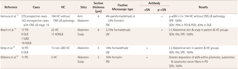
13. Ikemura M, Saito Y, Sengoku R, Sakiyama Y, Hatsuta H, Kanemaru K, et al. Lewy body pathology involves cutaneous nerves. J Neuropathol Exp Neurol. 2008; 67:945–953. PMID: 18800013.
crossref
14. Beach TG, Adler CH, Sue LI, Vedders L, Lue L, White Iii CL, et al. Multi-organ distribution of phosphorylated alpha-synuclein histopathology in subjects with Lewy body disorders. Acta Neuropathol. 2010; 119:689–702. PMID: 20306269.
15. Fumimura Y, Ikemura M, Saito Y, Sengoku R, Kanemaru K, Sawabe M, et al. Analysis of the adrenal gland is useful for evaluating pathology of the peripheral autonomic nervous system in Lewy body disease. J Neuropathol Exp Neurol. 2007; 66:354–362. PMID: 17483692.
crossref
16. Orimo S, Uchihara T, Nakamura A, Mori F, Kakita A, Wakabayashi K, et al. Axonal alpha-synuclein aggregates herald centripetal degeneration of cardiac sympathetic nerve in Parkinson’s disease. Brain. 2008; 131:642–650. PMID: 18079166.
17. Schneider SA, Boettner M, Alexoudi A, Zorenkov D, Deuschl G, Wedel T. Can we use peripheral tissue biopsies to diagnose Parkinson’s disease? A review of the literature. Eur J Neurol. 2016; 23:247–261. PMID: 26100920.
crossref
18. Pouclet H, Lebouvier T, Coron E, des Varannes SB, Rouaud T, Roy M, et al. A comparison between rectal and colonic biopsies to detect Lewy pathology in Parkinson’s disease. Neurobiol Dis. 2012; 45:305–309. PMID: 21878391.
crossref
19. Gibbons CH, Griffin JW, Polydefkis M, Bonyhay I, Brown A, Hauer PE, et al. The utility of skin biopsy for prediction of progression in suspected small fiber neuropathy. Neurology. 2006; 66:256–258. PMID: 16434668.
crossref
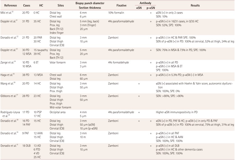
20. Miki Y, Tomiyama M, Ueno T, Haga R, Nishijima H, Suzuki C, et al. Clinical availability of skin biopsy in the diagnosis of Parkinson’s disease. Neurosci Lett. 2010; 469:357–359. PMID: 20026177.
crossref
21. Doppler K, Ebert S, Uçeyler N, Trenkwalder C, Ebentheuer J, Volkmann J, et al. Cutaneous neuropathy in Parkinson’s disease: a window into brain pathology. Acta Neuropathol. 2014; 128:99–109. PMID: 24788821.
crossref
22. Donadio V, Incensi A, Leta V, Giannoccaro MP, Scaglione C, Martinelli P, et al. Skin nerve α-synuclein deposits: a biomarker for idiopathic Parkinson disease. Neurology. 2014; 82:1362–1369. PMID: 24634456.
crossref
23. Doppler K, Weis J, Karl K, Ebert S, Ebentheuer J, Trenkwalder C, et al. Distinctive distribution of phospho-alpha-synuclein in dermal nerves in multiple system atrophy. Mov Disord. 2015; 30:1688–1692. PMID: 26175301.
crossref
24. Zange L, Noack C, Hahn K, Stenzel W, Lipp A. Phosphorylated α-synuclein in skin nerve fibres differentiates Parkinson’s disease from multiple system atrophy. Brain. 2015; 138:2310–2321. PMID: 26017579.
crossref
25. Haga R, Sugimoto K, Nishijima H, Miki Y, Suzuki C, Wakabayashi K, et al. Clinical utility of skin biopsy in differentiating between Parkinson’s disease and multiple system atrophy. Parkinsons Dis. 2015; 2015:167038. PMID: 25945280.
crossref
26. Wang N, Gibbons CH, Lafo J, Freeman R. α-Synuclein in cutaneous autonomic nerves. Neurology. 2013; 81:1604–1610. PMID: 24089386.
crossref
27. Gibbons CH, Garcia J, Wang N, Shih LC, Freeman R. The diagnostic discrimination of cutaneous α-synuclein deposition in Parkinson disease. Neurology. 2016; 87:505–512. PMID: 27385742.
crossref
28. Rodríguez-Leyva I, Chi-Ahumada EG, Carrizales J, Rodríguez-Violante M, Velázquez-Osuna S, Medina-Mier V, et al. Parkinson disease and progressive supranuclear palsy: protein expression in skin. Ann Clin Transl Neurol. 2016; 3:191–199. PMID: 27042679.
crossref
29. Donadio V, Incensi A, Piccinini C, Cortelli P, Giannoccaro MP, Baruzzi A, et al. Skin nerve misfolded α-synuclein in pure autonomic failure and Parkinson disease. Ann Neurol. 2016; 79:306–316. PMID: 26606657.
crossref
30. Donadio V, Incensi A, Cortelli P, Giannoccaro MP, Jaber MA, Baruzzi A, et al. Skin sympathetic fiber α-synuclein deposits: a potential biomarker for pure autonomic failure. Neurology. 2013; 80:725–732. PMID: 23390175.
crossref
31. Donadio V, Incensi A, Rizzo G, Capellari S, Pantieri R, Stanzani Maserati M, et al. A new potential biomarker for dementia with Lewy bodies: skin nerve α-synuclein deposits. Neurology. 2017; 89:318–326. PMID: 28667178.
32. Malek N, Swallow D, Grosset KA, Anichtchik O, Spillantini M, Grosset DG. Alpha-synuclein in peripheral tissues and body fluids as a biomarker for Parkinson’s disease-a systematic review. Acta Neurol Scand. 2014; 130:59–72. PMID: 24702516.
33. Takeda A, Hasegawa T, Matsuzaki-Kobayashi M, Sugeno N, Kikuchi A, Itoyama Y, et al. Mechanisms of neuronal death in synucleinopathy. J Biomed Biotechnol. 2006; 2006:19365. PMID: 17047300.
crossref
34. Chandra S, Gallardo G, Fernández-Chacón R, Schlüter OM, Südhof TC. Alpha-synuclein cooperates with CSPalpha in preventing neurodegeneration. Cell. 2005; 123:383–396. PMID: 16269331.
35. Breydo L, Wu JW, Uversky VN. Α-synuclein misfolding and Parkinson’s disease. Biochim Biophys Acta. 2012; 1822:261–285. PMID: 22024360.
crossref
36. Oueslati A. Implication of alpha-synuclein phosphorylation at S129 in synucleinopathies: what have we learned in the last decade? J Parkinsons Dis. 2016; 6:39–51. PMID: 27003784.
crossref
37. Fujiwara H, Hasegawa M, Dohmae N, Kawashima A, Masliah E, Goldberg MS, et al. Alpha-synuclein is phosphorylated in synucleinopathy lesions. Nat Cell Biol. 2002; 4:160–164. PMID: 11813001.
38. Spillantini MG, Crowther RA, Jakes R, Hasegawa M, Goedert M. Alpha-synuclein in filamentous inclusions of Lewy bodies from Parkinson’s disease and dementia with Lewy bodies. Proc Natl Acad Sci U S A. 1998; 95:6469–6473. PMID: 9600990.
39. Gelpi E, Navarro-Otano J, Tolosa E, Gaig C, Compta Y, Rey MJ, et al. Multiple organ involvement by alpha-synuclein pathology in Lewy body disorders. Mov Disord. 2014; 29:1010–1018. PMID: 24395122.
crossref
40. Gibbons CH, Wang N, Freeman R. Cutaneous alpha-synuclein from paraffin embedded autopsy specimens in Parkinson’s disease. J Parkinsons Dis. 2017; 7:503–509. PMID: 28582870.
crossref
41. Wang N, Gibbons C, Garcia J, Freeman R. Phosphorylated alpha synuclein within cutaneous autonomic nerves of patients with Parkinson’s disease (PD)-the implications of sample thickness on results (P1.107). Neurology. 2017; 88(16 Supplement):P1–P107.
42. Antelmi E, Donadio V, Incensi A, Plazzi G, Liguori R. Skin nerve phosphorylated α-synuclein deposits in idiopathic REM sleep behavior disorder. Neurology. 2017; 88:2128–2131. PMID: 28468843.
crossref
43. Corbillé AG, Letournel F, Kordower JH, Lee J, Shanes E, Neunlist M, et al. Evaluation of alpha-synuclein immunohistochemical methods for the detection of Lewy-type synucleinopathy in gastrointestinal biopsies. Acta Neuropathol Commun. 2016; 4:35. PMID: 27044604.
crossref
44. Beach TG, Corbillé AG, Letournel F, Kordower JH, Kremer T, Munoz DG, et al. Multicenter assessment of immunohistochemical methods for pathological alpha-synuclein in sigmoid colon of autopsied Parkinson’s disease and control subjects. J Parkinsons Dis. 2016; 6:761–770. PMID: 27589538.
crossref
Table 1

Summary of studies measuring cutaneous synuclein deposition in autopsied cases

jcn-15-135-i001
Reference Cases HC Sites Section thickness (μm) Fixative Microscope type Antibody Results
αSN p-αSN
Ikemura et al.13 279 prospective cases 194 HC without CNS LB pathology Arm 6 4% paraformaldehyde or 10% formalin + p-αSN (−) in 194 HC without CNS LB pathology
142 retrospective cases with CNS LB stage ≥II Abdomen FM SPE: 100%
SEN: 70% in PD & PDD; 40% in DLB
Beach et al.14 17 PD 23 HC Abdomen 6 3.75% formaldehyde + (−) Abdominal skin & scalp in patient & HC groups
9 DLB 17 ADNLB Scalp LM SEN: 0%, SPE: 100%
7 ILBD
19 ADLB
Gelpi et al.39 10 PD 13 non-LBD HC Abdomen 5 10% formaldehyde + (−) Abdominal skin in patient & HC groups
5 DLB LM SEN: 0%, SPE: 100%
Gibbons et al.40 11 PD 5 HC Abdomen 5 10% formalin + Greater deposition of αSN within pilomotor, sudomotor, & vasomotor nerve fibers in PD
Scalp FM SEN: 100%

αSN: alpha-synuclein, ADLB: Alzheimer's disease with Lewy bodies, ADNLB: Alzheimer's disease but no Lewy body pathology, DLB: dementia with Lewy bodies, FM: fluorescence microscope, HC: healthy controls, ILBD: incidental Lewy-body disease, LB: Lewy bodies, LBD: Lewy bodies disease, LM: light microscope, p-αSN: phosphorylated alpha-synuclein, PD: Parkinson's disease, PDD: Parkinson's disease dementia, SEN: sensitivity, SPE: specificity.

Table 2

Summary of studies measuring cutaneous synuclein deposition in samples from clinically diagnosed subjects

jcn-15-135-i002
Reference Cases HC Sites Biopsy punch diameter Section thickness Fixative Antibody Results
αSN p-αSN
Miki et al.20 20 PD 0 HC Distal leg 6 mm 10% formalin + αSN (+) in only 2 cases
Chest wall 6 μm SEN: 10%
Doppler et al.21 31 PD 35 HC Distal leg 5 mm (leg, back) 4% paraformaldehyde + p-αSN (+) in 16/31 cases, in 0/35 HC
Prox. leg 3 mm (finger) SEN: 52%, SPE: 100%
Back (Th12) 20 μm
Index finger
Donadio et al.22 21 PD 20 PAR Distal leg 3 mm Zamboni + p-αSN (−) in HC & PAR SPE: 100%
30 HC Distal thigh 10 μm SEN of p-αSN (+) in PD: 100% at cervical, 52% at thigh, 24% at leg
Cervical (C8)
Doppler et al.23 30 PD 15 taupathy Distal leg 5 mm 4% paraformaldehyde + SEN: 75% in MSA & 73% in PD, SPE: 100%
12 MSA 39 HC Prox. leg 20 μm
Back (Th12)
Zange et al.24 10 PD 6 ET Volar forearm 3 mm 4% formaldehyde + p-αSN (+) in all PD
10 MSA 3 μm p-αSN (−) in MSA & ET
SPE: 100%
Haga et al.25 38 PD 13 MSA Chest wall 6 mm Zamboni + p-αSN (+) in 5.3% PD; p-αSN (−) in MSA
Distal leg 60 μm
Wang et al.26 20 PD 14 HC Distal leg 3 mm Zamboni + αSN (+) associated with Hoehn & Yahr score, autonomic dysfunction
Distal thigh 50 μm SEN: 100%, SPE: 0%
Prox. thigh
Gibbons et al.27 28 PD 23 HC Distal leg 3 mm Zamboni + SEN: >90%, SPE: >90%
Distal thigh 50 μm
Prox. thigh
Mid-volar forearm
Rodríguez-Leyva et al.28 17 PD 10 PSP Occipital area 4 mm 4% paraformaldehyde + Higher αSN immunopositivity in PD
17 HC 5 μm
Donadio et al.29 16 PD 15 HC Distal leg 3 mm Zamboni + + αSN (+) in PD, PAF & HC; p-αSN (+) in only PD & PAF
14 PAF Distal thigh 50 μm (αSN) SEN of p-αSN (+) in PD: 100% at cervical, 75% at thigh, 31% at leg
Cervical (C8) 10 μm (p-αSN)
Donadio et al.30 9 PAF 12 AAN Distal leg 3 mm Zamboni + p-αSN (+) in all PAF
15 HC Distal thigh 10 m p-αSN (−) in HC & AAN
Cervical (C8) SEN: 100%, SPE: 100%
Donadio et al.31 18 DLB 13 AD Distal leg 3 mm Zamboni + p-αSN (+) in all DLB
6 FTD Distal thigh 10 μm p-αSN (−) in HC & other dementia cases
4 VD Cervical (C8) SEN: 100%, SPE: 100%
25 HC

αSN: alpha-synuclein, AAN: acquired autonomic neuropathy, AD: Alzheimer's disease, DLB: dementia with Lewy bodies, ET: essential tremor, FTD: frontotemporal dementia, HC: healthy controls, MSA: multiple system atrophy, p-αSN: phosphorylated alpha-synuclein, PAF: primary autonomic failure, PAR: parkinsonisms assumed not to have αSN deposits, PD: Parkinson's disease, prox.: proximal, PSP: progressive supranuclear palsy, SEN: sensitivity, SPE: specificity, VD: vascular dementia.

TOOLS
Similar articles