

 PDF
PDF ePub
ePub Citation
 Print
Print


INTRODUCTION
The incidence of adenocarcinoma of the esophagogastric junction (AEJ) has been increasing in Western countries [1]. In the East, AEJ has historically been relatively rare; however, an increasing incidence has recently been reported because of increasingly westernized dietary intake and decreased Helicobacter pylori infection [234]. A retrospective analysis showed that AEJ in Western countries was diagnosed at a more advanced stage and had a poorer prognosis compared with that in the East [2]. Furthermore, most cases of AEJ in Eastern countries are Siewert types II and III [56].
The thoracoabdominal approach is generally accepted in cases of Siewert type I AEJ, and the transhiatal approach is proposed as the method of choice for patients with Siewert type III tumors. However, there is controversy regarding whether the thoracoabdominal approach is superior to the transhiatal approach for the treatment of patients with Siewert type II cancer. The transhiatal approach, popularized by Orringer [7], has advantages in terms of pain and pulmonary complications compared with the thoracoabdominal approach. In contrast, the thoracoabdominal approach has the advantage of direct exposure of the lower mediastinal lymph nodes. In Japan, the JCOG 9502 trial, which compared the left-sided thoracoabdominal and transhiatal approaches for patients with gastric cancer or AEJ with esophageal invasion of 3 cm or less, showed that the left-sided thoracoabdominal approach did not increase survival and had higher surgical morbidity despite the better view of the lower mediastinal area obtained using this approach [8].
Despite the increasing popularity of laparoscopic distal gastrectomy, laparoscopic procedures are not yet widely accepted in total gastrectomy or proximal gastrectomy owing to their technical difficulty. However, laparoscopic surgery has been performed for AEJ with the accumulation of experience in certain specialized institutions. The theoretical advantage of the laparoscopic transhiatal approach (LA) for AEJ is that it provides a better surgical view compared to the open approach (OA). However, there are currently no randomized controlled trials comparing the laparoscopic approach with the open transhiatal approach, and few reports have focused on outcomes of the LA for AEJ. In the present study, we focused on the short- and long-term outcomes of the LA compared with the OA.
MATERIALS AND METHODS
Patients
The review of the electronic medical records from the Seoul National University Bundang Hospital for the period 2003–2015 allowed identification of 121 patients with AEJ who underwent transhiatal distal esophagectomy and gastrectomy with curative intent. We excluded 13 patients who had other malignancies or could not undergo R0 resection. The final analysis thus included 108 patients.
Surgical procedure
All patients underwent total gastrectomy with lymph node dissection, with the exception of 1 patient who underwent proximal gastrectomy. The patient with early gastric cancer underwent D1+ lymph node dissection, and those with advanced gastric cancer underwent D2 lymph node dissection based on the Japanese gastric cancer treatment guidelines [9].
In the LA, a 3- or 2-dimensional, flexible, high-definition laparoscope and an ultrasonic-activated device were used. Roux-en-Y esophagojejunostomy and jejunojejunostomy reconstruction were performed. The lower esophagus was divided using a laparoscopic purse-string clamp (Lap-jack; Eterne, Seongnam, Korea). The trocar incision in the left lower area was extended 4 to 5 cm transversely to retrieve the specimen and perform reestablishment of bowel continuity, extracorporeal jejunojejunostomy, and intracorporeal end-to-side esophagojejunostomy with a single-stapling technique using a circular stapler. In 4 cases, a transorally inserted anvil (OrVil™; Covidien Ltd., Mansfield, MA, USA) was used to achieve an adequate proximal resection margin. The lower esophagus was divided using a linear stapler, and a small hole was made in the corner of the staple line for passage of an OrVil™ connecting tube. Intracorporeal end-to-side esophagojejunostomy was accomplished using the hemi-double-stapling technique to prevent anastomotic stricture by the OrVil™ system.
In the OA, an upper midline incision was made from the xiphoid process to the umbilicus, and the open procedure was performed in the usual manner.
Postoperative evaluation
Based on pathologic reports, the tumors were classified according to the Siewert system, as follows: type I, 1 to 5 cm above the esophagogastric junction (EGJ); type II, 1 cm above to 2 cm below the EGJ; and type III, 2 to 5 cm below the EGJ. TNM staging was performed according to the guidelines of the American Joint Committee on Cancer classification, 7th edition. Postoperative complications, including wound infection, leakage, and intestinal obstruction, occurring within 30 days of surgery were evaluated based on the Clavien-Dindo classification. For analysis based on the World Health Organization classification, papillary, well-differentiated, and moderately differentiated types were classified as the differentiated group, and poorly differentiated, mucinous, and poorly cohesive types were classified as the undifferentiated group. Overall survival (OS) was calculated from surgery to death, and relapse-free survival (RFS) was defined as the time from surgery to the first recurrence or death from the same cancer and all treatment-related deaths.
Statistical analysis
The baseline characteristics of each group were compared using the χ2 or Fisher's exact test. The Kaplan-Meier method was used to estimate the survival rate, and differences between survival curves were analyzed using the log-rank test. To assess the confounding factors for survival, a multivariable Cox proportional hazards model was applied by incorporating the prognostic factors identified in the univariable log-rank test. All variables with P<0.2 in the univariable analysis were included in the multivariable analysis. A P-value threshold of 0.05 was considered statistically significant. All statistical analyses were performed with R software (R Foundation for Statistical Computing, Vienna, Austria; http://cran.r-project.org/).
Ethics statement
The Institutional Review Board of the Seoul National University Bundang Hospital (B-1808/484-117) approved the present study. All procedures followed were in accordance with the ethical standards of the responsible committees on human experimentation (institutional and national) and with the Helsinki Declaration of 1964 and later versions. This retrospective study was waived patient's informed consent.
RESULTS
Patient characteristics
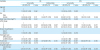
We included 108 patients with AEJ who underwent transhiatal distal esophagectomy and gastrectomy with curative intent. The clinicopathological characteristics of the patients are shown in Table 1, and the surgical outcomes are shown in Table 2. The LA and OA were utilized in 37 and 71 patients, respectively. Compared with the OA, the LA was associated with significantly shorter postoperative hospital stay (9 vs. 11 days, P=0.001), shorter proximal resection margins (3 vs. 7 mm, P=0.004), and extended operative times (240 vs. 191 min, P=0.001). No significant difference was found between the LA and OA in intraoperative blood loss (100 vs. 100 mL, P=0.392) or total number of harvested lymph nodes (54 vs. 51, P=0.889). Although there was a tendency for pathologic stage progression in the OA group, no significant difference was found between the 2 groups in stage or Siewert type (P=0.100 and 0.991, respectively).
Table 1
Clinicopathogical patient characteristics
Table 2
Surgical outcomes

Postoperative complications
The rate of surgical morbidity (grade≥II) for complications was not significantly different (8.1 vs. 23.9%, P=0.080) between the 2 groups. Table 3 shows the details of postoperative complications within 30 days in each group based on the Clavien-Dindo classification. There were 2 cases of anastomotic leakage in the OA group, but none in the LA group. Both patients recovered after percutaneous catheter drainage under radiologic intervention.
Table 3
Postoperative complications within 30 days
Survival outcomes
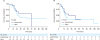
The median follow-up duration for all 108 patients was 34.5 months (range, 3–162 months). The 5-year OS rates were 81.8% and 50.7% for the LA and OA, respectively (P=0.024) (Fig. 1). The 3-year RFS rates were 77.3% and 46.4% for the LA and OA, respectively (P=0.009) (Fig. 2). In subgroup analysis of patients with stage III tumors, there were no significant differences in OS and RFS between the LA and OA groups (P=0.490 and 0.366, respectively) (Fig. 3). Regarding patients with stage I or II tumors, we could not analyze survival rates because there were no death or recurrence events in the LA group.
Fig. 1
OS rates according to the surgical approach. The 5-year OS rates were 81.8% for the laparoscopic transhiatal approach and 50.7% for the open approach (P=0.024).
OS = overall survival.
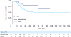
Fig. 2
RFS rate according to the surgical approach. The 3-year RFS rates were 77.3% for the laparoscopic transhiatal approach and 56.1% for the open approach (P=0.009).
RFS = relapse-free survival.
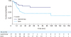
Fig. 3
OS and RFS rates for stage III patients. OS and RFS rates for stage III patients were not significantly different between the laparoscopic transhiatal approach and open approach groups (P=0.49 and 0.366, respectively). (A) Overall survival, (B) relapse-free survival.
OS = overall survival; RFS = relapse-free survival.
The factors associated with OS and RFS are listed in Table 4. In a univariable analysis for OS, patients with pathologic stage III and larger tumors, and those in the OA group, showed worse survival rates (hazard ratio [HR], 7.53; 95% confidence interval [CI], 1.03–55.20; P=0.047; HR, 1.14; 95% CI, 1.05–1.24; P=0.002; and HR, 2.50; 95% CI, 1.10–5.67; P=0.029; respectively). Multivariable analyses identified no independent prognostic factors for OS.
Table 4
Univariable and multivariable analysis of prognostic factors for OS and RFS
In a univariable analysis for RFS, female patients and patients with pathologic stage III and larger tumors showed worse survival rates (HR, 2.08; 95% CI, 1.17–3.70; P=0.012; HR, 9.65; 95% CI, 1.32–70.34; P=0.025; HR, 1.16; 95% CI, 1.08–1.26; P<0.001, respectively). Multivariable analyses revealed that female sex was the only independent prognostic factor for RFS (HR, 1.83; 95% CI, 1.01–3.32; P=0.046).
The number of patients with recurrence was 8 (21.6%) in the LA group and 29 (40.8%) in the OA group. The most common recurrence site was the peritoneum in both groups (Table 5). The pattern of recurrence was similar in the 2 groups.
Table 5
Recurrence pattern

DISCUSSION
The present study was conducted to evaluate the feasibility and safety of the LA for type II and III AEJ. Since the Japan Clinical Oncology Group (JCOG9502) revealed the advantage of the transhiatal approach compared to the thoracoabdominal approach for the treatment of type II and III AEJ, the transhiatal approach has been considered the optimal procedure. Considering that lower mediastinal lymph nodes might be difficult to visualize in the transhiatal approach, a laparoscopic view can overcome this drawback of the transhiatal approach.
In our institution, the indication for the laparoscopic procedure was initially confined to distal gastrectomy for clinical T1-T2 stage cancer without suspected lymph node metastasis. Based on experience, however, the indications were gradually extended to total gastrectomy for clinical T4 stage cancer. Consequently, the first LA for AEJ was performed in 2007.
Laparoscopic transhiatal mediastinal anastomosis after lower mediastinal lymph node dissection was first reported by Kinoshita et al. [10]. Previously, all reports on the LA had only described laparoscopic transhiatal esophagectomy and lymph node dissection with cervical anastomosis [11121314]. Compared to the thoracic approach, esophagojejunal anastomosis with the transhiatal approach is technically difficult. However, the development of stapling devices has allowed surgeons to safely perform esophagojejunal anastomosis in the mediastinum. Anastomotic leakage in the mediastinum is the most important and potentially life-threatening complication after esophagojejunal anastomosis. From a technical viewpoint, laparoscopic-enhanced visualization of the mediastinal space through the hiatus not only reduces the risk of hemorrhage or other complications, but also enables proper lymph node dissection in the lower mediastinum. These benefits of laparoscopy might reduce the postoperative complications and offer potential advantages for survival.
Recently, a Japanese group reported the safety and feasibility of the LA for Siewert type II AEJ [15]. The authors reported that the LA was associated with significantly reduced blood loss, but had longer operative times compared with the OA. The anastomotic leakage rate was almost the same in the 2 groups (approximately 4.5%). In China, Huang et al. [16] demonstrated that the LA was associated with better short-term outcomes, including less blood loss and shorter hospitalization periods for Siewert types II and III AEJ compared with the OA. However, no significant differences were detected in the rate and severity of postoperative complications in Huang's study. In the present study, no significant differences were observed between the postoperative complication rates, similar to the findings of Huang. However, anastomotic leakage only occurred in the OA group.
It is not known whether the appropriate number of lymph nodes can be retrieved in total gastrectomy using the LA [1718]. With regard to the lower mediastinal area, all lymph nodes can be visualized during dissection during the LA. Huang et al. demonstrated a significantly increased number of retrieved lymph nodes and superior survival rates for Siewert type II AEJ [16] with the LA compared to the OA. However, the number of harvested lymph nodes did not differ between the LA and OA in the present study. Furthermore, the LA did not independently affect the survival rate, a finding that might result from the relatively small sample size and earlier TNM stage of the LA group compared to the OA group.
Complete resection with negative margins is the most successful curative method in gastric cancer surgery. The safe length of resection margins has been reported in several studies [1920], and the Japanese gastric cancer treatment guidelines recommend a proximal margin of at least 2 cm for early gastric cancer and 3 cm (expansive growth type) or 5 cm (infiltrative growth type) for advanced gastric cancer [9]. However, other studies have reported that the length of free resection margins does not affect prognosis [212223]. Lee et al. [23] analyzed the correlation between the proximal margin and survival in 1,788 patients who had undergone curative surgery for gastric cancer. The authors reported that when a negative resection margin is pathologically confirmed, additional resections are not necessary, even in cases with proximal margins less than 0.5 cm. In the present study, the median length of proximal margins was 0.3 cm in the LA group. Regarding anvil-side esophageal tissue, the actual proximal margin was more than 1 cm. Since intraoperative frozen-section examinations were always performed at the authors' institution, the length of proximal margins in the present study was acceptable.
Although the current Japanese gastric cancer treatment guidelines recommend lower mediastinal lymph node dissection for patients with AEJ [9], the therapeutic effect of complete lower mediastinal lymph node dissection remains unclear. Hosoda et al. [24] compared patients with esophageal invasion of less than or equal to 3 cm and more than 3 cm to evaluate the therapeutic value of mediastinal lymphadenectomy. Lower mediastinal lymph node dissection showed benefits only in patients with esophageal invasion depth of more than 3 cm. Suh et al. [25] reported the role of mediastinal dissection using the validation index of recurrence. The authors demonstrated that routine complete mediastinal lymph node dissection was not essential in terms of recurrence in mediastinal lymph nodes. In the present study, we routinely performed lower mediastinal lymph node dissection for AEJ in the later study period. However, lower mediastinal lymph node dissection was not routinely performed in the early study period. Therefore, we could not evaluate the role of lower mediastinal lymph node dissection.
The current study has some limitations. First of all, its retrospective and single-institution design may have led to patient selection bias. Second, the sample sizes were very small and not well distributed between the groups. Lastly, this was a case control study; therefore, the surgical extent of the entire case series, particularly the extent of lymphadenectomy, was not standardized.
In conclusion, for patients with Siewert type II/III AEJ, the LA seems feasible and safe in comparison to the OA, not only with respect to the short-term but also with respect to the long-term oncologic outcomes. With respect to anastomotic leakage, the LA might have an advantage over the OA.
 XML Download