Journal List > J Vet Sci > v.20(2) > 1119450

Aubid, Liu, Vidal, and Hall: Isolation and culture of porcine primary fetal progenitors and neurons from the developing dorsal telencephalon

Abstract

The development of long-term surviving fetal cell cultures from primary cell tissue from the developing brain is important for facilitating studies investigating neural development and for modelling neural disorders and brain congenital defects. The field faces current challenges in co-culturing both progenitors and neurons long-term. Here, we culture for the first time, porcine fetal cells from the dorsal telencephalon at embryonic day (E) 50 and E60 in conditions that promoted both the survival of progenitor cells and young neurons. We applied a novel protocol designed to collect, isolate and promote survival of both progenitors and young neurons. Herein, we used a combination of low amount of fetal bovine serum, together with pro-survival factors, including basic fibroblast growth factor and retinoic acid, together with arabinofuranosylcytosine and could maintain progenitors and facilitate in vitro differentiation into calbindin 1+ neurons and reelin+ interneurons for a period of 7 days. Further improvements to the protocol that might extend the survival of the fetal primary neural cells would be beneficial. The development of new porcine fetal culture methods is of value for the field, given the pig's neuroanatomical and developmental similarities to the human brain.

INTRODUCTION

Comparative studies of the human and mouse cortex illustrate that the human cortex is unique and has higher mental and cognitive functions, which relate to language, movement, and sensation [1]. The differences in these functions are important evolutionary events that have led to the increased capabilities of the human species. Observed differences within the brain also extend to the cellular level where glia to neuron ratios have been shown to differ enormously between mouse and man [2].. Further differences at the transcriptomic level in the brain have also been reported, where molecular differences exist in neurodevelopmental processes such as cell migration and neural cell differentiation [3]. Some of these gene expression differences, such as those observed in glia are thought to even account for human specific neurodegenerative diseases, such as Alzheimer's disease [4]. Our interest and understanding of how we differ from other species, both at the neuroanatomical and functional level, remain high. Our added interest in medically intervening and treating neurological, neurodegenerative and fetal brain disorders has also meant that we continue to dissect and explore brain tissues so that we may understand them better at both the cellular and molecular level. Current research on the fetal brain, for example, has led to new findings for varying disorders such as anencephaly, holoprosencephaly, and congenital brain defects and causes lying behind them [5678].
It is however, very difficult to obtain fetal human tissue to study, and therefore researchers turn to model species to learn more about the developing human brain. Due to this reason, researchers have focused predominantly on rodent models, whereby tissue is easier to obtain. Considering, the mouse cortex and development differs both at the functional and cellular level [91011], more attention should be focused on using larger mammalian species. Cell culture systems have added considerable value to research, in that cells can be grown for extended periods and used and treated accordingly. Fetal cell culture research has however been predominantly performed in the mouse, but also successfully performed in other small mammals including chicks and rats [12131415].
There are no studies to date that have demonstrated the culture of primary fetal neurons from the fetal pig brain. There may be good reason to do so, considering the pig brain is highly similar anatomically to the human. It's gyrencephalic folding score is identical to the baboon (1.9) [16]. The pig is already a well-established model for brain imaging and neurosurgery, given its large brain size. Its larger brain can be imaged and is compatible in spatial resolution with PET scanners built for humans [1718]. During development, the pig brain also shows sexual dimorphisms and the large growth in brain volume postnatally is highly similar to humans [19]. Likewise, the cytoarchitecture of the telencephalon has been well described in the pig [20]. The piglet has also been shown to be an excellent model for studying brain and cognitive deficits associated with pre-term delivery [21]. Therefore, there is considerable value for developing methods that allow for the culture of primary fetal neurons from this species, that could help lead to new insight into, cortex growth in large mammalian species, cortex function in advanced species, as well as basic mechanisms in brain developmental disorders.
Culture of fetal brain however can be challenging and growth conditions to enhance neural cell survival have improved over the years, but many challenges still remain. For example, only short-term survival can be achieved for mouse immature cortex progenitors, which fail to proliferate in vitro [22]. Different adherent surfaces and serum free media appear to improve the survival and proliferation of murine radial glia-like cells [23] and a protocol established for rat prenatal cortical progenitors allowed cells to divide for approximately 14 cell divisions [15], however, these culture conditions are generally detrimental to differentiating neurons. Cultures may be performed as a monolayer, or in suspension, such that neurospheres may form from the isolated cortical cells. The latter has been performed using discarded fetal human cortex, however, again culturing the cells in vitro was limited to only two weeks [24]. Survival time of fetal neurons in vitro thus appears to be very limited. One study in a large mammalian model, the sheep, showed that cerebral cortex neurons could be cultured for 5 days in neural basal medium (NBM) supplemented with B-27 and GlutaMAX (Thermo Fisher Scientific, USA) [25]. Another study culturing primary cortical neurons from week 12–16 human brains in relatively simple media, including minimum essential media supplemented with 10% fetal bovine serum (FBS) allowed for maintenance of cells in vitro for up to 3 weeks [26]. Use of particular reagents such as arabinofuranosylcytosine (AraC) has proved to successfully help prevent the over-proliferation of astrocytes in the cultures [826] and the presence of relatively high concentrations of serum have a positive effect on the survival of neurons. Finding a balance therefore in culturing fetal progenitors and young neurons together, therefore remains a challenge, since culture conditions promote the differentiation of either progenitors or neurons, rather than both. In addition, there may be many important transitional signalling events that are lacking in the in vitro culture system that fails to support the long-term growth of the cells.
The objective of this study was to therefore develop a protocol that could be useful for the isolation, dissociation and culture of a range of different neural cell types in vitro from the porcine fetal brain. We wished to develop a protocol that could allow both short-term survival of progenitors but also allow for differentiation of these progenitors into neurons that could be used for a variety of different studies. We considered testing a protocol that might allow for the presence of both progenitors and neurons, using media and chemicals that provided some success from previous fetal cell culture studies in other species.

MATERIALS AND METHODS

Collection of fetal brains

Foetuses were collected following insemination of Duroc boar semen in Danish Landrace X Yorkshire crossed sows (two times within 24 h). Foetuses were collected at two time points for cell culture, i.e., at embryonic day (E) 50 and E60 (calculated upon first day of insemination). Foetuses were also collected for positive control immunohistochemistry experiments at E23, E50, E60, E70, and E80. To extract foetuses, professional personnel sacrificed pregnant sows at an abattoir and the uteri were transported to the laboratory in clean, plastic bags suspended in 37°C warm water. Upon arrival, uteri were opened using sterile surgical tools and the foetuses were found to be lifeless (passed away by asphyxia during the 45-min transport).
All chemicals were purchased from Sigma Aldrich (USA), unless specified otherwise. For fetal culture, 4 foetuses from both E50 and E60 respectively were placed into ice-cold HBSS containing 1 × Penicillin, Streptomycin, and Amphotericin solution and used for fresh dissections. For immunohistochemistry, the brains from 3 foetuses from each stage, were carefully dissected out of the cranium and placed directly into 4% paraformaldehyde (PFA) fixative in 0.15M phosphate buffered saline (PBS; pH 7.4; Thermo Fisher Scientific) and stored at room temperature (RT) for a minimum of 48 h. Following fixation, the brains were moved into PBS containing 0.01% sodium azide in PBS and stored on ice.

Dissection of fetal brains

Dissections were performed using aseptic technique with autoclaved dissection instruments. The fetal head was first dissected from the body. A horizontal cut was then made along the base of the cranium, running from the eye to the middle ear and the lower part of the head was removed. Warmed (65°C) 3% Agarose (BioNordika, Denmark) was then used to aid the attachment of the dorsal remaining fetal head to the petri dish. The fetal heads were then placed in a fridge set to 1°C to quickly cool the tissue and agarose for 15 min. This resulted in a firmer brain tissue during the regional brain dissection. For the dissection, the petri dish was placed on an ice bed and a medial incision was then made along the cranium and bone cutters were used to remove the outer layers, exposing the brain. A transverse cut at the posterior end of the head was performed to remove the cerebellum. Another transverse cut was performed to remove the anterior part of the brain. The remaining medial section of the brain including the medial thalamus was then further dissected using a stereomicroscope. The dorsal posterior and medial part of the brain containing the dorsal cingulate gyrus (referred to as dorsal telencephalon herein) was gently extracted from both brain hemispheres using a 200 micropipette and placed into a sterile 35 mm petri dish for further cell dissociation. Extraction of the dorsal telencephalon following removal of the whole foetus from the PBS on ice took approximately 25 min per foetus, but was performed in tandem, therefore the isolation of the brain tissue from 4 foetuses from 1 pregnancy took no more than 40 min.

Dissociation and culture of dorsal telencephalon

Cell dissociation was performed on tissue from individual foetuses using a papain dissociation method, according to the manufacturer's guidelines (Worthington Biochemical Corp., USA) with small modifications. Briefly, the tissue was digested for 30 min and inactivated with FBS. Prior to cell counting, the dissociated cells were resuspended in NBM (Thermo Fisher Scientific) containing 10% FBS. Cells were then counted using a haemocytometer and adjusted to a seeding density of 50,000 cells/cm2.
Dissociated cells were seeded at 50,000 cells/cm2 in 4-well Nunc dishes containing Poly-L-Ornithine coated, acid-treated glass coverslips and NBM containing 1% FBS, 1 B27, 20 ng/mL basic fibroblast growth factor (bFGF; PeproTech, USA), 1 × GlutaMax and 1 × Pen-Strep and culture for 48 h at 38.5°C. On day 2 of culture, medium was replaced with the same medium, with the exception that 25 mM AraC was added to inhibit over proliferation of astrocytes. Half the medium was replaced every 48 h thereafter until day 6 of culture when medium was then replaced with NBM A (Thermo Fisher Scientific) containing 1% FBS, 1 × B27, 20 ng/mL bFGF, 25 mM AraC and 10 mM retinoic acid (RA) to promote adult neuron growth. The cells were allowed to grow for up to 14 days in vitro, or alternately, fixed on D2, D7, and D14 (E50) and D2 and D7 (E60) in 4% PFA for 15 min and stored in PBS for later analysis.

Cryosectioning of fetal brains

Fetal brains to be used in immunohistochemistry were cryosectioned. First, brains were extracted from the cranium and placed in 30% sucrose solution for 48 h or until they sank in the solution. Brains were placed in plastic freezing moulds (Sakura; VWR, USA), coated with Tissue-Tek Optimal Cutting Temperature solution (Sakura; VWR) and snap frozen in liquid nitrogen cooled Hexane (VWR chemicals; VWR). Cryosections of 30 μm thickness were obtained in the frontal/coronal plane throughout the entire brain using a cryostat (Leica CM 1950; Leica Biosystems, Germany) and mounted on superfrost slides (VWR). Sections containing the telencephalon were selected for use and stored at −20°C until immunohistochemistry was performed.

Immunocyto- and Immuno-histochemistry

For the fetal cell monolayer cultures, immunocytochemistry was performed. Cells were first washed in PBS (5 min). Antigen retrieval was then conducted by adding boiling 0.01 M sodium citrate buffer (SCB; pH 6.0; VWR) for 3 min. Cells were washed in PBS (5 min) and then permeabilized in 0.1% Triton-X in PBS for 30 min. Cells were washed twice in PBS (5 min each) and blocked in 10% normal donkey serum (10% NDS) in PBS for 1 h. Cells were washed in PBS and primary antibodies were added (diluted in 10% NDS) and incubated at 4°C overnight. The cells were incubated with the following antibody combinations: glial fibrillary acidic protein/brain lipid-binding protein (GFAP/BLBP; 1:500 GFAP: DAKO, #Z0334/1:50 BLBP: Santa Cruz, #sc-374588) to detect radial glia; paired box protein 6/T-box brain protein 2 (PAX6/TBR2; 1:4,000 PAX6: Merck/Millipore, #AB2237/1:20 TBR2: R&D, #AF6166) to detect progenitors/intermediate progenitors; nuclear export signal (NES; 1:500 NES: Santa Cruz, #sc-21248) to detect progenitors and young neurons; calbindin 1/reelin (CALB1/RELN; 1:1,000 CALB1: Merck/Millipore, #AB1778/1:400 RELN: Merck/Millipore, #MAB5366) to detect neurons/interneurons. Following primary antibody incubation, cells were washed 3 × in PBS and then incubated for 1 h in corresponding secondary Alexa fluor antibodies (488 or 597) (Abcam, UK) raised in donkey, which were diluted at 1:200 in 10% NDS. Cells were washed 3 × in PBS and counterstained with 1 μg/mL Hoechst 33342 for 10 min and mounted in fluorescent mounting medium (DAKO, Denmark).
For the fetal brain immunohistochemistry, tissue sections were first warmed to RT for 1 h and then rehydrated in PBS (pH 7.4) for 10 min. Antigen retrieval was performed by boiling sections in SCB in a microwave for 5 min 3 times. Sections were cooled down to 4°C in PBS. Permeabilization was performed using 0.25% Triton-X in PBS for 10 min and sections were then washed 3 × in PBS. Blocking, incubation in primary and secondary antibodies, counterstaining and mounting was performed as described above. Negative controls were performed by omitting the primary antibody. Isotype controls were performed on fetal brain sections by adding the appropriate isotype (DAKO) instead of the primary antibody. Image acquisition was performed using the LEICA TCS SPE confocal microscope and Leica X software.

Neural cell counts and statistical analyses

Fetal cell cultures were semi-quantified to determine the proportion of surviving cells in culture using free software ImageJ (NIH, USA). First, images were converted to 16-bit files. The threshold tool was then used to demarcate the neuron soma. The analyse particle function was then used to annotate the number of soma and a cut-off score between 40–200 pixels was used to depict the soma size. The data from four foetuses from each time point, was then plotted as group graphs (plot summary or individual data graphs) showing the mean with standard deviation (SD) using PRISM. A 2-way analysis of variance (ANOVA) was performed to identify comparisons within treatments. A Tukey's multiple comparison test was used to determine differences in time and a paired two-tailed t-test was performed for comparing between two time points. Fetal cell cultures were also semi-quantified to determine the proportion of positive cells expressing different protein antigens. Images were analysed as separate layers in ImageJ as described previously. Interleaved bar graphs were created displaying the mean with SD and ordinary 1-way ANOVA or paired t-tests were performed to analyse for statistical differences. Statistical difference was found when p ≤ 0.05.

RESULTS

Establishment of neural developmental timing in the dorsal telencephalon

First, we evaluated 2 different times of porcine development, namely E50 and E60 to determine if we could identify neurogenesis and the presence of neural progenitor cells. Immunohistochemical analysis of the E50 and E60 porcine fetal brain showed initiation of neurogenesis, with the presence of a ventricular zone (VZ) containing GFAP positive cells at E50 that later became GFAP/BLBP positive at E60 (Fig. 1A). The VZ also expressed PAX6 positive radial glia at both stages of development (Fig. 1A). At the earlier stage of development (E50), we could also identify the subventricular zone (SVZ) from high expression of the intermediate progenitor cell marker, TBR2 that was reduced and lower in expression at E60 (Fig. 1A). We also evaluated an earlier stage of development, E23, and identified a VZ which expressed the neural progenitor marker NES (data not shown) suggestive that neurogenesis may also be occurring many days before E50. Together, this data suggested that both E50 and E60 were time points of active neurogenesis and would contain both radial glia (in the VZ) and intermediate progenitors (in the SVZ). It also confirmed that the antibodies worked well in the pig. We also tested our antibodies targeted to CALB and RELN on a later time point of development and found that at E70 of development, CALB positive neurons could be identified in COUP-TF interacting protein 2 (CTIP2) positive neurons throughout the different layers of the dorsal telencephalon (Fig. 1B). RELN is expressed and secreted by Cajal-Retzius cells (found in layer 1 [L1]) and expressed in interneurons. We found that in the E70 dorsal telencephalon, a strong expression of RELN was observed in L1 and that RELN+ cells could be observed in L4 which did not co-express CTIP2, which further helped to confirm an interneuron identity (Fig. 1B). This further validated the specificity of these neuron and interneuron antibody markers in pig tissue.
Fig. 1

Evaluation of neurogenesis in the developing fetal pig brain and assessment of antibodies in the porcine dorsal telencephalon. A VZ and SVZ could be identified at E50 of development by analysing markers that identify radial glia, GFAP/BLBP (VZ) and intermediate progenitors PAX6/TBR2 (SVZ) (A). A marker of more mature pyramidal neurons (CALB) was found to be expressed in E70 fetal dorsal telencephalon (B) that expressed CTIP2 (a neuron marker) and the interneuron/Cajal-Retzius cell marker RELN was also found to be highly expressed in L1 where the Cajal-Retzius cells reside and secrete RELN as well as in deeper L4 cells (B) presumably the interneurons which were CTIP2 negative.

GFAP/BLBP, glial fibrillary acidic protein/brain lipid-binding protein; PAX6/TBR2, paired box protein 6/T-box brain protein 2; CALB, calbindin; E, embryonic day; CTIP2, COUP-TF interacting protein 2; RELN, reelin; L, layer; VZ, ventricular zone; SVZ, subventricular zone.
jvs-20-e3-g001

Establishment of a primary cell culture method for culturing porcine fetal neurons

We decided to culture the dorsal telencephalon, since this was a relatively easy part of the cortex to remove from the foetuses (peripheral medial and posterior cortex) at these early stages of fetal development. We selected the papain dissociation method, since it has been successfully used to dissociate fetal cortex tissues prior [152527]. To ensure high progenitor and neuron cell survival, the first steps of resuspension were performed in high percentage (10% FBS) which was then reduced to 1% FBS for the culture period. We considered this might help not only promote neuron survival, but also promote progenitor cell survival. Dissociation of four foetuses per age (E50 and E60) was performed, yielding approximately 1.15 × 10⁶ total cells per foetus. We cultured the fetal cells separately, to determine later, whether differences might exist between foetuses. In the cell culture conditions, we included serum free components including B-27 to further support progenitor cell survival and a key growth factor was added, bFGF, which has a multifactorial role to promote neural stemness, proliferation and even induce neuronal differentiation [28]. We also included AraC in the culture to reduce the over-proliferation of astrocytes and also added RA on day 6 to promote neuron cell survival [29]. An overview of the cell culture protocol is shown in Fig. 2.
Fig. 2

Overview of the experimental procedures and cell culture conditions for establishing in vitro porcine fetal progenitors and neuron cultures.

AraC, cytosine arabinoside; FBS, fetal bovine serum; NBM, neural basal medium; bFGF, basic fibroblast growth factor; NBMA, neural basal medium A; RA, retinoic acid.
jvs-20-e3-g002

Fetal porcine primary neural cells can be cultured for 7 days with high survival

To test for the survivability of primary fetal tissue, we cultured dissociated cells in changing medium conditions that might promote the survival of both neural progenitors and neurons. Morphological assessment of E50 fetal cell cultures showed a good attachment of the seeded cells onto the coated glass coverslips 48 h after seeding (Fig. 3A). The cells had progenitor-like appearance and many had extending neurites. By 7 days after seeding the cells appeared to have thinned slightly and many had long extending neurites (Fig. 3A). However, counting of neural soma revealed no statistical differences in cell numbers (Fig. 3B), rather some cells had clustered. By 14 days after seeding, the number of cells had significantly declined in culture and neurites were no longer visible and cells appeared to be stressed, with small rounded morphology and many floating cells observed. This suggested that primary cells could be cultured for 7 days, but thereafter became difficult to maintain in the culture conditions used. We therefore repeated the experiment with E60 foetuses, but this time, ended the experiment at D7 (1 day after initiating neuron induction). Analyses of the neural cell survival showed no significant difference between D2 and D7 of culture (Fig. 3C).
Fig. 3

Neural cell culture and survival. The dorsal telencephalon could be cultured in vitro for 7 days before cultures began to thin (A). Neural cell survival was significantly reduced at day 14 from embryonic 50 stage foetuses. Neural cell survival was not significantly different between D2 and D7 in either culture of E50 or E60 fetal dorsal telencephalon (B, C).

E, embryonic day; PCC, porcine cingulate gyrus.
One asterisk denotes statistical significance at ≤ 0.05 and three asterisks denote significance at ≤ 0.001.
jvs-20-e3-g003

Porcine fetal cell cultures culture 7 days contain radial glia and intermediate progenitor cells

We then wanted to determine which neural cell types were present in the D7 cultures. We selected this time point, which demonstrated both the longest, and most viable time point to culture the porcine fetal cells. Herein, we repeated the culture of primary fetal tissue from E50 foetuses and performed immunocytochemistry to determine the proportion of progenitors versus neurons at D7 of culture. We expected since neurogenesis had initiated, that the tissue contained both progenitors and young neurons. We therefore used a combination of both anti-GFAP/anti-BLBP to detect radial glia (which co-express these markers), a combination of anti-PAX6/anti-TBR2 to detect dorsal telencephalon intermediate progenitors, anti-NES to detect either progenitors or young neurons and anti-CALB1/anti-RELN to detect pyramidal neurons and interneurons/deep layer neurons respectively. These combinations were tested on fetal tissue as described earlier to confer antibody reactivity in the pig, and to act as a positive control for the different antibodies.
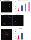
We found a subset of cells co-expressing GFAP and BLBP in the D7 cultures (Fig. 4A) The proportion of these was found to be relatively low at 16.43% (SD = 12.04; Fig. 4B). We then evaluated whether we could find intermediate progenitors from the dorsal telencephalon in the cultures. We could also detect a large number of co-expressing PAX6/TBR2 cells in the D7 culture with progenitor-like morphology suggesting that intermediate progenitors with both markers could be found (Fig. 4C). Individual assessment of PAX6 and TBR2 expression showed a slightly higher number of TBR2+ progenitors existed in the culture, but this was not shown to be significantly higher than PAX6+ cells (Fig. 4B). We then assessed whether the common marker of neural progenitors (which also demarcates young neurons) could be used to identify such populations in the culture. Here, we found a high proportion (almost all) of the cells in the D7 culture labelling positive for NES (Fig. 4C), indicative of both progenitors and neurons being labelled. Together, this revealed a high proportion of cells were of radial glia and intermediate progenitor origin.
Fig. 4

Neural cell cultures at D7 consisted of a mixture of progenitors and neurons. Radial glia expressing GFAP/BLBP were present (A) and quantified (B). Intermediate progenitors co-expressing PAX6/TBR2 were also found in the cultures (B, C), as well as early neurons/progenitors (NES) (B, C). Despite observing an increased trend, no significant difference was observed between the proportions of intermediate progenitors were the radial glia (B). A proportion of neurons expressing the principal neuron marker, CALB1 and neurons/interneurons expressing RELN could also be found in the cultures (D, E) and no significant difference could be observed in the proportion of these markers in the cell cultures (E).

GFAP/BLBP, glial fibrillary acidic protein/brain lipid-binding protein; PAX6/TBR2, paired box protein 6/T-box brain protein 2; NES, nuclear export signal; CALB, calbindin; RELN, reelin.
jvs-20-e3-g004

Porcine fetal cultures contain a smaller proportion of neurons and a larger proportion of RELN+ neural cells

To assess whether the 7-day culture could result in the induction of young neurons (which are not present in the E50/E60 brain), we performed analyses for the pyramidal neuron marker CALB1. We found CALB1 neurons in culture with a mean proportion of 26.15% in the cultures (Fig. 4D and E). The expression of CALB1 was surprisingly strong compared to the weak in vivo expression at E70 indicating a more mature cell phenotype may have been acquired. In addition, to identify whether other subpopulations may exist in the culture we analysed the cells for the expression of RELN (expressed in Cajal-Retzius cells, deeper pyramidal neurons and interneurons) and found interestingly that some RELN neurons co-expressed CALB1 (suggesting that these may be deeper layer pyramidal neurons, or potentially typical of those seen in the ventral telencephalon) (Fig. 4D and E). A larger proportion of RELN+ neural cells were found in the culture, indicative of either surviving Cajal-Retzius cells, interneurons or potentially also progenitors (since more than 50% of the population were RELN positive (Fig. 4E). Together, this indicated that pyramidal neurons and other neural cell types expressing RELN were present in the D7 fetal cell cultures.

DISCUSSION

Our study revealed that neurogenesis in the pig brain is present at E50 and E60 of development and is an optimal time to isolate fetal cells for establishing fetal cell cultures. The VZ and SVZ are present, containing radial glia and intermediate progenitors. At present, no studies have previously investigated the timing in onset of neurogenesis in this model species. The mouse is a well-documented model of neural development [1130], however, given the functional and transcriptomic differences between mouse and man, we believe the pig may be a more suitable model to use. We found that it was relatively easy to obtain fetal material from pig production farmers and believe that further exploration of the pig cortex's higher functions and transcriptome, may help to promote its use in neuroscience.
We have developed a protocol that results in a fast and easy isolation of brain tissue using macroscopic dissection from the fetal pig brain. Given the structural fragility of the tissues, careful dissection and harvesting of tissue is required and we found it an advantage to cool the tissue to very low temperatures to increase the viscosity of the fetal tissue, which improved the dissection procedure considerably. The protocol results in good survival of the neural cells. It is important that the brain is allowed to remain in the developing cranium as long as possible to maintain its structure, since our preliminary dissection attempts showed that removal of the brain resulted in complete loss of structure and gross mass (data not shown).
Our study revealed that we were able to obtain a heterogeneous neural cell culture of predominantly neural progenitors including radial glia and intermediate progenitors. We were also able to induce neural differentiation into pyramidal neurons present in the later developing fetal brain, either through supporting in vitro spontaneous differentiation, or either through induction by use of RA. This is promising in itself. However, we were unable to support the survival of the neural cells in culture beyond 7 days. Our research is aligned with other studies showing that it is difficult to extend the culture period of fetal neurons in vitro [143132]. Despite the incorporation of novel reagents, such as RA in combination with other prosurvival factors such as bFGF, we still could not support survival long-term in the dish. The presence of FBS (albeit the low concentration in the media) may also have been detrimental to the progenitors. Addition of neural supportive reagents such as brain-derived neurotrophic factor and glial cell line-derived neurotrophic factor earlier in the culture may have been beneficial for the survival of young newborn neurons. It is also unclear whether reagents and chemicals isolated from different species can be used to support the primary porcine fetal neurons.
Cell survival could have been affected by species-specific differences, which hamper the effectiveness of the added chemicals or molecules. For example, the bFGF used was of human origin and might not have been as effective on the porcine cells compared to porcine origin bFGF. There is little data to actually support whether bFGF may cross-react with other species, however, the PeproTech website claims that their recombinant human bFGF cross-reacts with pig. Experiments focused on optimizing cell survival by testing the addition and subtraction of varying factors are still needed, but we were optimistic from these studies, that we could at least find a diverse range of cell types in the cultures by day 7.
To summarize, we have developed a dissection and dissociation method, which leads to a high number of surviving neural cells from the porcine fetal cortex. We are the first to initiate primary fetal brain cultures from this species and show that neurogenesis has initiated already at E50 and E60 of development. We were able to establish primary fetal neuron cultures from the dorsal telencephalon that could survive well for at least 7 days in vitro and which consisted of a mix of radial glia, intermediate progenitors and a smaller proportion of neurons. Further research is required to improve the culture conditions which can promote both the proliferation and differentiation of progenitors into their respective neuron sub-types so that culture conditions can better mimic neurogenesis in the developing brain.

Notes

Funding This research has received funding from the Innovation Fund Denmark supported project, BrainStem (a stem cell center of Excellence in Neurology) and from the Danish Council for Independent Research (DFF-7017-0071).

Conflict of Interest No conflict of interest exists for any of the authors of this manuscript.

Author Contributions

  • Conceptualization: Hall V.

  • Data curation: Hall V.

  • Formal analysis: Aubid NN, Liu Y, Vidal JMP.

  • Methodology: Aubid NN.

  • Project administration: Hall V.

  • Resources: Hall V.

  • Writing - original draft: Hall V.

  • Writing - review & editing: Aubid NN, Liu Y, Vidal JMP.

References

1. Monuki ES, Walsh CA. Mechanisms of cerebral cortical patterning in mice and humans. Nat Neurosci. 2001; 4:Suppl. 1199–1206.
crossref
2. French L, Tan PP, Pavlidis P. Large-scale analysis of gene expression and connectivity in the rodent brain: insights through data integration. Front Neuroinform. 2011; 5:12.
crossref pmid pmc
3. Masjosthusmann S, Becker D, Petzuch B, Klose J, Siebert C, Deenen R, Barenys M, Baumann J, Dach K, Tigges J, Hübenthal U, Köhrer K, Fritsche E. A transcriptome comparison of time-matched developing human, mouse and rat neural progenitor cells reveals human uniqueness. Toxicol Appl Pharmacol. 2018; 354:40–55.
crossref pmid
4. Miller JA, Horvath S, Geschwind DH. Divergence of human and mouse brain transcriptome highlights Alzheimer disease pathways. Proc Natl Acad Sci U S A. 2010; 107:12698–12703.
crossref pmid pmc
5. Aubry MC, Aubry JP, Dommergues M. Sonographic prenatal diagnosis of central nervous system abnormalities. Childs Nerv Syst. 2003; 19:391–402.
crossref pmid
6. Edwards L, Hui L. First and second trimester screening for fetal structural anomalies. Semin Fetal Neonatal Med. 2018; 23:102–111.
crossref pmid
7. Gondré-Lewis MC, Gboluaje T, Reid SN, Lin S, Wang P, Green W, Diogo R, Fidélia-Lambert MN, Herman MM. The human brain and face: mechanisms of cranial, neurological and facial development revealed through malformations of holoprosencephaly, cyclopia and aberrations in chromosome 18. J Anat. 2015; 227:255–267.
crossref pmid pmc
8. Nagaraj UD, Lawrence A, Vezina LG, Bulas DI, duPlessis AJ. Prenatal evaluation of atelencephaly. Pediatr Radiol. 2016; 46:145–147.
crossref pmid
9. Chen W, Xia X, Song N, Wang Y, Zhu H, Deng W, Kong Q, Pan X, Qin C. Cross-species analysis of gene expression and function in prefrontal cortex, hippocampus and striatum. PLoS One. 2016; 11:e0164295.
crossref
10. DeFelipe J, Alonso-Nanclares L, Arellano JI. Microstructure of the neocortex: comparative aspects. J Neurocytol. 2002; 31:299–316.
pmid
11. Molnár Z, Clowry G. Cerebral cortical development in rodents and primates. Prog Brain Res. 2012; 195:45–70.
crossref pmid
12. Matsushita T, Fujihara A, Royall L, Kagiwada S, Kosaka M, Araki M. Immediate differentiation of neuronal cells from stem/progenitor-like cells in the avian iris tissues. Exp Eye Res. 2014; 123:16–26.
crossref pmid
13. Pozzi D, Ban J, Iseppon F, Torre V. An improved method for growing neurons: Comparison with standard protocols. J Neurosci Methods. 2017; 280:1–10.
crossref pmid
14. Sensenbrenner M, Deloulme JC, Gensburger C. Proliferation of neuronal precursor cells from the central nervous system in culture. Rev Neurosci. 1994; 5:43–53.
crossref pmid
15. Xu SY, Wu YM, Ji Z, Gao XY, Pan SY. A modified technique for culturing primary fetal rat cortical neurons. J Biomed Biotechnol. 2012; 2012:803930.
crossref
16. Mota B, Herculano-Houzel S. Response to comments on “cortical folding scales universally with surface area and thickness, not number of neurons”. Science. 2016; 351:826.
crossref
17. Sauleau P, Lapouble E, Val-Laillet D, Malbert CH. The pig model in brain imaging and neurosurgery. Animal. 2009; 3:1138–1151.
crossref pmid
18. Winterdahl M, Audrain H, Landau AM, Smith DF, Bonaventure P, Shoblock JR, Carruthers N, Swanson D, Bender D. PET brain imaging of neuropeptide Y2 receptors using N-11C-methyl-JNJ-31020028 in pigs. J Nucl Med. 2014; 55:635–639.
crossref pmid
19. Conrad MS, Dilger RN, Johnson RW. Brain growth of the domestic pig (Sus scrofa) from 2 to 24 weeks of age: a longitudinal MRI study. Dev Neurosci. 2012; 34:291–298.
crossref pmid pmc
20. Bjarkam CR, Glud AN, Orlowski D, Sørensen JC, Palomero-Gallagher N. The telencephalon of the Göttingen minipig, cytoarchitecture and cortical surface anatomy. Brain Struct Funct. 2017; 222:2093–2114.
crossref pmid
21. Radlowski EC, Conrad MS, Lezmi S, Dilger RN, Sutton B, Larsen R, Johnson RW. A neonatal piglet model for investigating brain and cognitive development in small for gestational age human infants. PLoS One. 2014; 9:e91951.
crossref
22. Rubio A, Belles M, Belenguer G, Vidueira S, Fariñas I, Nacher J. Characterization and isolation of immature neurons of the adult mouse piriform cortex. Dev Neurobiol. 2016; 76:748–763.
crossref pmid
23. Markó K, Kohidi T, Hádinger N, Jelitai M, Mezo G, Madarász E. Isolation of radial glia-like neural stem cells from fetal and adult mouse forebrain via selective adhesion to a novel adhesive peptide-conjugate. PLoS One. 2011; 6:e28538.
crossref
24. Lu J, Delli-Bovi LC, Hecht J, Folkerth R, Sheen VL. Generation of neural stem cells from discarded human fetal cortical tissue. J Vis Exp. 2011; (51):2681.
crossref
25. Reddy RC, Amodei R, Estill CT, Stormshak F, Meaker M, Roselli CE. Effect of testosterone on neuronal morphology and neuritic growth of fetal lamb hypothalamus-preoptic area and cerebral cortex in primary culture. PLoS One. 2015; 10:e0129521.
crossref
26. Fu W, Ruangkittisakul A, MacTavish D, Baker GB, Ballanyi K, Jhamandas JH. Activity and metabolism-related Ca2+ and mitochondrial dynamics in co-cultured human fetal cortical neurons and astrocytes. Neuroscience. 2013; 250:520–535.
crossref pmid
27. Hashimoto A, Onodera T, Ikeda H, Kitani H. Isolation and characterisation of fetal bovine brain cells in primary culture. Res Vet Sci. 2000; 69:39–46.
crossref pmid
28. Yang H, Xia Y, Lu SQ, Soong TW, Feng ZW. Basic fibroblast growth factor-induced neuronal differentiation of mouse bone marrow stromal cells requires FGFR-1, MAPK/ERK, and transcription factor AP-1. J Biol Chem. 2008; 283:5287–5295.
crossref pmid
29. Jacobs S, Lie DC, DeCicco KL, Shi Y, DeLuca LM, Gage FH, Evans RM. Retinoic acid is required early during adult neurogenesis in the dentate gyrus. Proc Natl Acad Sci U S A. 2006; 103:3902–3907.
crossref pmid pmc
30. Dwyer ND, Chen B, Chou SJ, Hippenmeyer S, Nguyen L, Ghashghaei HT. Neural stem cells to cerebral cortex: emerging mechanisms regulating progenitor behavior and productivity. J Neurosci. 2016; 36:11394–11401.
crossref pmid pmc
31. Barnea A, Roberts J. An improved method for dissociation and aggregate culture of human fetal brain cells in serum-free medium. Brain Res Brain Res Protoc. 1999; 4:156–164.
crossref pmid
32. Yu L, Vásquez-Vivar J, Jiang R, Luo K, Derrick M, Tan S. Developmental susceptibility of neurons to transient tetrahydrobiopterin insufficiency and antenatal hypoxia-ischemia in fetal rabbits. Free Radic Biol Med. 2014; 67:426–436.
crossref pmid
TOOLS
ORCID iDs

Yong Liu
https://orcid.org/0000-0002-0574-2877

Juan Miguel Peralvo Vidal
https://orcid.org/0000-0002-1283-8156

Vanessa Jane Hall
https://orcid.org/0000-0002-3598-2214

Similar articles