

 PDF
PDF Citation
 Print
Print


INTRODUCTION
Enterococcus spp. colonizing the intestinal tract of humans and animals comprise an essential part of the microbiota and are recognized as important opportunistic pathogens causing nosocomial infections of the urinary tract, endocarditis, bacteremia, and central nervous system infections [12]. Related to the use of glycopeptides in humans and livestock as medicines or growth promoters, in addition to intrinsic resistance to a number of antibiotics such as β-lactams and aminoglycosides, enterococci detected in humans and livestock have shown antibiotic resistance resulting from mutation or acquisition of foreign genetic material. The latter includes the transfer of the vancomycin-resistant gene via plasmids and transposons [34]. Therefore, the emergence of drug-resistant enterococci has been monitored not only in hospitals and clinics but also in the community [5].
Antibiotics have not been administered to laboratory mice and rats by breeding companies for an extended period, resulting in a low incidence of drug resistance in such populations. Furthermore, no correlation has been found between the emergence of drug resistance in laboratory animals and those in humans or livestock [6]. However, detection of drug-resistant Enterococcus faecalis and Enterococcus faecium that show resistance to tetracycline (TE) or erythromycin (E) in addition to β-lactams and aminoglycosides has been reported in laboratory mice and rats [789]. Recently, antibiotics have been used to control bacterial infections such as Rodentibacter species (“Pasteurella pneumotropica”) in laboratory animals in the animal facilities of academic institutions [1011]. Therefore, monitoring for the emergence of antibiotic-resistant bacteria including Enterococcus spp. in laboratory animals may be required in laboratory animal facilities.
Our previous study showed that low-level vancomycin-resistant Enterococcus spp. (VRE; 5 Enterococcus casseliflavus and 19 Enterococcus gallinarum isolates possessing vanC2/3 and vanC1 genes, respectively) were frequently detected in laboratory mice, including immunodeficient strains, obtained from commercial mouse breeding companies in Japan [12]. Our previous study also indicated that some of these VRE isolates showed resistance to other antibiotics, such as E, ciprofloxacin (CIP), or TE. In the present study, the drug sensitivities of these previously identified VRE isolates against the antibiotics are investigated in detail, and the related resistant genes partially identified. The results provide information on the antibiotic resistance profiles, other than their intrinsic resistances, of enterococci isolated in laboratory mice.
MATERIALS AND METHODS
VRE isolates from laboratory mice and Enterococcus type strains
VRE were isolated from fresh feces of laboratory mice supplied from 4 different commercial breeding companies between October 2011 and February 2012. Twenty-four isolates were identified (5 E. casseliflavus and 19 E. gallinarum) as reported previously [12] and as summarized in Table 1. These isolates possessed the vanC2/3 and vanC1 vancomycin-resistant genes, respectively, and exhibited low-level resistance to vancomycin with minimal inhibitory concentration (MIC) levels of no more than 12 μg/mL. The E. faecalis ATCC 29212 (JCM 7783), E. casseliflavus ATCC 25788 (JCM 8723), and E. gallinarum ATCC 49573 (JCM 8728) provided by the Japan Collection of Microorganisms (RIKEN BRC, Japan), which is participating in the National BioResource Project of the MEXT, Japan, were used as controls.
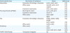
Table 1
Isolates of low-level vancomycin resistant Enterococcus species and type strain used in this study

![]()
Antibiotic susceptibility
Antibiotic susceptibilities to E 15, CIP 5, norfloxacin 10 (NOR 10), TE 30, minocycline 30 (MI 30), and doxycycline 30 (DX 30) were examined by using Sensi-Disc disc diffusion testing (Becton Dickinson, USA). The MICs for E, CIP, and TE for each Enterococcus isolate were determined by using the E-test (bioMérieux, France). These tests were performed as described previously [12]. Briefly, bacterial suspensions in phosphate-buffered saline were adjusted to a turbidity of 0.5 McFarland standard, spread onto Mueller-Hinton agar (NISSUI, Japan), and incubated for 24−48 h at 37°C under aerobic conditions. After incubation, the sensitivities were graded as sensitive, intermediate or resistant based on measurement of the diameter of the inhibition zone. MICs were derived based on where the edge of the inhibition ellipse area intersected the test strip.
Sequencing analysis of the quinolone resistance-determining region (QRDR)
In order to detect mutations within the QRDR in 2 subunits of both DNA gyrase (gyrA and gyrB) and topoisomerase IV (parC and parE), polymerase chain reaction (PCR) and sequencing analysis were performed using the specific primer pairs summarized in Table 2. Briefly, after amplification of the QRDR by PCR, the PCR products were cloned into pCR 2.1-TOPO vectors (Invitrogen, USA) and introduced into Escherichia coli DH5α cells. Sequencing analysis of the QRDR from purified plasmids of competent cells was performed by using ABI PRISM BigDye Terminator v3.1 Cycle Sequencing Kits (Applied Biosystems, USA).
Table 2
Primers used to amplify the QRDR of Enterococcus isolates and analyze sequence

![]()
Detection of antibiotic-resistant genes
Macrolide- [1314], fluoroquinolone- [151617], TE- [1418], and multidrug- [192021] resistant genes and transposon integrase genes [14] were detected by PCR using specific primers for each reference cited, as summarized in Table 3. All reactions were performed in a final reaction volume of 20 μL with bacterial cells as a template, as well as 10 μL of EmeraldAmp PCR Master Mix (Takara Bio, Japan), and 1 μM of each primer. The PCR reactions were performed as described in each cited reference, and products were visualized on 1.5% agarose gels. Positive and suspicious PCR products were sequenced using each specific primer pair and were verified by comparison to other sequences in the National Center for Biotechnology Information database (https://blast.ncbi.nlm.nih.gov/Blast.cgi).
Table 3
PCR detection of antimicrobial resistance and transposon integrase genes in this study
| Antimicrobial class | Mechanism of resistance | Genes | References |
|---|---|---|---|
| Macrolides | Prevention of binding to ribosome | ermA, ermB, ermC | [14] |
| Efflux pump | msrA/B, msrA, msrC | [14] | |
| mefA/E | [13] | ||
| Enzymatic inactivation | ereA, ereB, mphA | [13] | |
| Fluoroquinolones (PMQR) | Blocking the action | qnrA, qnrB, qnrS | [15] |
| Enzymatic inactivation | aac6′-Ib | [17] | |
| Efflux pump | oqxA, oqxB | [16] | |
| qepA | [17] | ||
| TEs | Prevention of binding to ribosome | tet(M), tet(O), tet(S), tet(W) | [14] |
| tet(T), tet(Q) | [18] | ||
| Efflux pump | tet(K), tet(L) | [14] | |
| Multidrug | Efflux pump | acrB | [21] |
| emeA | [19] | ||
| norA, norB, norC, mepA | [20] | ||
| Tn916-Tn1545 family | Transposon integrase | int | [14] |
![]()
RESULTS
E-resistant VRE isolates from laboratory mice
All 19 E. gallinarum isolates and type strain E. gallinarum ATCC 49573 were sensitive to E. Although the type strain of E. casseliflavus (ATCC 25788) showed intermediate resistance to E, 1 of the 5 E. casseliflavus isolates indicated E resistance at a MIC of 8 μg/mL (Table 4). This E-resistant VRE isolate was detected in 1 of the 2 tested mouse strains supplied by company 2, and this isolate also indicated resistance to fluoroquinolones as described below. The macrolide-resistant genes ermA, ermB, ermC, msrA/B, msrA, msrC, mefA/E, ereA, ereB, and mphA, and multidrug-resistant genes acrB, emeA, norA, norB, norC, and mepA (summarized in Table 3) were not detected in this E. casseliflavus isolate (Table 4). In addition, the transposon integrase gene (int) associated with the transfer of macrolide-resistant genes was not detected in the E. casseliflavus E-resistant isolate.
Table 4
Summary of antimicrobial resistance profiles of VRE isolates from laboratory mice
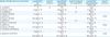
E, erythromycin; CIP, ciprofloxacin; NOR, norfloxacin; TE, tetracycline; MI, minocycline; DX, doxycycline; MIC, minimal inhibitory concentration for each antibiotics; S, sensitive; I, intermediate; R, resistant; NI, not identified; QRDR, quinolone resistance-determining region.
*The upper and lower lines were represented as the results of Sensi-Disc and MIC tests, respectively.
![]()
Fluoroquinolone-resistant VRE isolates from laboratory mice
The susceptibility of all 24 enterococcal isolates and the 2 type strains to the fluoroquinolone antibiotics CIP and NOR was between intermediate and resistant based on Sensi-Disc test scores. The MICs of all isolates for CIP were determined and 2 isolates, 1 each of E. casseliflavus and E. gallinarum, had MICs of 32 and 6 μg/mL, respectively, indicative of apparent resistance (Table 4). The E. casseliflavus isolate showing apparent resistance to fluoroquinolones also indicated resistance to E as described above. The MIC of the other E. casseliflavus isolate scored as resistant by Sensi-Disc was ≤ 3 μg/mL indicating intermediate resistance to CIP. The 2 apparent resistant and 1 intermediate resistant isolates were obtained from different mouse strains supplied by company 2 and company 4 (Table 4). To clarify the mechanism of resistance to fluoroquinolones, the amino acid (AA) sequences of the QRDR of these 3 isolates at positions 69−110 AA of gyrA, 415−460 AA of gyrB, 58−106 AA of parC, and 421−470 AA of parE, which correspond to the AA positions of E. faecalis V583 (accession# AE016830), were analyzed and compared with those of each type strain and the sensitive isolates. However, no AA substitutions were detected in the QRDR of the 3 fluoroquinolone-resistant isolates. In addition, the plasmid-mediated quinolone resistance (PMQR) determinant genes qnrA, qnrB, qnrS, aac6’-Ib, oqxA, oqxB, and qepA, and the multidrug-resistant genes acrB, emeA, norA, norB, norC, and mepA (summarized in Table 3) were not detected in these 3 fluoroquinolone-resistant VRE isolates (Table 4).
TE-resistant VRE isolates from laboratory mice
All 24 isolates were tested for resistance to the TE antibiotics TE, MI, and DX, by using Sensi-Disc discs. Two of the 24 VRE isolates, both E. gallinarum, indicated resistance to TE and intermediate resistance to MI and DX, while the type strain showed resistance to both TE and MI. One of the 2 TE-resistant E. gallinarum isolates was obtained from mice supplied by company 2 while the other was from company 4. Neither isolate was resistant to macrolides or fluoroquinolones (Table 4). The MICs for TE of the 2 isolates were 48 and 64 μg/mL and the MIC for the E. gallinarum type strain was 96 μg/mL, indicating high-level resistance. To identify the TE-resistant gene, tet genes and the multidrug-resistant genes acrB, emeA, norA, norB, norC, and mepA (summarized in Table 3) underwent PCR in the TE-resistant E. gallinarum isolates and the resistant E. gallinarum type strain. Although multidrug-resistant genes were not detected, tet(O) and tet(S) genes were identified in the 2 TE-resistant isolates and the type strain, respectively (Table 4). The detected tet(O) gene fragment, approximately 720−730 base pairs (bp), in the 2 isolates, showed 99% sequence identity with those of E. faecalis (GenBank accession No. NG048262 and AY660532), Actinobacillus spp. (NG048264), Streptococcus spp. (LC131132, FR691055, and NG048265), Campylobacter spp. (JQ613156 and CP002030), and Clostridium spp. (GQ240298). The tet(S) gene fragment, approximately 700 bp, detected in the E. gallinarum type strain showed a sequence identity of 99% with those of E. faecalis (GenBank accession No. JN208881, AM039489, and AM039490), E. faecium (GQ900487), Streptococcus spp. (KX077886, NG048275, and NG048276), and Listeria monocytogenes (NG048273 and JX865374). The transposon integrase gene was not detected in the 2 E. gallinarum isolates or in the E. gallinarum type strain possessing the TE-resistant gene.
DISCUSSION
In this study, the resistance levels to antibiotics other than glycopeptides were determined in detail for 24 Enterococcus strains that had been isolated from commercially available mouse strains showing intrinsic resistance to vancomycin as reported previously [12]. The results in the present study showed resistance to macrolides, fluoroquinolones, and TEs in 5 of the 24 isolates. In addition, the mechanisms of resistance to each antibiotic agent in those 5 isolates were investigated and the TE-resistant gene, tet(O), was identified in each of the TE-resistant isolates. No resistant genes to macrolides and fluoroquinolones were detected in the macrolide- and fluoroquinolone-resistant isolates. In each commercial breeding companies that provided mice used in this study, antibiotics had not been used to eliminate pathogens in their laboratory mice [12]. Our results show that antibiotic resistance was not consistently observed among the mice obtained from the same breeding company. In addition, although the drug-resistant bacteria were detected in laboratory mice [89], the emergence of drug resistance in laboratory animals has shown no correlation with emergences in humans or livestock [6]. Therefore, these antibiotic-resistant genes may be possessed intrinsically in the Enterococcus isolates or maintained for generations among the microbiota, including enterococci, of the mouse strain.
Although a macrolide-resistant gene, ermB, has frequently been detected in macrolide-resistant enterococci in livestock, erm genes, including ermA and ermC, have only been detected in isolates that were highly resistant to macrolides [2223]. In this study, 1 isolate showed low-level resistance to E with a MIC of 8 μg/mL. In addition to the erm genes, no PCR fragment of other macrolide- or multidrug-resistant genes were obtained. The macrolide-resistant isolate in this study also indicated apparent resistance to fluoroquinolones with a MIC of 32 μg/mL for CIP. Other known or unknown factors might be associated with both the macrolide and fluoroquinolone resistance in this isolate.
Three VRE isolates, including one showing resistance to E, indicated resistance to fluoroquinolones. The antimicrobial effects of quinolones are the result of inhibition of 2 subunits of DNA gyrase (gyrA and gyrB) or of DNA topoisomerase IV (parC and parE) [2425]. The fluoroquinolone resistance in gram-positive bacteria is associated with AA substitutions in the QRDR of gyrA and gyrB, and parC and parE [26]. However, no AA substitutions were detected in the QRDR of the 3 fluoroquinolone-resistant isolates when compared with those of sensitive isolates and each Enterococcus spp. type strain. In addition to the QRDR and multidrug-resistant genes, another fluoroquinolone-resistant factor has been identified among the PMQR genes [26]. However, no PCR fragments of the expected molecular size indicating PMQR and multidrug-resistant genes were detected in the 3 fluoroquinolone-resistant isolates in this study. Since the novel mutations in the QRDR and mechanisms associated with quinolone resistance have been clarified [26], another known or unknown mechanism may be involved in the fluoroquinolone resistance of the Enterococcus isolates.
Ribosomal protection genes tet(O) and tet(S) were detected in the 2 E. gallinarum isolates and in the E. gallinarum type strain, respectively, indicating apparent resistance to TE. The tet(O) and tet(S) genes were not commonly detected in the other E. gallinarum isolates, and the resistant isolates were not detected in the other mouse strains obtained from the same breeding company. Although the Tn916-Tn1545 transposon family gene associated with transferable tet genes was not detected in the 2 isolates in this study, the tet(O) gene has been detected on conjugative plasmids and is transferable via plasmids in enterococci [2728]. Therefore, Enterococcus isolates from laboratory mice may spread the tet(O) genes horizontally, either intra- or inter-species. On the other hand, although the tet(O) gene confers resistance to both 1st generation (TE) and 2nd generation (MI and DX) TEs [27], our 2 Enterococcus isolates possessing the tet(O) gene showed apparent resistance only to TE, with only intermediate resistance to MI and DX. The DNA sequence identities of the tet(O) genes detected in our 2 isolates were not a complete match to the deposited DNA sequences in GenBank; furthermore, the detection of mosaic TE resistance genes has been reported [29]. Therefore, the tet(O) genes detected in this study may be classified as members of a new class of resistance gene.
Antibiotics have been used to control specific bacterial pathogens in laboratory mice in the animal facilities of academic institutions but not in the mouse populations of breeding companies [1011]. The use of antibiotics induces the proliferation of drug-resistant bacteria in a mouse colony and those bacteria may be spread to the other colonies by the users or caretakers of the mice. The data presented in this study may remind researchers and animal facility managers of the need to consider the existence of antibiotic-resistant bacteria in a laboratory mouse colony when antibiotics are used in laboratory mice.
 XML Download