Journal List > J Vet Sci > v.20(2) > 1119441

Kim, Park, Bae, and Park: Evolutionary relationship analysis of Middle East respiratory syndrome coronavirus 4a and 4b protein coding sequences

Abstract

The 4a and 4b proteins of the Middle East respiratory syndrome coronavirus (MERS-CoV) have been described for their antagonism on host innate immunity. However, unlike clustering patterns of the complete gene sequences of human and camel MERS-CoVs, the 4a and 4b protein coding regions did not constitute species-specific phylogenetic groups. Moreover, given the estimated evolutionary rates of the complete, 4a, and 4b gene sequences, the 4a and 4b proteins might be less affected by species-specific innate immune pressures. These results suggest that the 4a and 4b proteins of MERS-CoV may function against host innate immunity in a manner independent of host species and/or evolutionary clustering patterns.

Human infection with the Middle East respiratory syndrome coronavirus (MERS-CoV), a positive-sense single-stranded RNA virus of the family Coronaviridae, was first reported in 2012 [1]. Since then, more than 2,200 laboratory-confirmed cases have been described with an estimated 35.5% case-fatality rate [2]. Middle East countries appeared to be mainly affected by the life-threatening pathogenicity of the virus, but some exported cases were also reported in Asia, Europe, and the United States of America [13]. Given zoonotic transmission of the virus from dromedary camels to humans and its lethality in humans [1], vaccines and/or antivirals should be prepared, but no approved medical countermeasures are available, yet.
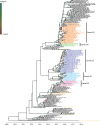
In line with the pathogenic mechanisms reported with MERS-CoV [4], such as acute respiratory symptoms and local immune responses, viral antagonism against host innate immune responses may be of great significance in terms of the pandemic potential and host range restriction of the virus. Viral antagonism had been already described for MERS-CoV [5], and, of the MERS-CoV proteins, protein 4a was suggested responsible for its role against RNA-dependent protein kinase-mediated antiviral immunity and activation of retinoic acid-inducible gene I and Melanoma Differentiation-Associated protein 5 of host cells [67]. The antagonistic effects of MERS-CoV protein 4b were also demonstrated against nuclear factor-κB-dependent immunity and RNase L activation of host cells [89]. As shown for influenza and Zika viruses [1011], the species-specific effects of viral antagonism of MERS-CoV on host innate immunity can be revealed by analysis of the evolutionary relationship of the 4a and 4b genes between human and camel MERS-CoV strains. To this end, we investigated the evolutionary history and rates of MERS-CoV 4a and 4b protein coding regions as well as those of MERS-CoV complete gene sequences using a time-framed Bayesian inference method (BEAST package, v1.10.0; BEAST, New Zealand) [12] with individual settings of molecular clock (for complete gene, lognormal relaxed clock model and for 4a and 4b coding regions, strict clock model), nucleotide substitution (based on the estimated results of best-fit substitution model using jModeltest, v2.1.10: for complete gene, GTR + I + γ; for 4a, TN93 + I; and for 4b, TN93 + γ) [13], and Markov chain Monte Carlo runs (2 × 10⁸ chain length with every 2 × 10⁵ iterations after a 10% burn-in). The phylogenetic relationship of MERS-CoV complete gene sequences was reconstructed using a total of 260 sequences (human strains, n = 126 and camel strains, n = 134; sequence sets are available upon request) that were downloaded from the Virus Variation Resource database of National Center for Biotechnology Information (https://www.ncbi.nlm.nih.gov/genome/viruses/variation/), and their maximum clade credibility trees were visualized using FigTree (v1.4.3; Figtree Systems Pty, Australia). Given the phylogenetic relationship of the complete gene sequences, most camel sequences constituted 5 different genetic groups (camel G1–G5), except for some distantly-related sequences, and the camel G2, G3, and G4 strains appeared to evolve exclusively from human strains. Transmission of MERS-CoV between humans and camels appeared to be identified only in the camel G1 and G5 groups (Fig. 1).
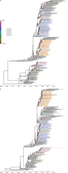
Unlike the phylogenetic clustering patterns of the complete gene sequences (Fig. 1), human and camel strains appeared to constitute common genetic groups in the 4a and 4b phylogenetic trees (Fig. 2). Neither human-specific nor camel-specific clustering patterns were observed in the 4a and 4b phylogenetic trees. Rather, the camel G2, G3, and G4 sequences, which constituted the camel-only phylogenetic groups in the complete gene sequence analysis (Fig. 1), were mixed with other camel sequences of different phylogenetic groups and with human sequences, and, from camel G1 to G5, all camel sequences were grouped together with human sequences (Fig. 2). These groupings may imply little or no species-specific antagonism of the human and camel MERS-CoV 4a and 4b proteins against host innate immune responses. Of course, not phylogenetic placements between different host viruses but certain genetic determinants may determine the viral antagonistic mechanism(s) in hosts. The other viral proteins or genetic mutations on the other viral proteins may also compensate for a lack of sufficient viral antagonism [14]. However, results of serological and epidemiological analyses indicate dromedary camels are one of the susceptible hosts of MERS-CoV, even though disease severity of MERS is quite different between humans and camels [1], and, given that the interaction mechanism(s) between viral surface protein and cellular receptors of hosts may also determine host range restriction [15], the effects of species-specific viral antagonism on host innate immunity might not be associated with the sustained dissemination of MERS-CoV in humans. Rather, a lack of sufficient viral antagonism of MERS-CoV in humans may be associated with the transmission of the virus from camels to humans, as indicated by the almost 2-fold increased evolutionary rate (1.42 × 10⁻³ substitutions/site/year) of the complete gene sequences of human and camel MERS-CoV strains (Table 1), compared with the previous study result (0.74 × 10⁻³ substitutions/site/year) reported by Kim et al. [3], which assessed only the human strain sequences. In contrast, the evolutionary rates of the 4a (1.89 × 10⁻³ substitutions/site/year) and 4b (1.44 × 10⁻³ substitutions/site/year) protein-coding regions of human and camel MERS-CoV strains were estimated to be almost in similar ranges with those (1.26 × 10⁻³ and 1.27 × 10⁻³ substitutions/site/year for the 4a and 4b protein coding regions, respectively) reported by Kim et al. [3] (Table 1), which may suggest the absence of large immune-mediated pressure differences between human and camel MERS-CoV 4a and 4b proteins and no need for the adaptation of camel MERS-CoV strains prior to zoonotic dissemination to humans. As mentioned above, the effects of other viral proteins and molecular determinants of immune evasion mechanisms of MERS-CoVs on the zoonotic and reverse zoonotic transmission between humans and camels should be investigated further using in vitro and in vivo studies.
In this study, we investigated the evolutionary history and rates of the 4a and 4b protein coding sequences of human and camel MERS-CoV strains. By demonstrating that neither species-specific nor group-specific clustering patterns were observed in the 4a and 4b protein coding regions, we suggest that the antagonistic mechanism of MERS-CoV against host innate immunity might be mediated in a manner independent of host species and/or evolutionary clustering patterns. Given the disease severity and consequent public health threats of MERS, our results have expanded the knowledge of molecular evolution patterns and virus-host interactions of MERS-CoVs and provide information useful in the development of MERS-CoV vaccines.

Figures and Tables

Fig. 1

Phylogenetic relationship of the complete genes of human and camel MERS-CoV strains. The complete gene sequences of human and camel MERS-CoV strains were evaluated for their evolutionary relationships. Human strain sequences were colored black, whereas camel sequences were colored differently based on their individual distinct groups (camel G1, magenta; G2, sky blue; G3, purple; G4, light green; and G5, orange). The other camel strains not included in the camel G1–G5 groups were colored cantaloupe. The color of each node indicates its posterior probability score (0.0025–1).

MERS-CoV, Middle East respiratory syndrome coronavirus.
jvs-20-e1-g001
Fig. 2

Phylogenetic relationship of the 4a and 4b coding regions of human and camel MERS-CoV strains. The 4a (A) and 4b (B) coding region sequences of human and camel MERS-CoV strains were evaluated for their evolutionary relationships. Human strain sequences were colored black, and camel sequences were colored differently based on their corresponding complete gene sequence group, as indicated and as in Fig. 1. The color of each node indicates its posterior probability score (0.0013–1).

MERS-CoV, Middle East respiratory syndrome coronavirus.
jvs-20-e1-g002
Table 1

Estimated root years and evolutionary rates of the complete genes and the 4a and 4b coding regions of human and camel MERS-CoV strains

jvs-20-e1-i001
Gene (coding region) tMRCA* (yr) Evolutionary rate (10−3 substitutions/site/yr)
Mean 95% HPD Mean 95% HPD
Complete gene 2009.37 2006.35–2011.46 1.42 (0.74) 1.19–1.72
4a 2009.84 2006.67–2011.65 1.89 (1.26) 0.98–3.02
4b 2010.18 2008.17–2011.83 1.44 (1.27) 0.94–2.01
Values are presented as median (interquartile range) or number (%).
MERS-CoV, Middle East respiratory syndrome coronavirus; tMRCA, time to the most recent common ancestor; HPD, highest posterior density.
*The time of most recent common ancestor; Lower and upper limits of 95% highest probability density; Estimation results reported by Kim et al. [3] are indicated in parentheses.

ACKNOWLEDGMENTS

We appreciate the efforts of all the authors who provided the complete genome sequences of human and camel MERS-CoV strains with the database of National Center for Biotechnology Information (NCBI).

Notes

Funding This study was supported by grants of the National Research Foundation of Korea (grant No. NRF-2016M3A9B6916830) and the Korea Health Technology R&D Project through the Korea Health Industry Development Institute (KHIDI), funded by the Ministry of Health & Welfare, Republic of Korea (grant No. HI15C2870).

Conflict of Interest The authors declare no conflicts of interest.

Author Contributions

  • Conceptualization: Park MS.

  • Data curation: Kim JI, Park MS.

  • Formal analysis: Kim JI, Park S, Bae JY.

  • Funding acquisition: Park MS.

  • Supervision: Park MS.

  • Validation: Kim JI, Park MS.

  • Visualization: Kim JI, Park S, Bae JY.

  • Writing - original draft: Kim JI.

  • Writing - review & editing: Kim JI, Park MS.

References

1. World Health Organization. WHO MERS Global Summary and Assessment of Risk [Internet]. World Health Organization: c2018. updated August 2018. cited September 10, 2018. Available from: http://www.who.int/csr/disease/coronavirus_infections/risk-assessment-august-2018.pdf?ua=1.
2. World Health Organization. MERS Situation Update [Internet]. World Health Organization: c2018. updated September 2018. cited September 10, 2018. Available from: http://applications.emro.who.int/docs/EMROPub_2018_EN_20487.pdf.
3. Kim JI, Kim YJ, Lemey P, Lee I, Park S, Bae JY, Kim D, Kim H, Jang SI, Yang JS, Kim H, Kim DW, Nam JG, Kim SS, Kim K, Myun Lee J, Song MK, Song D, Chang J, Hong KJ, Bae YS, Song JW, Lee JS, Park MS. The recent ancestry of Middle East respiratory syndrome coronavirus in Korea has been shaped by recombination. Sci Rep. 2016; 6:18825.
crossref pmid pmc
4. van den Brand JM, Smits SL, Haagmans BL. Pathogenesis of Middle East respiratory syndrome coronavirus. J Pathol. 2015; 235:175–184.
crossref pmid
5. Kindler E, Jónsdóttir HR, Muth D, Hamming OJ, Hartmann R, Rodriguez R, Geffers R, Fouchier RA, Drosten C, Müller MA, Dijkman R, Thiel V. Efficient replication of the novel human betacoronavirus EMC on primary human epithelium highlights its zoonotic potential. MBio. 2013; 4:e00611–e00612.
crossref
6. Rabouw HH, Langereis MA, Knaap RC, Dalebout TJ, Canton J, Sola I, Enjuanes L, Bredenbeek PJ, Kikkert M, de Groot RJ, van Kuppeveld FJ. Middle East respiratory coronavirus accessory protein 4a inhibits PKR-mediated antiviral stress responses. PLoS Pathog. 2016; 12:e1005982.
crossref
7. Siu KL, Yeung ML, Kok KH, Yuen KS, Kew C, Lui PY, Chan CP, Tse H, Woo PC, Yuen KY, Jin DY. Middle East respiratory syndrome coronavirus 4a protein is a double-stranded RNA-binding protein that suppresses PACT-induced activation of RIG-I and MDA5 in the innate antiviral response. J Virol. 2014; 88:4866–4876.
crossref pmid pmc
8. Canton J, Fehr AR, Fernandez-Delgado R, Gutierrez-Alvarez FJ, Sanchez-Aparicio MT, García-Sastre A, Perlman S, Enjuanes L, Sola I. MERS-CoV 4b protein interferes with the NF-κB-dependent innate immune response during infection. PLoS Pathog. 2018; 14:e1006838.
crossref
9. Thornbrough JM, Jha BK, Yount B, Goldstein SA, Li Y, Elliott R, Sims AC, Baric RS, Silverman RH, Weiss SR. Middle East respiratory syndrome coronavirus NS4b protein inhibits host RNase L activation. MBio. 2016; 7:e00258.
crossref
10. Tripathi S, Balasubramaniam VR, Brown JA, Mena I, Grant A, Bardina SV, Maringer K, Schwarz MC, Maestre AM, Sourisseau M, Albrecht RA, Krammer F, Evans MJ, Fernandez-Sesma A, Lim JK, García-Sastre A. A novel Zika virus mouse model reveals strain specific differences in virus pathogenesis and host inflammatory immune responses. PLoS Pathog. 2017; 13:e1006258.
crossref
11. Turnbull ML, Wise HM, Nicol MQ, Smith N, Dunfee RL, Beard PM, Jagger BW, Ligertwood Y, Hardisty GR, Xiao H, Benton DJ, Coburn AM, Paulo JA, Gygi SP, McCauley JW, Taubenberger JK, Lycett SJ, Weekes MP, Dutia BM, Digard P. Role of the B allele of influenza A virus segment 8 in setting mammalian host range and pathogenicity. J Virol. 2016; 90:9263–9284.
crossref pmid pmc
12. Suchard MA, Lemey P, Baele G, Ayres DL, Drummond AJ, Rambaut A. Bayesian phylogenetic and phylodynamic data integration using BEAST 1.10. Virus Evol. 2018; 4:vey016.
crossref
13. Posada D. jModelTest: phylogenetic model averaging. Mol Biol Evol. 2008; 25:1253–1256.
crossref pmid
14. Aydillo T, Ayllon J, Pavlisin A, Martinez-Romero C, Tripathi S, Mena I, Moreira-Soto A, Vicente-Santos A, Corrales-Aguilar E, Schwemmle M, García-Sastre A. Specific mutations in the PB2 protein of influenza a virus compensate for the lack of efficient interferon antagonism of the NS1 protein of bat influenza A-like viruses. J Virol. 2018; 92:e02021-17.
crossref
15. Widagdo W, Raj VS, Schipper D, Kolijn K, van Leenders GJ, Bosch BJ, Bensaid A, Segalés J, Baumgärtner W, Osterhaus AD, Koopmans MP, van den Brand JM, Haagmans BL. Differential expression of the Middle East respiratory syndrome coronavirus receptor in the upper respiratory tracts of humans and dromedary camels. J Virol. 2016; 90:4838–4842.
crossref pmid pmc
TOOLS
ORCID iDs

Jin Il Kim
https://orcid.org/0000-0001-8833-2138

Sehee Park
https://orcid.org/0000-0002-4799-2241

Joon-Yong Bae
https://orcid.org/0000-0002-2483-4208

Man-Seong Park
https://orcid.org/0000-0002-7427-486X

Similar articles