

 PDF
PDF ePub
ePub Citation
 Print
Print


Introduction
Maxillary antroliths are calcified bodies found in the maxillary antrum, known to be formed as the result of mineral salt deposition around a nidus within the antral cavity. The condition has been referred to using various names, such as rhinoliths, antral rhinoliths, antral stones, and antral calculi, and the term ‘maxillary antrolith’ was first suggested by Bowerman1 in 1969 to describe these entities and to differentiate them from nasal stones. Maxillary antroliths are uncommon and usually asymptomatic, and most cases are discovered incidentally on routine radiographic examinations.2 Rarely, clinical symptoms such as pain and discharge may be noted.3 In order to provide appropriate treatment and to avoid unnecessary treatment, the clinical significance of incidental antroliths requires clarification.
On radiographs, antroliths are observed as radiopaque masses of various size and shape, and they are usually accompanied by maxillary sinus inflammation involving antral mucosal thickening and fluid.456 Periapical, panoramic, or other plain radiographs have been reported to be of limited help in detecting antroliths.6 Two-dimensional radiographs, including panoramic views, could miss small antroliths due to projection effects and superimpositions of anatomic structures.3 In contrast, computed tomography (CT) and cone-beam computed tomography (CBCT) can clearly depict the localization and characteristics of antroliths, as well as the associated inflammation.6 Even though CT is considered to be the gold standard in paranasal sinus imaging, its cost and high radiation dosage limit its application.78 However, CBCT can provide valuable information on maxillary sinus inflammation and antroliths without excessive radiation exposure.
The purpose of this study was to evaluate the prevalence of maxillary antroliths and their clinical and radiographic characteristics in Korean adult dental patients using CBCT.
Materials and Methods
This study was approved by the institutional review board (PNUDH-2018-031). The study population comprised 13,946 adult patients (6,016 males and 7,930 females) aged between 20 and 79 years. CBCT images were taken of all patients for diagnosis and treatment planning in relation to orthodontics, endodontics, dental implants, maxillofacial surgery, or other pathologies. Patients who had undergone surgery involving the maxillary sinus or with a chief complaint of sinus symptoms were excluded from the study.
CBCT scans were obtained using a DCT Pro (Vatech, Kihung, Korea) with a voxel size of 0.3 mm. The exposure parameters for each scan in this study were a field of view measuring 20×19 cm, 90 kVp, 4–5 mA, and a 24 s exposure time. The images were reconstructed using a high spatial frequency reconstruction algorithm, and the acquired image data consisted of a 14-bit scale with a 0.57 mm×0.57 mm×0.57 mm voxel size. The images were displayed using the Ez3D2009 software (Vatech, Kihung, Korea) in the coronal plane on the monitor with the settings of 2048×2560 image matrices, a 10-bit viewable gray scale, and 145.9-ft-lambert luminescence.
Two oral and maxillofacial radiologists with more than 10 years' experience independently reviewed the CBCT images for maxillary antroliths. The brightness and contrast of the images were freely adjustable. In cases of disagreement between the 2 examiners' findings, the results were further discussed and a consensus was reached.
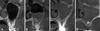
Each examiner was asked to determine the presence or absence of an antrolith and the degree of inflammation of the relevant sinus. The degree of maxillary sinus inflammation was graded as follows: 1, mild (less than one-third opacification of the sinus); 2, moderate (from one-third to two-thirds opacification of the sinus); or 3, severe (greater than two-thirds opacification of the sinus). Additionally, any dental cause of sinus inflammation was assessed. The size of the antroliths was measured as maximum width and height on the CBCT coronal image by 1 examiner and repeated 1 week later to evaluate reproducibility. Antroliths were classified by shape into 3 types based on their dimensions: punctate (3 mm or less in both height and width), linear (3 mm or less in height and more than treble that in width), and amorphous (over 3 mm in both width and height) (Fig. 1).
SPSS version 23 (SPSS Inc, Armonk, NY, USA) was used to analyze the data, and interobserver agreement for antrolith detection was evaluated by calculating the kappa coefficient.9 The chi-square or Fisher exact test was used to compare the prevalence of antroliths by sex, age, and side, and the Kendall tau-b was used to assess the relationships of antrolith multiplicity, shape, and size to the degree of sinus inflammation. P values of <0.05 were considered to indicate statistical significance. The reproducibility of dimensional measurements was analyzed by calculating the coefficient of variation.
Results
The intraobserver agreement in detecting antroliths was almost perfect (k=0.90). A total of 138 (0.99%) of the 13,946 patients (6,016 males and 7,930 females) showed an antrolith in at least 1 sinus. The prevalence was 0.78% and 1.15% in male and female patients, respectively (Table 1). Of these 138 patients, 18 (8 males and 10 females) presented a bilateral manifestation, which resulted in a total of 156 affected sinuses (0.56%). The prevalence was 0.44% on the right side and 0.68% on the left side (Table 2). There were no statistically significant differences between male and female patients, the right and left sides, or among age groups (P>0.05).
Table 3 shows the distribution of antroliths by multiplicity. Of the 156 affected sinuses, 36 (23.1%) had more than 1 antrolith; 28 sinuses contained 2 antroliths, 4 sinuses contained 3 antroliths, 3 sinuses contained 4 antroliths, and 1 sinus contained 7 antroliths. The only statistically significant association in this study was found between the presence of multiple antroliths and the degree of sinus inflammation (P<0.05). Only 8 of the affected sinuses showed definite dental origin: 3 oro-antral fistulas due to tooth extraction, 2 periodontal causes, 1 endodontic cause, 1 implant, and 1 root in the sinus.
Table 4 shows the distribution of antroliths by shape and size according to the degree of inflammation. Among the 207 antroliths, 110 were classed as punctate, 65 as linear, and 32 as amorphous. Amorphous antroliths were relatively frequently observed in moderately or severely inflamed sinuses, but no statistically significant association was found. The dimensions of the antroliths varied from 1 mm2 to 91 mm2 (average, 10.2±15.5 mm2), although 71.5% were small, at less than 10 mm2. No statistically significant difference was observed between size and degree of sinus inflammation. The coefficient of variation was 6.7%.
Discussion
Antroliths are pathological calcifications that form as a result of mineral salt deposition around an organic nucleus in the maxillary sinus.61011 The pathogenesis of antrolith formation is not clearly understood, but long-standing and fungal infections, poor sinus drainage, and the presence of foreign bodies are predisposing factors.61213 Antroliths may be formed by the precipitation of calcium salts around a nidus or concentrated mucus.6 Such a nidus is usually endogenous in origin, such as tooth or bony fragments, blood, pus, mucus, or fungi, but can occasionally be of exogenous origin, such as cotton, paper, dental implants, and gutta-percha points.121415161718 In this study, we found 1 root remnant and 1 implant within the sinuses; in those cases, it was clear that the oro-antral fistula formed by the passage of the root and implant caused sinus inflammation. However, the root and implant did not seem to act as a nidus in those cases because the antroliths were found at a distance from them.
CBCT is known to be an effective method of identifying sinus opacification, and can give valuable information on maxillary sinus inflammation without excessive radiation exposure.19 We found the same held true for antroliths, as the CBCT scans straightforwardly depicted antroliths as dense, irregular, but well-defined masses.
In this study, the intraobserver agreement was almost perfect (k=0.90), and the size measurements varied by only a few millimeters. One discrepancy was observed in differentiating an antrolith from a very small antral exostosis combined with inflammation, and another in distinguishing an antrolith from condensed mucus where a very small spot of slightly higher density than the surrounding inflammation was found. Otherwise, there were no difficulties in identifying antroliths in this study, although previous research has reported that they must be differentiated from supernumerary teeth, root fragments, osteoma, complex odontoma, mature cementoma, periapical condensing osteitis, buccal exostosis, foreign bodies, and even neoplasms.20
The prevalence of antroliths in this study was 0.99% for patients and 0.56% for all sinuses, and no age or sex predilections for antroliths were found, in accordance with previous reports.56 In a study of 500 CBCT examinations of dental patients, Lana et al.21 found antroliths in 16 (3.2%) patients and 18 (1.8%) sinuses. Elsewhere, Rege et al.22 reported that 3.2% of the sinuses reviewed in 1,113 CBCT examinations presented an antrolith. In paranasal sinus CT examinations, Nass Duce et al.6 found antroliths in 3 of 1,957 patients (0.15%).
Prevalence can vary according to the population sample and type of imaging method. For example, patients with a history of dental problems seem to develop antroliths more frequently than other populations, particularly those who have undergone tooth extraction6 or endodontic treatment.2151023 It has been reported that the close proximity of the sinus floor to dental structures may predispose patients to oro-antral irritation, followed by sinus inflammation during dental procedures152324 and that root fragments or endodontic filling material in the sinus may serve as central nidi for antrolith formation.5625 CBCT allows the straightforward detection of both antrolith and maxillary sinus inflammation. This predisposition may explain the relatively high prevalence of antroliths in studies using CBCT to examine a dental population, including the present investigation.622
Antroliths are formed in the context of long-standing sinusitis, and it has been assumed that antrolith formation depends on the severity, duration, and frequency of sinus inflammation. However, sinus inflammation can be asymptomatic for long periods and often goes unrecognized, meaning that clarifying the correlation between clinical features of sinus inflammation and antrolith formation is not straightforward. For this reason, only the severity of sinus inflammation was included in the analysis; the only statistically significant relationship in this study was found between antrolith multiplicity and severity of inflammation, indicating that severely inflamed sinuses tend to form multiple antroliths.
Antroliths can be of any size, but most examples in this study were small; however, since they grow with time, their shape and size could be affected by the duration of sinus inflammation. Unfortunately, information on the duration of inflammation could not be obtained in this cross-sectional study. The two largest antroliths were found in mildly inflamed sinuses, suggesting that severity is not the main factor in their growth. Mild sinus inflammation is very common even in the general population,26 which helps explain the tendency of antroliths to develop in the sinus floor. Linear antroliths were most often observed in mildly inflamed sinuses, indicating that they had grown along the sinus floor.
Most antroliths are asymptomatic and are incidentally discovered through routine radiological examinations.20 Larger examples may present symptoms such as pain, nasal obstruction, and discharge, depending on whether there is a co-existing infection of the involved sinus.6 Surgical removal is considered the treatment of choice, but is only recommended for large antroliths,10 and should be performed together with appropriate treatment of the infection.6
Careful assessment of the maxillary sinus is required before and after the endodontic treatment of upper posterior teeth, the surgical removal of tooth root, or any other procedures involving the sinus floor, such as sinus lift for an implant.
In conclusion, CBCT is an effective imaging modality for identifying antroliths and co-existing sinus inflammation. According to these results, antroliths are not particularly common in the Korean dental population and most are very small and asymptomatic; periodic check-ups therefore appear to be the primary choice of treatment for antroliths.


 XML Download