

 PDF
PDF ePub
ePub Citation
 Print
Print


Introduction
Bisphosphonates, which are inhibitors of osteoclastic bone resorption,1 are useful for the treatment of osteoporosis and bone metastases.23456 However, they are also implicated in the onset of medication-related osteonecrosis of the jaw (MRONJ).178 Patients with MRONJ are often referred to a larger hospital for the evaluation of bisphosphonate-induced changes in the jaws, along with the differential diagnosis of other diseases of the jaws, such as osteoradionecrosis (ORN) or osteomyelitis.9
ORN is a pernicious complication of radiotherapy for head and neck carcinomas.10 The most common cause of ORN is radiation arteritis,111213 which leads to the onset of a hypocellular, hypovascular, and hypoxic environment. Jaws with MRONJ or ORN must be evaluated before any medical procedure is performed.9
Tc-99m hydroxymethylene diphosphonate (HMDP) scintigraphy is capable of demonstrating physiologic changes in bone, and it has been shown that scintigraphy is effective for detecting MRONJ.14 Furthermore, multiple imaging modalities, such as Tc-99m HMDP scintigraphy, computed tomography (CT), and magnetic resonance (MR) imaging, are useful for detecting MRONJ.8 However, to the best of our knowledge, the imaging features of MRONJ and ORN on scintigraphy, CT, and MR imaging have not been presented in the literature. The aim of this study was to evaluate the Tc-99m HMDP scintigraphy, CT, and MR imaging findings of osteonecrosis in the mandible, especially ORN and MRONJ.
Materials and Methods
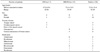
Thirteen patients with MRONJ and 7 patients with ORN in the mandible underwent Tc-99m HMDP scintigraphy, CT, and MR imaging at Radiology, The Nippon Dental University Niigata Hospital from July 2013 to December 2017. Table 1 characteristics of the patients with ORN and MRONJ.
The images were acquired using a 16-MDCT apparatus (Aquilion TSX-101A; Canon Medical Systems, Otawara, Japan), a 1.5-T MR imaging system (EXCELART Vantage MRT-2003; Canon Medical Systems, Otawara, Japan), and a SNC-5100R scintigraphy apparatus (Shimadzu, Kyoto, Japan) with Tc-99m HMDP (Clear Bone Injection; Nihon Medi-Physics, Tokyo, Japan), following our institutional protocol.8141516
The findings of Tc-99m HMDP scintigraphy, CT, and MR imaging of ORN and MRONJ were evaluated by 2 oral radiologists. Scintigraphy was used to analyze areas of increased uptake. CT was used to evaluate osteolytic changes of the jaws, sequestrum separation, and periosteal bone proliferation. In MR imaging, T1-weighted images (T1WI), T2-weighted images (T2WI), short inversion time inversion recovery images (STIR), diffusion-weighted images (DWI), and apparent diffusion coefficient (ADC) maps were obtained.
The associations of scintigraphy, CT, and MR imaging findings with MRONJ and ORN were analyzed using the chi-square test with the Pearson exact test. P values<0.05 were considered to indicate statistical significance.
Results
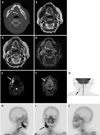
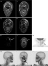
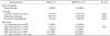
The bone scintigraphy, CT, and MR imaging findings are compared between MRONJ and ORN in Table 2. Thirteen patients with MRONJ and 7 patients with ORN in the mandible showed low signal intensity on T1WI and ADC mapping, high signal intensity on STIR and DWI, and increased uptake on scintigraphy. Periosteal bone proliferation on CT was observed in 69.2% of patients with MRONJ (9 of 13) versus 14.3% of patients with ORN (1 of 7) (P=0.019). Figures 1 and 2 show images of MRONJ and ORN, respectively.
Discussion
Imaging findings are unclear both in the early stages of ORN and when the disease is advanced.111213 Although the radiological findings are nonspecific, they do appear to play a role in the management of MRONJ.17 In this study, we have presented the characteristic imaging findings of MRONJ and ORN on bone scintigraphy, CT, and MR imaging.
Tc-99m HMDP scintigraphy is an effective diagnostic tool for detecting bone changes, and has a higher sensitivity than that of radiography.14 Many authors have reported that scintigraphy showed increased uptake at sites affected by MRONJ.51718 In our study, all cases (MRONJ and ORN) showed positive findings on bone scintigraphy. Arce et al.3 reported that the presence of increased uptake in the bone on Tc-99m HMDP depends on osteoblastic activity and skeletal vascularity. We therefore suggest that scintigraphy shows areas of increased uptake at the sites affected by mandibular diseases.
Bisdas et al.9 reported that CT was useful for detecting predominantly osteolytic diseases and sclerotic lesions in the jaws, with or without periosteal bone proliferation. In our study periosteal bone proliferation on CT was observed in 69.2% of patients with MRONJ (9 of 13) versus 14.3% of patients with ORN (1 of 7) (P=0.019). Therefore, we propose that periosteal bone proliferation is a distinctive characteristic of MRONJ on CT.
MR imaging is useful for the diagnosis of bone marrow disease, and necrosis is usually detected as areas of decreased signal on T1WI.13 Krishnan et al.2 reported that the earliest MR imaging finding of MRONJ was the loss of the normal T1 hyperintensity of fatty marrow in the mandible. In our study, all cases (ORN and MRONJ) showed low signal intensity on T1WI and high signal intensity on STIR. Driessen et al.19 reported that DWI demonstrated consistently high accuracy and high negative predictive value for head and neck cancer. Bonello et al.20 showed that DWI could provide information on microstructural tumor characteristics. Previous studies have reported that DWI and ADC maps were useful for the differential diagnosis of oral and maxillofacial lesions.1516 In our study, all cases (ORN and MRONJ) showed high signal intensity on DWI, and low signal intensity on ADC maps. We therefore consider that DWI and ADC maps reflect the histopathological differences between malignant tumors and inflammatory diseases in the mandible.
Regarding the reasons underlying the different imaging characteristics of MRONJ and ORN, we propose that periosteal bone proliferation on CT in association with ORN is rare because ORN is caused by radiation arteritis.111213
All cases of MRONJ and ORN in our study showed low signal intensity on T1WI and ADC maps, high signal intensity on STIR and DWI, and increased uptake on bone scintigraphy. We suggest that the increased uptake on bone scintigraphy may be correlated with MR imaging findings, especially those on DWI and ADC maps, although the sample was relatively small in this study.
In conclusion, this study presented the characteristic imaging findings of MRONJ and ORN on bone scintigraphy, CT, and MR imaging. Our results suggest that CT can be effective for detecting MRONJ and ORN.
 XML Download