

 PDF
PDF ePub
ePub Citation
 Print
Print


INTRODUCTION
Tumor microenvironments play a fundamental role in tumor generation and progression via interaction with tumor cells [1]. Recently, several studies have reported the importance of tumor microenvironment in breast cancer [23456]. The tumor microenvironment comprises of inflammatory cells, macrophages, extracellular matrix, endothelial cells, and fibroblasts. It affects cancer progression and influences the clinical outcome. Macrophages play an essential role in inflammation, homeostasis, host defense, tissue repair and remodeling, and cancer progression [7]. Tumor-associated macrophages (TAMs) are a major component of the tumor microenvironment and associated with poor prognosis by promoting tumor progression [35]. The main role of TAMs is promotion of tumor growth and metastasis [5]. Macrophages can polarize and exhibit M1 or M2 phenotype [8]. M1 macrophages have a pro-inflammatory function and activate T helper type 1 T cells to induce a lethal T-cell response against pathogens, resulting in tumoricidal activity as well as increased hypoxia [9]. M2 macrophages potentially generate anti-inflammatory cytokines [1]. In addition, M2 type macrophages promote tissue repair and wound healing, and are involved in angiogenesis and tumor progression [1011]. An increased number of polarized M2 macrophages in oral squamous cell carcinoma is associated with lymph node (LN) metastasis [12]. Several immunohistochemical markers are available to evaluate TAMs. CD68 is known as a pan-macrophage marker and facilitates identification of both M1 and M2 macrophages [3]. In breast cancer, a higher degree of CD68+ macrophage infiltration was associated with poor prognosis [5] and was an independent prognostic marker for reduced cancer specific survival in tumor stroma (TS) [3]. CD163 is a scavenger receptor and a highly specific marker for M2 macrophages [13]. Infiltration of CD163+ macrophages into TS of breast cancer is clinically relevant. It is positively correlated with higher histological grade, larger mass/size, Ki-67 proliferating index, hormone receptor negativity, triple-negative/basal-like breast cancer, and inversely correlated with luminal A breast cancer [3]. CD11c is considered a marker of M1 macrophages [14]. To our knowledge, CD11c is yet to be evaluated as a TAM marker in primary breast cancer. In this study, we evaluated the association between the expression of various TAM markers and clinicopathological parameters using tissue microarray (TMA) of 367 invasive breast cancer (IBC) patients. In addition, we evaluated the relationship between clinicopathological parameters and the degree of expression of CD163, CD11c, and CD68 in IBC tissues. Furthermore, the localization of TAMs in TS and tumor nest (TN) in breast cancer was used to determine their prognostic relevance in IBC [3].
METHODS
Patients and clinical data selection
A total of 367 primary IBC patients who underwent surgery at Keimyung University Dongsan Medical Center between 2000 and 2007 were recruited. None of the patients had received preoperative chemotherapy or radiotherapy. All tissues were fixed in 10% buffered formalin and embedded in paraffin. Patient and tumor characteristics, including patient age at the time of diagnosis, tumor size, LN status, histologic grade, and follow-up data, were determined from patients' medical records and pathology reports. Most of the clinicopathological parameters are listed in Table 1. Tumor stage was determined according to the 7th American Joint Committee on Cancer criteria [15]. Overall survival (OS) was defined as the time interval between the date of cancer diagnosis and the date of death from any cause or death related to IBC. Disease free survival (DFS) was defined as the number of months from surgical resection to the development of documented relapse, including recurrence or distant metastasis. TMA blocks were obtained from Keimyung University Human Bio-resource Bank, Korea. The requirement for informed consent from the patients was waived by the ethics committee; this study was approved by the Institutional Review Board at Keimyung University Dongsan Medical Center (DSMC No. 2018-02-007).
Table 1
Mean values of macrophages (CD68+, CD11c+, or CD163+) in TS and TN and relevant clinicopathologic features of the patients
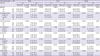
TS = tumor stroma; TN = tumor nest; SD = standard deviation; LN = lymph node; ER = estrogen receptor; PR = progesterone receptor; HER2 = human epithelial growth factor receptor 2; NA = not available.
*Independent t-test.
![]()
TMA construction
Before TMA construction, all tissues were histopathologically studied on hematoxylin and eosin stained slides by a breast pathologist, based on the 2012 World Health Organization Classification [16]. After reviewing the slides, TMAs were constructed using 2 representative cores, each measuring 2.0 mm in diameter, taken from paraffin blocks, and were arrayed in a new recipient block using a manual device (Unitma, Seoul, Korea). In general, the cores were selected from the peripheral aspect of the tumor without hemorrhage or necrosis.
Immunohistochemistry
Immmunohistochemistry (IHC) staining was performed using the automated Benchmark platform (Ventana Medical Systems, Tucson, USA), according to the manufacturer's instructions. Four-micrometer-thick TMA sections were mounted onto slides and deparaffinized followed by antigen retrieval using cell conditioning solution and stained with the UltraView™ Universal DAB detection kit (Ventana Medical Systems). The following primary antibodies were used in this study: estrogen receptor (ER; pre-diluted; Ventana Medical Systems), progesterone receptor (PR; pre-diluted; Ventana Medical Systems), human epidermal growth factor receptor 2 (HER2; pre-diluted; Ventana Medical Systems), Ki-67 (1:200; DAKO Co., Carpinteria, USA), CD68 (1:400; Santa Cruz Biotechnology, Dallas, USA), CD11c (1:100; Abcam, Cambridge, USA), and CD163 (1:200; Thermo Fisher Scientific, Waltham, USA).
Assessment of IHC staining
Two pathologists (H.S.J. and S.Y.K) analyzed the IHC stained slides in a blinded manner. In case of any discrepancy, the analysis was discussed, and the histological examination was repeated. IHC staining of ER and PR was evaluated according to the American Society of Clinical Oncology (ASCO)/College of American Pathologists (CAP) guidelines. The guidelines recommend classification of all cases with at least 1% receptor-positive cells [17]. HER2 expression was also scored according to the ASCO/CAP guidelines [18], which include 4 scores ranging from 0 to 3: no staining or incomplete, faint/barely perceptible membrane staining in ≤10% of invasive tumor cells (score 0); incomplete, faint/barely perceptible membrane staining in >10% of invasive tumor cells (score 1); incomplete and/or weak to moderate circumferential membrane staining in >10% of invasive tumor cells or complete, intense, circumferential membrane staining in ≤ 10% of invasive tumor cells (score 2); and complete, intense, and circumferential membrane staining in > 10% of invasive tumor cells (score 3). A score of 3 for HER2 was considered as positive. HER2 status of equivocal (score 2) cases was determined using silver in situ hybridization. Quantification of Ki-67 was performed by counting nuclear stained cells among 1000 tumor cells [19]. We set a 14% cut-off of Ki-67 for all statistical analyses. In terms of distribution of the intrinsic subtypes of IBC according to IHC, IBC was classified into 5 subtypes: luminal A (ER+ and/or PR+, HER2−, Ki-67 <14%); luminal B, HER2(−) (ER+ and/or PR+, HER2−, and Ki-67 ≥14%); luminal B, HER2 (+) (ER+ and/or PR+, HER2+); HER2-enriched (ER−, PR−, and HER2+); and triple-negative (ER−, PR−, and HER2−) [20]. The expression of CD68, CD11c, and CD163 was determined by counting the number of positive macrophages. After reviewing TMA slides containing cores from each case, a field of maximum intensity of expression was selected. In each case, 3 visual fields with the highest infiltration density of positive macrophages were selected using ImageScope (Aperio Technologies, Vista, USA), yielding a final magnification of 400× (1 mm2). Number of stained macrophages was counted in the 3 visual fields to obtain an average. Only cells with monocytoid/macrophage-like morphology were counted. For statistical evaluation, the number of positive cells was divided into lower and higher groups based on the cut-off points. CD68+, CD11c+, or CD163+ macrophages were counted in the TN and TS, separately. We analyzed the correlations between number of macrophages (CD68+, CD11c+, or CD163+) and the clinicopathological parameters. Additionally, we assessed the impact of CD68+, CD11c+, or CD163+ macrophages in TS and TN in IBC.
Statistical analysis
Statistical analyses were performed using SPSS version 20.0 (IBM Corp., Armonk, USA) or R 3.4.3 (R Development Core Team, Vienna, Austria, https://www.R-project.org). The independent t-test was used to identify the correlation between the number of macrophages (CD68+, CD11c+, or CD163+) and clinicopathological parameters. The independent t-test was also used to identify association between the number of macrophages (CD68+, CD11c+, or CD163+) and molecular subtypes. MaxStat (MaxStat Software, Jever, Germany), a maximal χ2 method in R 3.4.3 was used to identify optimal cut-off points for the number of CD68+, CD11c+, and CD163+ macrophages. Optimal cut-off points of the number of CD68+, CD11c+, and CD163+ macrophages in OS were as follows: CD68 in TS: 17.8, CD68 in TN: 33, CD11c in TS: 75, CD11c in TN: 1, CD163 in TS: 21, and CD163 in TN: 1.67. The cut-off points of the number of CD68+, CD11c+, and CD163+ macrophages in DFS were as follows: CD68 in TS: 17.8, CD68 in TN: 33, CD11c in TS: 75, CD11c in TN: 10, CD163 in TS: 20, and CD163 in TN: 1.67. Kaplan-Meier analysis and log rank tests were used to demonstrate differences in DFS and OS based on CD68, CD11c, and CD163 expression. Cox proportional hazard models were used to determine the hazard ratios (HR) for death from breast cancer or other causes based on CD68, CD11c, and CD163 expression in both uni- and multivariable analysis.
RESULTS
Clinical characteristics
Among 367 patients, 319 (86.9%) were invasive ductal carcinoma, not otherwise specified; 11 (3.0%) were invasive lobular carcinoma; and 37 (10.1%) were a variety of other histological types of carcinomas (5 mucinous, 5 micropapillary, 4 apocrine, 4 metaplastic, 4 medullary, 3 neuroendocrine, 2 cribriform, 2 papillary, 2 solid papillary, 2 tubular, and 4 mixed carcinoma). We classified 134 patients (36.5%) as luminal A, 97 patients (26.4%) as luminal B (HER2−), 53 patients (14.4%) as luminal B (HER2+), 29 patients (7.9%) as HER2-enriched, and 54 patients (14.7%) as triple-negative based on the results of IHC. Median age was 49 years (range 24–84 years). One hundred five patients (28.6%) received mastectomy and 262 patients (71.4%) underwent breast conserving surgery. After surgery, 281 patients (76.6%) received chemotherapy and 153 patients (41.7%) underwent radiation therapy. Of the 256 patients (69.6%) receiving endocrine therapy, 99 patients (26.9%) were treated with tamoxifen only. During the follow-up, 61 patients (16.6%) died and 76 patients (20.7%) had recurrence or metastasis. The median follow-up time was 9.3 years (range 0.1–17.1 years) for all patients.
Clinical significance of infiltration
The CD68+, CD11c+, and CD163+ expression in TN and TS was determined for all the 367 samples. CD68+, CD11c+, and CD163+ macrophages were detected in both TS and TN of IBC (Figure 1). The relationship between the number of macrophages (CD68+, CD11c+, or CD163+) and clinicopathological features is shown in Table 1. High numbers of TAMs (CD68+, CD11c+, or CD163+) in both TS and TN were associated with higher histological grade, higher Ki-67 proliferating index, ER negativity, and PR negativity. High numbers of macrophages (CD11c+ or CD163+) in TS were associated with a larger tumor size. Age, LN metastasis, and HER2 status showed no significant differences.
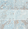 | Figure 1Representative images of IHC analysis of TAM markers in IBC. (A) CD68+ macrophages are mostly present in TS (IHC for CD68, 400× magnification). (B) CD68+ macrophages are present in both TS and TN (IHC for CD68, 400× magnification). (C) CD11c+ macrophages are mainly present in TS (IHC for CD11c, 400× magnification). (D) CD11c+ macrophages are mostly present in TN (IHC for CD11c, 400× magnification). (E) CD163+ macrophages are chiefly present in TS (IHC for CD163, 400× magnification). (F) CD163+ macrophages are present in both TS and TN (IHC for CD163, 400× magnification).IBC = invasive breast cancer; IHC = immunohistochemistry; TAM = tumor-associated macrophage; TS = tumor stroma; TN = tumor nest.
|
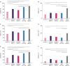
Infiltration densities of all the TAM markers were significantly different among different molecular subtypes (Figure 2). In TS, the luminal A showed significantly lower number of CD68+ macrophages (42.72 ± 27.69) than all other molecular subtypes (p < 0.05) (Figure 2A). In TN, the triple-negative showed significantly higher number of CD68+ macrophages (26.67 ± 33.10) than all other molecular subtypes (p < 0.05) (Figure 2B). In TS, luminal A showed significantly lower number of CD11c+ macrophages (44.63 ± 31.74) than luminal B (HER2−) (62.24 ± 36.04, p < 0.001), HER2-enriched (67.56 ± 33.53, p = 0.001), and triple-negative (77.74 ± 35.94, p < 0.001). The number of positive cells in triple-negative (77.74 ± 35.94) was significantly higher than luminal B (HER2−) (62.24 ± 36.04, p = 0.012) and luminal B (HER2+) (54.53 ± 34.23, p = 0.001) (Figure 2C). In TN, triple-negative showed significantly higher number of CD11c+ macrophages (20.11 ± 29.14) than luminal A (7.51 ± 9.49, p = 0.003), luminal B (HER2+) (10.08 ± 12.23, p = 0.023), and HER2-enriched (7.97 ± 9.40, p = 0.007). The number of positive cells in luminal A (7.51 ± 9.49) was significantly lower than luminal B (HER2−) (12.57 ± 14.50, p = 0.003) (Figure 2D). In TS, luminal A showed a significantly lower number of CD163+ macrophages (33.36 ± 25.78) than all other molecular subtypes (p < 0.05) (Figure 2E). In TN, the triple-negative showed a significantly higher number of CD163+ macrophages (17.72 ± 23.97) than all other molecular subtypes (p < 0.05) (Figure 2F).
Prognostic significance of CD68+, CD11c+, and CD163+ macrophages
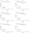
We found that infiltration of higher number of CD68+ macrophages in TS was not correlated with OS, but infiltration in TN was correlated with unfavorable OS (Figure 3A and B). Infiltration of higher number of CD11c+ macrophages in TS was correlated with favorable OS, but infiltration in TN was not correlated with OS (Figure 3C and D). Like CD68+ macrophages, infiltration of higher number of CD163+ macrophages in TS was not correlated with OS, but infiltration in TN was correlated with unfavorable OS (Figure 3E and F).
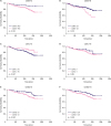 | Figure 3The OS curves according to the infiltration density of CD68+ macrophages in TS (A) and TN (B), CD11+ macrophages in TS (C) and TN (D), and CD163+ macrophages in TS (E) and TN (F) in IBC.OS = overall survival; TS = tumor stroma; TN = tumor nest; IBC = invasive breast cancer.
|
Additionally, the infiltration of higher number of CD68+ macrophages in TS was not correlated with DFS, but infiltration in TN was correlated with unfavorable DFS (Figure 4A and B). Infiltration of higher number of CD11c+ macrophages in TS was correlated with favorable DFS, but infiltration in TN was not correlated with OS (Figure 4C and D). Like CD68+ macrophages, infiltration of higher number of CD163+ macrophages in TS was not correlated with DFS, but infiltration in TN was correlated with unfavorable DFS (Figure 4E and F).
 | Figure 4Disease free curves based on the infiltration density of CD68+ macrophages in TS (A) and TN (B), CD11+ macrophages in TS (C) and TN (D), and CD163+ macrophages in TS (E) and TN (F) in IBC.TS = tumor stroma; TN = tumor nest; IBC = invasive breast cancer.
|
Univariate Cox regression analyses revealed that PR positivity, HER2 overexpression, higher Ki-67 proliferating index (≥ 14%), larger tumor size, LN metastasis, and CD68+ or CD163+ macrophages in TN were independent prognostic factors for OS. HER2 overexpression, higher Ki-67 proliferating index (≥ 14%), larger tumor size, LN metastasis, CD11c+ macrophages in TS, and CD68+ or CD163+ macrophages in TN were independent prognostic factors for DFS (Table 2).
Table 2
Univariate Cox regression analyses for OS and DFS

OS = overall survival; DFS = disease free survival; HR = hazard ratio; CI = confidence interval; ER =estrogen receptor; PR =progesterone receptor; HER2 = human epidermal growth factor receptor 2; LN =lymph node; HG = histologic grade; TS = tumor stroma; TN = tumor nest.
![]()
Multivariate analyses revealed that larger tumor size, CD11c+ macrophages in TS, and CD163+ macrophages in TN were an independent prognostic factor for DFS. In addition, HER2 overexpression and LN metastasis were independent prognostic factors for both OS and DFS (Table 3).
Table 3
Multivariate Cox regression analyses for OS and DFS

OS = overall survival; DFS = disease free survival; HR = hazard ratio; CI = confidence interval; PR = progesterone receptor; HER2 = human epidermal growth factor receptor 2; LN = lymph node; TS = tumor stroma; TN = tumor nest; NA = not available.
![]()
DISCUSSION
In this study, we found that a high degree of TAM infiltration in IBC was associated with adverse clinical and pathological prognostic parameters, such as high histological grade, large tumor size, ER negativity, PR negativity, and high Ki-67 proliferating index. We found that the high expression of TAMs in breast cancer correlated with negative hormone receptor status. These results were similar to those of previous studies, which reported that high expression of CD68 or CD163 was associated with hormone receptor negativity [2346]. We found no significant association between HER2 status and TAMs. Several studies have disputed the association between macrophage infiltration and HER2 status. A few studies have reported that TAMs are associated with HER2-positivity [24]. Other studies showed no association between TAMs and HER2 status [36]. We found that the higher number of TAMs in IBC correlated with a higher expression of tumor proliferation marker, Ki-67, as reported previously [6]. Our study showed that luminal A breast cancer contained fewer CD68+, CD11c+, and CD163+ cells when compared with triple-negative breast cancer. This result corroborated the results of a previous study suggesting that the triple-negative/basal-like breast cancer contained more CD163+ or CD68+ macrophages in TS than in luminal A breast cancer [3].
Programmed cell death 1 (PD-1) is a receptor that regulates the activation and apoptosis of inflammatory cells. PD-ligand 1 (PD-L1) binds to PD-1 leading to a reduced immune response of the tumor cells. In breast cancer, PD-L1 expression is associated with LN metastasis, higher histological grade, ER negativity, and triple-negative breast cancer [21]. PD-L1 expression has been associated with poor prognosis in various human cancers [22]. In triple-negative breast cancer, TAMs regulate the activity of anti-PD-1 and PD-L1 agents by secreting cytokines [23].
Macrophage polarization into M1 or M2 phenotypes leads to varied cytokine secretion and function. M1 macrophages generate interleukin (IL)-12 and tumor necrosis factor with lethal antimicrobial and antitumor effects in cells. M2 macrophages produce cytokines, including IL-10, IL-1 receptor antagonist type II, and IL-1 decoy receptor. They regulate the immune response and adaptive immunity and promote angiogenesis and tissue repair. TAMs are functionally similar to M2 macrophages due to the secretion of IL-10 and transforming growth factor-beta (TGF-β). By expressing characteristics of M2 macrophages, TAMs promote tumor growth and metastasis [8].
CD68 is best known as a pan-macrophage marker and commonly used as a marker for TAMs [5]. Previous studies have reported that higher CD68+ macrophage infiltration in IBC is associated with larger tumor size and unfavorable prognosis [35]. It was suggested that IBC interacts with TAMs and induces an immune response that supports tumor growth. Infiltration of CD68+ macrophages correlated with poor prognosis in breast, cervix, and bladder carcinoma, but with favorable prognosis in prostate, lung, and brain tumors [24]. These results are probably due to similar CD68 expression by both M1 and M2 macrophages. In our study, the infiltration of CD68+ macrophages in TN showed poor prognosis and infiltration into TS showed a trend of unfavorable prognosis.
CD163 is the optimal marker for M2 macrophages [13]. High numbers of CD163+ macrophages in breast cancer are associated with rapid proliferation, poor differentiation, ER negativity, and ductal histological type [25]. In this study, the higher numbers of CD163+ macrophages in TS or TN were associated with poor prognosis, high histological grade, larger tumor size, high Ki-67 proliferating index, ER negativity, and PR negativity. In pancreatic cancer, the increased number of CD163+ macrophages within the tumor invasive front was associated with LN metastasis and poor prognosis [26]. High-grade tumor may secrete higher levels of monocyte colony stimulating factor, IL-10, and/or TGF-β, which results in the high number of CD163+ M2 macrophages [25]. Considering that M2 macrophages play a role in tumor progression by producing vascular endothelial growth factor and extracellular matrix remodeling proteins [12], the increased number of CD163+ macrophages might explain an unfavorable prognosis.
Tumor infiltrating mature dendritic cells have been known to be associated with a favorable prognosis; however, immature dendritic cells are not associated with a favorable prognosis [27]. CD11c is overexpressed in myeloid- and monocyte-associated dendritic cells, in natural killer cells, macrophages, and even a few activated B and T cells [28]. In our study, the infiltration of CD11c+ macrophages in both TS and TN was associated with high histological grade, high Ki-67 proliferating index, ER negativity, and PR negativity. Further, the infiltration of CD11c+ macrophages in TS was associated with favorable OS and DFS. Interestingly, the CD11c+ macrophages in TS were associated with large tumor size. However, infiltration of high number of CD11c+ macrophages in TS was an independent predictor of favorable OS or DFS. In gastric cancer, high expression of CD11c reduced the risk of death and relapse compared with patients showing a low expression, except for tumor size [27]. Consequently, CD11c expression may be a potential indicator of tumor size.
In our patients, the infiltration of CD68+ or CD163+ macrophages in TN correlated with clinicopathological features and was associated with unfavorable OS and DFS, while the infiltration of CD11c+ macrophages in TS was associated with favorable OS and DFS. In melanoma, the infiltration of CD68+ TAMs in TN correlated with poor OS and DFS [29]. In endometrial cancer, the infiltration of CD68+ cells in TN showed positive correlation with decreased recurrence [30]. Considering these results, it was suggested that when examining TAMs in malignant tumors, localization should also be considered apart from their mere presence. However, 1 breast cohort study showed that CD68+ macrophages in the TS, but not in the TN, were correlated with clinicopathological features or survival [3]. Although our result showed that the infiltration of TAMs in TN appears to be associated with patient outcomes in IBC, further evaluation is still needed.
We found that increased abundance of TAMs including M2 macrophages are associated with tumor progression in IBC. The infiltration of CD68+ or CD163+ macrophages in TN suggested unfavorable prognosis for breast cancer patients, whereas CD11c+ macrophages in TS lead to favorable prognosis. In addition to simply analyzing the degree of TAM infiltration, it is also important to analyze the location of TAMs as a prognostic marker. Finally, the lack of a reliable M1 macrophage marker appears to be a challenge in studies investigating macrophages.
 XML Download