

 PDF
PDF ePub
ePub Citation
 Print
Print


INTRODUCTION
CD9 is a member of the tetraspanin family which is involved in a variety of biological processes such as cell adhesion, motility, and proliferation due to its interactions with integrins, growth factor receptors, transmembrane proteins, and signaling molecules [1]. It has 4 distinct and functionally important regions: the extracellular domain which consists of a large extracellular loop and a small extracellular loop; the transmembrane domain; palmitoylation site; and a cytoplasmic domain comprising of a short inner loop, and N-terminal and C-terminal tails [12]. The extracellular loops mediate specific interactions with other proteins, the transmembrane domains serve as sites for tetraspanin-tetraspanin interactions to stabilize the individual tetraspanins, and the C-terminal cytoplasmic tail interacts with the intracellular signaling molecules, such as protein kinase C and phosphatidylinositol 4-kinase (PI4K) [12]. CD9 is widely expressed in various normal and cancer tissues [3]. Previous studies have reported conflicting results on the prognostic value of CD9 expression in different types of cancer [456789101112]. A prior study focusing on the histological type of breast cancer [13], the invasive carcinoma of no special type (invasive carcinoma no special type [NST]) revealed that CD9 was overexpressed in the tumor cells (TCs) compared to the non-neoplastic mammary epithelial cells, and the degree of expression varied between the patients. In addition, CD9 expression was associated with clinicopathological features linking to aggressive tumor behavior and correlating to poor prognosis in breast cancer patients.
Invasive lobular carcinoma (ILC) is a special type of breast cancer that accounts for 5%–15% of all invasive breast cancers worldwide [14]. ILC is characterized by non-cohesive TCs that are individually dispersed or arranged in a single file linear pattern with loss of expression of the cell-cell adhesion molecule, E-cadherin. It depicts different molecular profiles, metastatic patterns, and clinical behaviors compared to the invasive carcinoma NST. ILC has clinicopathological features for favorable prognosis such as low grade, estrogen receptor (ER)-positive, human epidermal growth factor receptor 2 (HER2)-negative, and low proliferation. However, previous studies have reported either a worse prognosis for ILC compared to the invasive carcinoma NST or no significant difference between the two histological types [14]. Owing to the relatively rare cases of ILC (2%–5%) in Korea compared to all invasive cancers [131516], the clinical focus of biomarkers for breast cancer has been mostly investigating the invasive carcinoma NST. Therefore, in this study, we aimed to investigate the prognostic significance of CD9 expression in patients with ILC and confirm the collective prognostic importance of CD9 expression in breast cancer regardless of the histological subtype.
METHODS
Case selection
A total of 141 ILCs surgically resected between 2001 and 2013 were collected from the pathology archives of our institution. Tumor representative blocks were selected for immunohistochemistry (IHC) after reviewing the corresponding hematoxylin and eosin (H&E) slides. 113 cases with complete clinicopathological data and respective tumor blocks were included in this study. All the patients had received standard radiotherapy or adjuvant systemic therapy (hormone therapy or chemotherapy) after surgery. Clinicopathological information such as age, tumor size, lymph node (LN) status, lymphovascular invasion (LVI), histologic grade, Ki-67 labeling index, and survival data, including recurrence or metastasis were obtained from the pathological reports and patients' medical records. Overall survival (OS) was defined as the period from the date of surgical resection until the date of death due to any cause or the last follow-up. Disease-free survival (DFS) was defined as the period from the date of surgical resection until the date of detection of locoregional recurrence, distant metastasis, death, or the last follow-up. This study was approved by the Institutional Review Board of Yeungnam University Hospital (YUMC 2017-09-037), which waived the requirement for the informed consent of the patients.
Immunohistochemistry
CD9 expression was evaluated by IHC on the whole-mount slides using a rabbit monoclonal antibody for the C-terminal cytoplasmic domain of CD9 (clone EPR2949, 1:1,200 dilution; Abcam, Cambridge, UK). IHC was performed using the automated Benchmark® platform (Ventana Medical Systems, Tucson, USA). We elucidated CD9 immunoreactivity in TCs after considering the intensity as well as the extent of staining. Intensity was evaluated in the form of score as follows: negative (0), weak positive (1), moderately positive (2), or strong positive (3). The extent of staining was quantified as the percentage of positive TCs showing membranous-to-cytoplasmic staining: 0% (0), 1%–25% (1), 26%–50% (2), 51%–75% (3), or > 75% (4). The final CD9 immunoreactivity score (IS) was determined by multiplying the intensity score and the extent score (range, 0–12). For statistical analyses, we divided the cases into positive or negative based on the mean IS (negative, IS < 4 and positive, IS ≥ 4).
IHC for ER, progesterone receptor (PR), HER2, and Ki-67 was performed at the time of diagnosis. ER and PR were considered to be positive if there was a nuclear immunoreactivity in at least 1% of the TCs in the tissue sample [17]. HER2 was confirmed to be positive only on the intensity score of protein overexpression (3+). For five cases with equivocal immunostaining (2+), silver in situ hybridization (INFORM HER2 DNA probe kit; Ventana Medical Systems) was performed, and three of them showed gene amplification according to the current guidelines [18]. The Ki-67 labeling index was expressed as the percentage of positive cells among at least 500 tumor cells.
Statistical analysis
Statistical analyses were performed using the SPSS statistical software for Windows (version 23.0, IBM Co., Armonk, USA). The χ2 test or Fisher's exact test was used for evaluating the correlation of CD9 expression with the clinicopathological data of the patient. OS and DFS curves were plotted using the Kaplan-Meier method and the log-rank test was used to analyze the significance of differences in survival. Cox proportional hazards model was used for comparing the hazard ratios (HRs) in the univariate as well as multivariate analyses. Adjusted HRs and the associated 95% confidence intervals (CIs) were estimated for each variable. Significant variables identified in the univariate analysis were further analyzed in the multivariate analysis by backward stepwise selection method. All the tests were two-sided, and a p-value of < 0.05 was considered to be statistically significant.
RESULTS
Patient demographics
For the 113 cases, the median age at diagnosis was 48 years (range, 27–88 years). Tumor sizes among the patients ranged from 0.3 to 9.5 cm (mean, 2.6 ± 1.8 cm). 56 (49.6%) patients had an invasive tumor ≤ 2 cm, and the remaining 57 (50.4%) patients had an invasive tumor > 2 cm. Axillary LN metastasis was found in 38 (33.6%) patients, and LVI was found in 41 (36.3%) patients. The histologic grade 1was observed in 24 (21.2%) patients, grade 2 in 70 (62%) patients, and grade 3 in 19 (16.8%) patients. A total of 40 (35.4%) patients underwent breast conserving surgery while 73 (64.6%) patients underwent mastectomy. The surgical margins were free from tumor in patients who underwent breast conserving surgery. During a median follow-up of 62 months (range, 7–187 months), recurrence was observed in 12 (10.6%) patients. The first recurrence was loco-regional which was seen in 2 (1.8%) patients and distant metastasis was noted in 10 (8.8%) patients. At the last follow-up, 9 (8.0%) deaths were reported.
Correlations between CD9 expression and clinicopathological parameters
Non-neoplastic mammary epithelial cells were observed to be negative or weakly positive for CD9 expression. Stromal inflammatory cells exhibited moderate-to-strong immunoreactivity for CD9. Immunoreactivity intensities for CD9 in TCs varied between cases, however were similar in individual cases (Figure 1). The distribution of CD9 ISs was as follows: 0 in 40 (35.4%) cases, 1 in 1 (0.9%) case, 2 in 16 (14.2%) cases, 3 in 8 (7.1%) cases, 4 in 30 (26.5%) cases, 6 in 2 (1.8%) cases, 8 in 15 (13.3%) cases, and 9 in 1 (0.9%) case. There was no case with an IS of 12. Positive CD9 expression (IS ≥ 4) was observed in 48 (42.5%) cases.
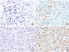 | Figure 1Representative immunohistochemical results for CD9 expression in invasive lobular carcinoma. (A, B) Negative CD9 expression in non-neoplastic mammary glands and positive CD9 expression in plasma cells infiltrated within lobules. (C, D) Diffuse and strong CD9 expression in tumor cells. A and C, hematoxylin and eosin stain; B and D, immunohistochemistry for CD9; magnification of all figures, ×400.
|
Positive CD9 expression was not significantly correlated with the reported clinicopathological variables of the patient including age, tumor size, LN metastasis, LVI, histologic grade, ER or PR expression, HER2 status, and Ki-67 labeling index (Table 1).
Table 1
Patient characteristics according to CD9 expression in invasive lobular carcinoma
![]()
Prognostic significance of CD9 expression
Patients with CD9 expression had shorter DFS (p = 0.014) and tended to have worse OS (p = 0.051) compared to patients without CD9 expression (Figure 2). In addition to CD9 expression status, multivariate analysis including tumor size, LN status, and LVI was performed. The analysis revealed that CD9 expression in TCs was an independent marker for DFS (HR, 3.745; 95% CI, 1.007–13.926; p = 0.049), along with LVI (Table 2), in patients with ILC.
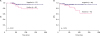 | Figure 2Kaplan-Meier survival curves according to CD9 expression in patients with invasive lobular carcinoma. (A) OS. (B) DFS.OS = overall survival; DFS = disease-free survival.
|
Table 2
Univariate and multivariate analyses of clinicopathological variables affecting disease-free survival in invasive lobular carcinoma

![]()
DISCUSSION
We observed CD9 expression in 42.5% of patients with ILC and high CD9 expression was associated with a poor clinical outcome. We showed that CD9 expression in TCs was an independent prognostic marker for DFS in patients with ILC. In our previous study considering breast cancer samples of varied histological subtypes, majority of them were invasive carcinoma NST. The study had revealed that patients with CD9 expression had shorter DFS compared to patients without CD9 expression. In addition, CD9 expression was more frequent in invasive carcinoma NST and micropapillary subtypes however less frequent in mucinous, tubular, medullary, and papillary subtypes which are considered to be associated with a favorable prognosis [13]. As rare cases are observed with some special histological subtypes, we could not perform subgroup analysis for prognostic significance of CD9 expression based on the histological subtypes in the previous study. Our hypothesis was to highlight CD9 expression as of prognostic importance in breast cancer regardless of histological subtype. Consequently, we collected more cases of ILC which is the second most common histological subtype of breast cancer, and observed that CD9 expression can be correlated with poor clinical outcome in patients with ILC too.
In the previous study which consisted of many invasive carcinoma NST cases, CD9 expression was significantly associated with LN metastasis, LVI, histologic grade 3, and HER2-positive [13]. However, we did not observe the same trends in this study with ILC samples. No correlation between CD9 expression and the histologic grade or HER2 status was observed possibly due to the infrequency of ILC cases with histologic grade 3 or HER2-positive in this study. In addition, the relatively small number of ILC patients in this study population may have led to failure in observing statistically significant correlation between CD9 expression, and LN metastasis or LVI. Despite the statistical data, it has been observed that CD9-positive ILC patients tend to have LN metastasis and LVI more frequently.
Our findings of correlation between CD9 expression in TCs and an unfavorable prognosis are consistent with some of the previous studies [910111920]. CD9 expression is associated with an advanced tumor stage, LN metastasis, LVI, and poor prognosis in gastric and esophageal cancers [91011]. The role of CD9 in breast cancer invasion and metastasis might explain the correlation between CD9 expression in TCs and worse prognosis of breast cancer patients in our study. CD9 crosslinking can induce matrix metalloproteinase 2 (MMP2) transcription by activating intracellular signaling molecules such as PI4K and Src homology 2, which result in increased invasiveness of TCs [19]. In addition, CD9 has been known to interact with transforming growth factor-α which is protected from cleavage and leads to persistent epidermal growth factor receptor (EGFR) activation in epithelial cells [1920]. Altered cellular proliferation induced by activated EGFR signaling may also contribute to the decreased survival of patients with CD9-expressing tumors. In our future study, we plan to investigate the correlation between CD9 expression and the expression of these CD9-interacting molecules and study their prognostic significance. Many in vitro and in vivo studies have been published in support with our results of this study. One such study shows that CD9-deficient breast cancer cells (MDA-MB-231) exhibit decreased invasion into a layer of multipotent mesenchymal stromal cells in vitro, and CD9 knockdown inhibited tumor growth and metastasis of MDA-MB-231 in mouse xenograft [21]. Kischel et al. [22] reported that CD9 was overexpressed in osteotropic breast and prostate cancer cell lines and when the osteotropic cancer cell xenografts were treated with anti-CD9 antibodies in vivo, the rate of bone destruction was significantly decreased.
Earlier studies have reported that decreased CD9 expression correlates with the tumor progression in breast cancer, and esophageal and oral squamous cell carcinomas [4567]. The reasons for such contradictory results remain to be elucidated. However, whether CD9 promotes or inhibits tumor progression probably depends on its interacting molecules. High CD9 expression is accompanied by high-level integrin expression that strengthens the adhesion of the TCs to the extracellular matrix. Such a CD9 complexed with fibronectin-bound integrins can interfere with MMP2 transcription [19]. In addition, we used an antibody that recognized the C-terminal cytoplasmic tail of CD9 while in previous studies, other researchers used an antibody against the extracellular loop of CD9. CD9 antibodies targeting different functional domains could be another explanatory reason for the difference in the clinical significance of CD9 expression between the previous studies and our study.
Our previous as well as present study support that CD9 may interact with specific molecules to promote invasion and metastasis of breast cancer cells regardless of the histological subtype [13]. We found that CD9 expression showed no association with the predicted conventional tumor characteristics which indicates that CD9 expression in TCs confers additional importance in defining a subgroup of ILC patients with poor prognosis. This understanding is further supported by the finding that CD9 expression in TCs is a prognostic factor for ILC independent of tumor size, LN status, and LVI by multivariate analysis.
Longo et al. [23] reported that CD9 was localized at the TC-endothelial cell contact area during the active transmigration of TCs across endothelial cell monolayers grown on 3-dimensional collagen matrices. They also found that the treatment with anti-CD9 monoclonal antibodies specifically inhibited the transendothelial migration of melanoma cells. Kohmo et al. [24] reported that CD9 expression was increased in chemoresistant small cell lung cancer (SCLC) and inhibition of CD9 by monoclonal antibodies or small interfering RNA induced apoptosis of CD9-expressing chemoresistant SCLC cells. These findings suggest that CD9 could be useful as a therapeutic target for cancer treatment. However, further studies are required to determine the potential of anti-CD9 monoclonal antibodies to inhibit tumor growth and improve the clinical outcome in breast cancer patients with CD9 expression.
In conclusion, we suggest that CD9 expression is associated with worse prognosis of patients with ILC by univariate and multivariate analyses. Therefore, CD9 expression could be a useful prognostic biomarker in patients with ILC. We plan to expand this study involving similar investigations in other histological subtypes of breast cancer.
 XML Download