

 PDF
PDF ePub
ePub Citation
 Print
Print


INTRODUCTION
The dawn of rapid palatal expansion (RPE) dates back to 1860, with Angell cited as the founding father. His claim, however, was perceived with much resistance as many disputed the validity and capability of maxillary separation. It was not until the advent of radiology that RPE re-emerged in the United States.1 In 1956, a German scientist named Korkhaus visited the department of orthodontics at the University of Illinois. His cephalometric records of cases treated using RPE provoked the curiosity of colleagues, such as Brodie and Haas, and ultimately led to the reintroduction of RPE in the country.2
Since then, much has been learned about the biology of the midpalatal suture, the concomitant skeletal and dental effects, and the various indications of palatal expansion in correcting a malocclusion. However, as its popularity in correcting transverse maxillary deficiency began to increase, so did interest in the long-term stability of the procedure.
Previous studies have evaluated the long-term stability of RPE by using various measurement techniques ranging from manual measurement of dental casts3 to plain film radiographic techniques45 to digital imaging.6 However, more sophisticated techniques for evaluating morphological changes in the dentofacial complex have been developed. The advent of the surface laser scanner and companion software has made it possible to generate three-dimensional (3D) images from plaster casts and, through various viewing functions, offer greater accuracy and precision in measurement. The purpose of this study was to assess the long-term maxillary and mandibular arch width stability of RPE followed by fixed edgewise appliances. One experienced practitioner treated the entire sample at his private practice, and the arch dimensions were analyzed over the longest known period of time reported in the literature.
MATERIALS AND METHODS
Subjects
This study was approved by the Saint Louis University Institutional Review Board (protocol #22020). All patients and parents provided written informed consents.
The sample consisted of 197 paired dental casts obtained from 67 patients (53 female and 14 male) treated by a single practitioner. To be included in the study, the pretreatment and posttreatment dental casts had to be available, the pretreatment dental casts had to be obtained under 18 years of age, and all the cases had to be treated using Haas-type RPE and subsequent non-extraction edgewise appliance therapy. All patients exhibited posterior crossbites and/or narrow maxillary arches (i.e., ≤ 31 mm maxillary intermolar width, as suggested by McNamara et al.6).
Sample size calculation for 80% power and a 5% significance level was performed with the Epi Info® 7 software (Centers for Disease Control and Prevention, Atlanta, GA, USA) by using the parameters from a previous study.6 This calculation showed that the available sample was sufficient.
The patients underwent a standardized protocol of Haas-type RPE with two turns a day (0.25 mm per turn) until the expansion screw reached 11 to 14 mm. The desired expansion was achieved when the mandibular arch was completely contained by the maxillary arch. The Haas expander was retained on the teeth as a passive retainer for an average of 3 months. After expansion, all patients received full maxillary and mandibular fixed standard edgewise appliances. The retention protocol after orthodontic treatment consisted of an upper removable appliance and a lower fixed lingual retainer from canine to canine, worn for approximately 6.5 years.
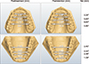
Dental casts were obtained at three observation times: pretreatment (T1), after expansion and fixed appliance therapy (T2), and at long-term recall (T3). The mean age was 12 years 3 months ± 2 years 5 months at T1, 17 years 0 months ± 3 years 11 months at T2, and 27 years 11 months ± 6 years 2 months at T3. The mean duration of the T1–T2 and T2–T3 periods was 4 years 10 months ± 3 years 6 months and 11 years 0 months ± 5 years 5 months, respectively.
Data collection
The dental casts were digitized using the R700™ inoffice model scanner (3Shape, Copenhagen, Denmark). Three scans were performed using the R700™ for each set of dental models: the full maxillary model, the full mandibular model, and the models together in occlusion. The software used a best-fit algorithm to automatically fit the individual full scans with the occlusion scan to produce on-screen digital models with an accurate occlusion.7
Landmark acquisition
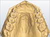
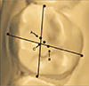
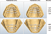
Ortho Analyzer™ (3Shape) was used to compute the midpoints and thus identify the landmarks on the 3D models. A point was placed on the distal, facial, mesial, and lingual surfaces of the canines, premolars/primary molars, and first permanent molars in the same arch (Figure 1). These points were selected in accordance with the guidelines established by Moyers et al.8 to determine the geometric center of each tooth, i.e., the tooth centroid (Figure 2). This point provided a more valid measurement of arch width because it eliminated the effect of tooth rotation. The midpoints and landmarks were not recorded if the teeth were in the process of eruption before the height of the contours of the four outer surfaces (mesial, distal, facial, and lingual) were visible.
Measurements
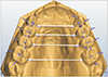
The Ortho Analyzer™ software was also used to measure arch width at the following teeth: primary canines/permanent canines, first primary molars/first premolars, second primary molars/second premolars, and first permanent molars (Figure 3). Arch width was evaluated using two sets of measurements: from the lingual point of a given tooth to the same point on its antimere, and between the centroid of a tooth and its antimere as described by Moyers et al.8
Error of method
To test the reliability of the model measurements, a random number generator (www.random.org)9 was used to select 10% of the sample for re-evaluation. Intraclass correlation was estimated using Cronbach's alpha. Reliability is commonly considered “adequate” when the intraclass correlation coefficients are equal to or greater than 0.80. For all variables, the reliability coefficients were found to be greater than 0.80. Therefore, all data were reliable.
Statistical analysis
Maxillary pretreatment (T1) comparisons between the treated subjects and corresponding reference data provided by Moyers et al.8 were performed using Student's t-test for independent samples.
Comparisons between the treatment time points of the treated subjects were performed using Student's t-test for dependent samples. The following statistical comparisons were performed: evaluation of treatment changes (T2–T1), evaluation of postretention changes (T3–T2), and evaluation of overall changes (T3–T1).
For each subject, the age- and sex-specific z scores were calculated for arch width by using the reference data reported by Moyers et al.8 The z scores provided the subjects' deviations (in standard units) from values expected for untreated subjects and obviated dimensional adjustments due to arch changes that normally occurred between the mixed and permanent dentitions. The z scores were then compared using Student's t-test for dependent samples. The following statistical comparisons were performed: z score evaluation of treatment changes (T2–T1), z score evaluation of postretention changes (T3–T2), and z score evaluation of overall changes (T3–T1).
RESULTS
Maxillary arch
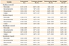
At T1, the maxillary arch widths of the treated patients were significantly narrower than the corresponding dental arch widths of the established reference data. Maxillary arch widths increased significantly (p < 0.05) during treatment (T2–T1) (Table 1 and Figure 4). At the centroid level, intercanine widths increased the least (3.18 ± 2.47 mm) and inter-second premolar widths increased the most (7.40 ± 2.56 mm). Lingual arch widths followed a similar pattern, increasing the most at the second premolars (7.10 ± 2.43 mm) and the least at the canines (1.56 ± 2.60 mm). The ratio of the centroid width increase to the corresponding lingual width increase, which provides a rough measure of tipping, was the greatest for the canines (2.0:1) and the least for the second premolars (1.0:1). The z scores, adjusted for age and sex, showed significant treatment-related increases in arch width at the centroid (Table 2 and Figure 5).
With the exception of intermolar width, maxillary arch widths decreased significantly postretention (T3–T2) (Table 1 and Figure 4). Width decreases ranged from 9% of the treatment-related increase at the second premolars to 27% of the treatment-related increase at the canines. Compared to untreated reference data (Table 2 and Figure 5), with the exception of the first molar, arch width significantly decreased more than was expected posttreatment. Arch width at the first molar decreased more than expected; however, the difference was not statistically significant.
When considering the net change of treatment (T3–T1), maxillary arch widths at both the centroid and lingual levels increased significantly (Table 1 and Figure 4). At the centroid level, intercanine widths increased the least and inter-second premolar widths increased the most. The z scores were greater than expected and significant for the overall observation period (Table 2 and Figure 5).
Mandibular arch
Mandibular arch widths also increased significantly during treatment (T2–T1) (Table 1 and Figure 4). Intercanine widths increased the least (1.66 ± 1.49 mm), and inter-second premolar widths increased the most (3.07 ± 2.20 mm). Lingual arch widths increased the most at the second premolars (3.02 ± 2.54 mm) and the least at the canines (0.58 ± 1.70 mm). The ratio of the centroid-to-lingual-width expansion was the least for the second premolars (1.0:1) and the greatest for the canines (2.9:1). The z scores showed that all mandibular arch width dimensions increased during treatment significantly more than expected for the untreated controls (Table 2 and Figure 5).
With the exception of intermolar width, mandibular arch widths decreased significantly over the postretention period (T3–T2) (Table 1 and Figure 4). Width decreases ranged from 12% of the treatment increase at the first premolar to 35% at the canine. With the exception of the intermolar width, the z scores showed a greater than expected decrease in arch widths, but the differences were not statistically significant (Table 2 and Figure 5).
Despite postretention relapse, mandibular arch widths at both the centroid and lingual levels increased significantly in the overall observation period (T3–T1) (Table 1 and Figure 4). At the centroid level, intercanine widths increased the least and intermolar widths increased the most. The z scores showed that throughout the observation period, mandibular arch width increases were significantly greater than those in the untreated controls (Table 2 and Figure 5).
DISCUSSION
The present longitudinal study assessed arch width changes that occurred in patients treated using RPE followed by edgewise appliances. The data from these treated patients were then compared to established reference data via z scores to obviate the growth changes that normally occur between the mixed and permanent dentitions. Therefore, the z scores were used as a means to compare our results to a “normal” population (reported by Moyers et al.8), since we were unable to evaluate a matched untreated control group throughout such a long period. An important point to consider when evaluating the results of this study is that pretreatment records were often taken during the mixed dentition. Thus, the measurements were not recorded from the time deciduous teeth were shed to full eruption of the permanent successors. Moreover, the single clinician who treated this sample often used Kloehn cervical headgear or a protraction chin cup in addition to edgewise appliances.10 Posttreatment arch width measurements may have been influenced by the use of extraoral forces via the headgear.
When analyzing the literature, a direct comparison of the outcomes of this study can be performed appropriately with the results reported by Moussa et al.,3 McNamara et al.,6 and Lima et al.11 (Table 3). With the exception of Lima et al.,11 these authors used a treatment protocol that was very similar to the one evaluated in the present investigation. All of the studies used a tissue-borne device for RPE (the Haas expander) that was applied to the maxillary arch. Moussa et al.3 and McNamara et al.6 followed RPE with a subsequent phase of edgewise appliances, whereas Lima et al.11 used only RPE (no subsequent orthodontic intervention). All of the studies used a similar retention protocol. The long-term stability of RPE is still under investigation in the literature,1213 but it is important to highlight that the present study presents a large and controlled sample for an extended postretention assessment.
Treatment with RPE followed by fixed appliances, T2–T1, produced significantly greater increments in all the variables for maxillary and mandibular arch widths when compared to the reference data. This finding is consistent with the treatment effects reported by McNamara et al.6 Maxillary expansion was greater in the posterior than anterior aspect of the arch, as previously reported with RPE therapy.4 Although some tipping of the buccal segments occurred, much of the expansion involved translation of the teeth, reflecting an orthopedic effect.
Postretention evaluation (T3–T2), with the exception of the first permanent molars, showed significant maxillary and mandibular arch width relapse. This finding is consistent with normal occlusal development as reported by Moyers et al.8 From age 6 to 17 years, maxillary and mandibular intermolar widths naturally increase 5–6 mm and 3–4 mm, respectively.8 However, when comparing the relapse to established reference data, the z score was only significant for the maxillary first premolar, second premolar, and canine. With the exception of the intermolar width, the mandibular z scores showed a greater than expected decrease in arch widths, but the differences were not statistically significant. McNamara et al.6 found no significant differences in the postretention changes, with the exception of maxillary intercanine widths, which showed significantly greater decreases in the treated group, and of mandibular intermolar arch width (both centroid and lingual), which presented greater increases in the treated group. While the present study found mandibular intermolar arch width to increase postretention, the difference was not significant when compared to the established reference data. Relapse differences between the present study and McNamara et al.6 may be due to control group selection and data collection. McNamara et al.6 used the records from two growth study groups (University of Michigan Elementary and Secondary School Growth Study and the University of Groningen Growth Study) to establish the control sample, whereas the present study used a single growth study (University of Michigan Elementary and Secondary School Growth Study8).
Despite postretention relapse, treatment changes for the overall observation period, when compared to the established reference data, showed significant increases for maxillary and mandibular arch widths. This finding is consistent with the overall changes reported by McNamara et al.,6 yet different from those reported by Lima et al.11 and Moussa et al.3 Lima et al.11 found no change in mandibular intercanine width after RPE, whereas Moussa et al.3 found an increase; however, that treatment increase was offset by relapse resulting in an insignificant net change.
CONCLUSION
Significant increases were observed in both maxillary and mandibular arch width dimensions following treatment with RPE and full fixed appliances. Although many arch width dimensions decreased after orthodontic treatment, the subsequent net gains persisted for all measurements when evaluated at an average of 11 years posttreatment.


 XML Download