

 PDF
PDF Citation
 Print
Print


INTRODUCTION
Three-dimensional (3D) volume models consist of pixel information of two-dimensional (2D) medical images with spacing information between the 2D images. Based on the pixel and spacing information, volume models enable the observation of structures in a body not only on classical horizontal, coronal, and sagittal planes for the diagnosis of common diseases,1 but also on various oblique planes for specific clinical fields, such as a probe's eye and trajectory view on the magnetic resonance images (MRIs) in deep brain stimulation surgery,23 neuro-ocular plane on computed tomographs (CTs) of ophthalmology,45 and ultrasonographic view for heart and liver diagnoses.67 Furthermore, volume models of MRIs can be used for volumetric analysis of a structure for the diagnosis of various diseases89 and for human simulation purposes, such as phantoms for electromagnetic dosimetry.1011
Most volume models for medicine are usually produced from MRIs and CTs with a gray color and low resolution. Nevertheless, real-color volume models from real-color sectioned images (> 24 bits color) are naturally superior to grayscale volume models (< 16 bits gray),1213 since the sectioned images are superior to MRIs.141516 One reason why only gray color volume models can be produced is because the software can read only digital imaging and communications in medicine (DICOM) format, which includes information of a patient, scanning methods, etc.8917
In some projects, however, real-color volume models are made from real-color sectioned images: in Germany, VOXEL-MAN from real-color sectioned images of Visible Human Project (VHP),1218 and in the United States, virtual dissection table of Anatomage from real-color sectioned images of Visible Korean.13 On the other hand, VOXEL-MAN had low resolution and cannot show the oblique plane. Moreover, owing to the small number of segmented structures, it was difficult to identify detailed anatomical structures. In case of the Anatomage Table, its expensive price limited the utilization by students with low economic status. Crucially, both projects did not release the volume rendering methods from the sectioned images.
The aim of this study was to release a real-color volume model along with a segmented volume model and its production methods. The real-color volume models with high resolution and detailed segmentation will enhance the understanding of sectional anatomy by medical students and doctors. Another aim is to inform the purpose of the oblique planes in various medical fields. For this study, real-color sectioned images and segmented images with 297 structures from Visible Korean were reconstructed by volume modeling.
METHODS
Preprocessing of sectioned images
Horizontal sectioned images of a human male head had been prepared in a previous study.19 The original voxel size of the images (0.1 × 0.1 × 0.1 mm3) was too small to be handled by a personal computer with Intel Core i9-9900K 3.6 GHz, 48 GBytes main memory, and NVIDIA 8 GBytes graphic memory. Therefore, the voxel size was increased to 0.5 × 0.5 × 0.5 mm3 on Adobe Photoshop CC version 2015 (Adobe Systems Inc., San Jose, CA, USA). One of every five sectioned images were selected to align the images at 0.5 mm intervals.
The file format of the sectioned images was converted using Photoshop as follows. Using the skin segmentation data, the embedding agent surrounding the head in the images was removed and the pixel size was resized from 0.1 × 0.1 mm2 to 0.5 × 0.5 mm2. The tagged image file format format of the images was converted to DICOM format. In the conversion process, the study times of the DICOM header should be different from each other by more than one second for further alignment process. Therefore, a pause of 1 second was inserted between each image in the action tool.
Real-color DICOM files were aligned using Dicom Browser (http://nrg.wustl.edu/software/dicom-browser).20 Using a script tool in the Dicom Browser, the instance number in the header of each DICOM file was changed automatically to serial time in its own study time. After changing the instance number, the study time and study date in the DICOM files were unified automatically to produce images considered as a single study in a further reconstructing process.
Manipulation of volume model
Using freeware MRIcroGL including dcm2nii (http://www.mccauslandcenter.sc.edu),21 the aligned DICOM files were reconstructed by volume modeling and saved as a neuroimaging informatics technology initiative (NIFTI) file to produce a real-color volume model.
Using auto-batch processing by the commands in Scripting tool of MRIcroGL, the oblique planes could be captured automatically. All commands in Scripting tool could be found in MRIcroGL. In the commands, the clipazimuthelevation command, the values of the depth (depth from the surface, 0 to 1, by 0.001), azimuth (0 to 360), and elevation (−180 to 180) tools can be adjusted by the user to produce the desired planes. The planes were captured and saved as bitmap (BMP) format using the savebmp command.
Using the identical process to produce a real-color volume model, a segmented volume model of the head was produced based on the segmented images of 297 head structures. Among the 297 segmented structures, the cerebellum, brainstem, putamen, and globus pallidus were selected in this study to produce individual segmented volume models.
RESULTS
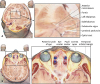
Real-color DICOM files (dimension, 414 × 412 × 469; file size, 106 MB) and a real-color volume model (voxel size, 0.5 × 0.5 × 0.5 mm3; dimension, 414 × 412 × 469; file size, 98.3 MBytes; file format, nii.gz) of a human head were produced from horizontal sectioned images (pixel size, 0.1 × 0.1 mm2; intervals, 0.1 mm) of Visible Korean. In the real-color volume model, three classical planes and oblique planes of a real color and real shape in a body could be shown vividly unlike MRIs (Fig. 1A). A segmented volume model was made (voxel size, 0.5 × 0.5 × 0.5 mm3; dimension, 414 × 412 × 469; file size, 35.4 MBytes; file format, nii.gz) from the segmented images (pixel size, 0.1 × 0.1 mm2; intervals, 0.5 mm) of Visible Korean. In the segmented volume model, the accurate boundary of a real shape in the body could be shown (Fig. 1B). The real-color volume model revealed the actual color of the anatomical structures, while the segmented volume model showed the precise location and outline of the structures. Experts can utilize a real-color volume model solely, while novice students can utilize the segmented volume model together.
 | Fig. 1Real color volume model and segmented volume model. Classical three (horizontal, coronal, and sagittal) sectional planes on a real-color volume model (A) and segmented volume model (B) from Visible Korean.
|
Using MRIcroGL, the volume models can be observed in various methods as follows.
First, after selecting Display - Multi planar, by dragging the mouse pointer on one of the classical planes (horizontal, coronal, or sagittal planes), the other two planes can be displayed continuously in real time from superior to inferior, from anterior to posterior, or from left to right. The classical planes can be shown on the volume model (Fig. 2).
 | Fig. 2Classical horizontal, coronal, and sagittal planes and the cutout volume models. (A) The midpoint anterior and posterior commissural planes at the horizontal, coronal, and sagittal 0 mm are displayed on a classical two-dimensional viewer. (B) Real-color volume model. (C, D) The hippocampus can be found, in which a plane passes the bilateral ear helix, bilateral external acoustic meatus, and medial eyebrow.
|
Second, by sectioning the real-color volume model, the 3D location of the structures can be understood precisely. For example, the hippocampus is located lateral to the diencephalon and inferior to the lateral ventricle (Fig. 2C). On the other hand, it is difficult to know the location of the hippocampus on the surface of the head, which is important for planning surgery. Using the Cutout tool, the hippocampus can be found on three planes in the real-color volume model that pass the bilateral top of the ear helix (horizontal −7 mm), bilateral opening of the external acoustic meatus (coronal −7 mm), and medial eyebrow (sagittal −30 mm). The cutout region can be dug shallowly or deeply using the depth tool (Fig. 3A).
 | Fig. 3Azimuth and elevation on a coordinate system in a real-color volume model. (A) In the case of combination of azimuth, elevation, and depth in a real-color volume, the number of cases that the planes are made are 129,600,000. (B) Oblique planes of the azimuth from 180° to 270° at every 15°, elevation from 0° to 90° at every 15°, and a depth of 0.5 are displayed.
|
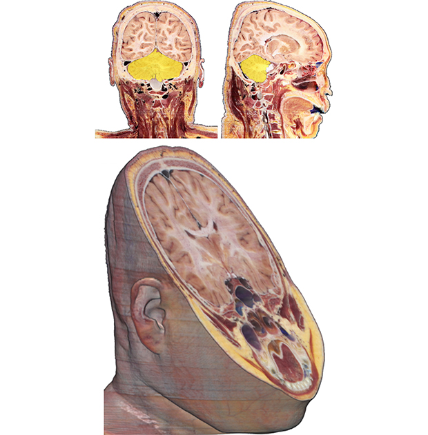
Third, by adjusting the azimuth and elevation tools, any oblique plane can be displayed in real time on a volume model. At the oblique plane, deeper or more superficial planes can be shown continuously by adjusting the depth tool. In the case of a combination of azimuth and elevation tools with depth tools, the number of cases that the planes are made are 129,600,000 (1,000 × 360 × 360) (Fig. 3A). The volume model with the oblique plane can be rotated freely (Fig. 3B, Supplementary Video). Furthermore, the oblique planes are useful aids for teaching deep brain surgery or ophthalmology because a surgeon needs to interpret the oblique planes vertical to the electrode of deep brain surgery3 (Fig. 4A) or neuro-ocular plane45 (Fig. 4B).
 | Fig. 4Oblique planes for clinical education. (A) Subthalamus, which is the target of deep brain surgery, is identified on the probe's eye view for a deep brain stimulation and (B) on the neuro-ocular plane for ophthalmology.
|
Fourth, using the overlay tool, the segmented volume models of each individual structure can be overlapped with not only the classical sectional planes (Fig. 5A), but also the real-color volume model (Fig. 5B). Individual segmented volume models of the cerebrum (voxel size, 1.0 × 1.0 × 1.0 mm3; file size, 1,612 KBytes), cerebellum (file size, 367 KBytes), brainstem (file size, 287 KBytes), thalamus (file size, 288 KBytes), putamen (file size, 243 KBytes), and globus pallidus (file size, 234 KBytes) as well as segmented volume model of 297 structures, can be produced. In the overlapped volume model, the location and outline of the structure can be elucidated easily.
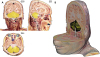 | Fig. 5Real-color volume model overlapped with a segmented volume model. (A) From the overlapped volume model, the cerebellum is indicated on classical three planes and (B) on the cutout volume model.
|
The real-color volume model of this study has higher quality and more vivid color than the Anatomage Table13 and VHP22 in MRIcroGL. It is because the voxel size of this study is smaller (0.5 × 0.5 × 0.5 mm3), while the dimensions of this study is larger (414 × 412 × 469), respectively, even if the volume models of this study and Anatomage Table uses identical sectioned images of Visible Korean. Furthermore, the models of this study are free but Anatomage Table costs approximately 80,000 US dollars (Fig. 6).13
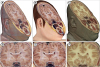 | Fig. 6Oblique planes on the volume models of other studies. Volume models of this study (A) with its expanded view of the brain (D) is superior to Anatomage Table (B, E) in terms of resolution, even though the two models from identical sectioned images of Visible Korean. The volume model of this study (A, D) is superior to the model of Visible Human Project (C, F) in both resolution and color.
|
DISCUSSION
The volume models from real-color sectioned images are beneficial in medical education, research, and clinical practice for the following reasons. First, the real-color volume models can be sectioned in the horizontal, coronal, and sagittal planes in real time (Figs. 1-3, and 5), which are helpful in learning the classical planes of MRIs and CTs. Second, the real-color classical or oblique planes and the volume model can be displayed together, to allow a clear understanding of the location and shape of various anatomical structures (Figs. 1-5). Third, the oblique planes of the volume models can be compared with the oblique planes of various clinical fields (Fig. 4).
Real-color sectioned images are an optimal material to reconstruct the volume model because of the benefits of real color. The volume modeling technique from MRIs already exist on most MRI software, including MRIcroGL. Therefore, the volume model from real-color sectioned images can be made theoretically using the volume modeling technique. Nevertheless, there are four obstacles that need to be overcome when making the volume model.
First, most MRI software recognizes only DICOM format, including the header information (patient, scanning methods, and so on). On the other hand, the sectioned images were in BMP format. Fortunately, in Photoshop CC, any image format can be converted to DICOM without header information.
Second, most MRI software reconstructs volume models based on the DICOM header information. To reconstruct the volume model properly, the study time and study date in the header should be unified and the instance number should be in consecutive order. Despite this requirement, the BMP format of the sectioned images does not have any header information like DICOM. In recent MRI processing freeware,2021 the DICOM header information could be changed artificially or input automatically.
Third, most MRI software operates volume models with small size. The volume model of a full voxel size of the sectioned images cannot be handled on a typical personal computer. Therefore, the file size of sectioned images was decreased by increasing the sectioning interval and pixel size. The voxel size can be improved with advances in the performance of personal computers.
Fourth, the manufacturers of the existing real-color volume models, such as VOXEL-MAN12 and Anatomage Table13 software, did not release all source codes or production methods. Even if their policy is open-source, the common user cannot produce a real-color volume model because it involves complicated programming. Furthermore, the software is quite expensive. In this study, the volume models and the making methods were released free of charge for researchers who wish to produce real-color volume models from their color images.
The real-color volume models may function as free and optimal tools for learning sectional anatomy. One can holistically understand the horizontal, coronal, and sagittal planes, by observing oblique planes between the orthogonal planes. To achieve user-friendly interface, captured images of the volume model can be put into browsing software.23 The volume models of this study and surface models of the very subject can compensate each other.24
The volume models can be used to interpret the medical images of diverse medical fields. The examples will be the observation of the hippocampus (Fig. 2C and D),25 probe's eye view during deep brain stimulation surgery (Fig. 4A),3 and intraoperative ultrasound of the brain (Fig. 4).26 The structures on the oblique planes will be labeled to function as an atlas for the sectional anatomy. So as to introduce the production technique of the volume models, the sectioned images of head were chosen as a representative in this study. In further study, the technique will be applied to sectioned images of various regions and other subjects from humans to animals.15192728 The volume models of this study can be downloaded from the authors' homepage (http://anatomy.co.kr) free of charge or registration. Interested researchers may utilize this technique to their own sectioned images.29 By contacting the authors, the image data and detailed production method can be freely obtained.
 XML Download