Abstract
Purpose
To report the long-term outcome after surgical repair of a full-thickness macular hole (FTMH) in a patient with retinitis pigmentosa (RP).
Case summary
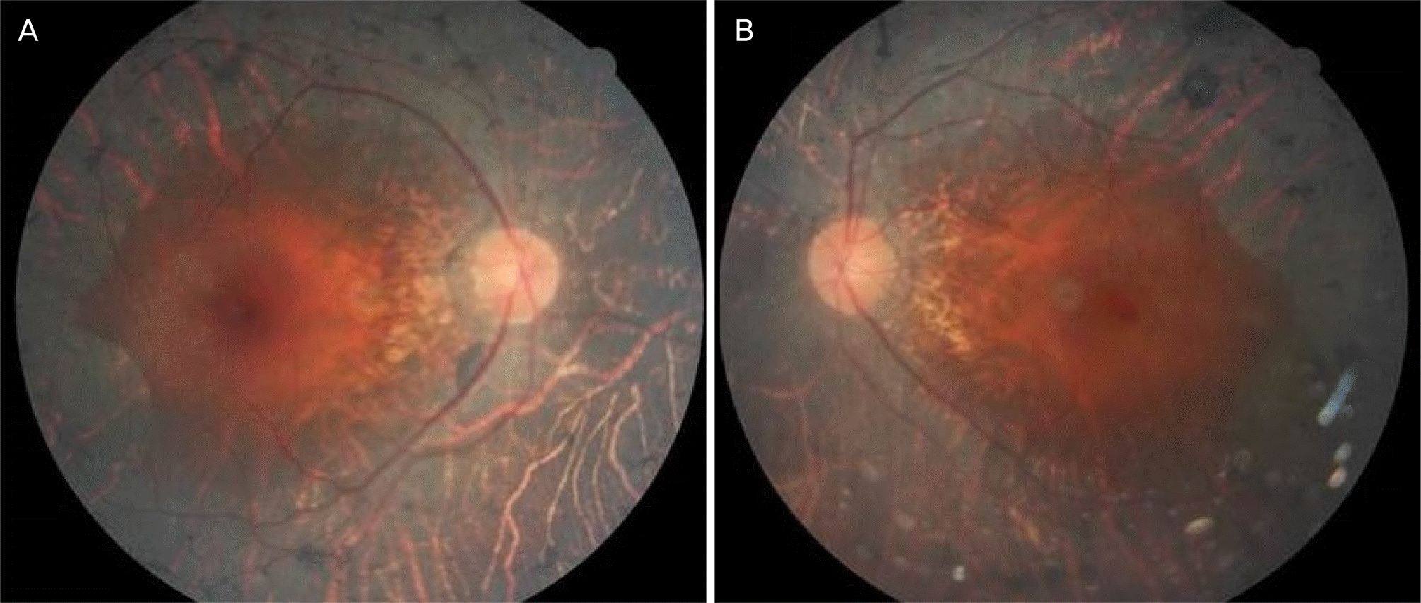
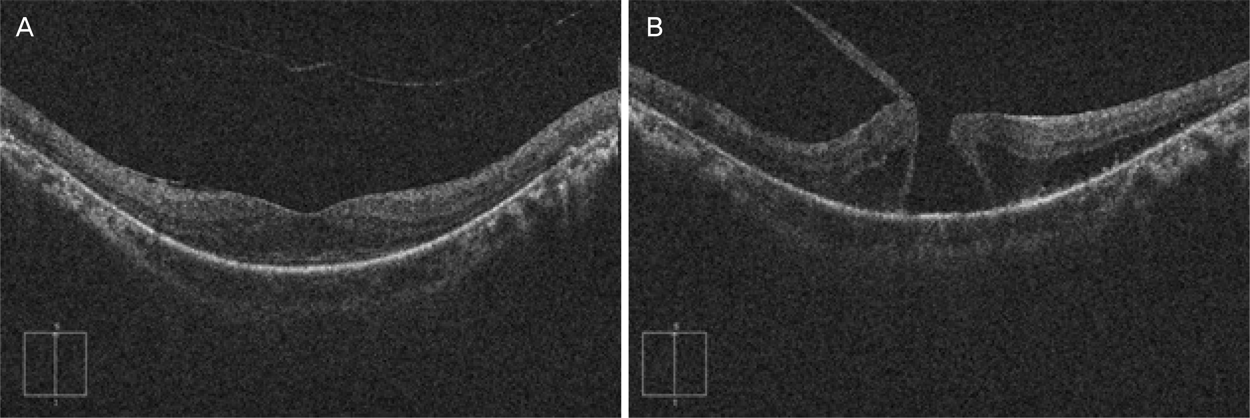
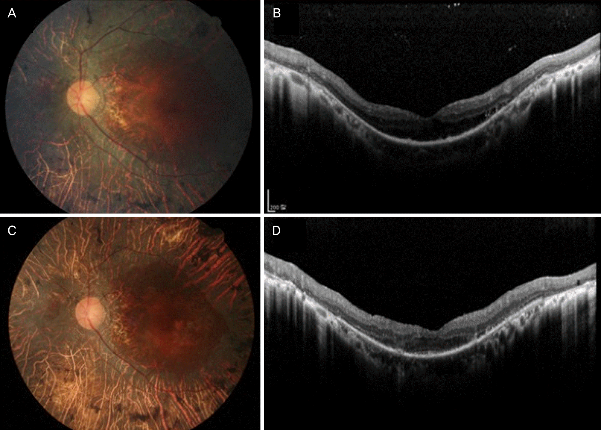
A 55-year-old male who had been diagnosed with retinitis pigmentosa in both eyes 5 years earlier presented with decreased visual acuity in his left eye over the last 6 months. On examination, his Snellen best-corrected visual acuity (BCVA) was 1.0 in the right eye and 0.3 in the left eye. Slit-lamp examination of the anterior segment was remarkable only for posterior chamber intraocular lenses in each eye. Fundus examination demonstrated extensive bony spicule-like pigmentation in the midperipheral region in both eyes and a FTMH with approximately one-third disc diameter in the left eye. The optical coherence tomography (OCT) findings confirmed a FTMH with a surrounding cuff of intraretinal fluid and vitreomacular traction in the left eye. The patient underwent 23-gauge pars plana vitrectomy (PPV) with indocyanine green-assisted internal limiting membrane peeling and gas tamponade. One week postoperatively, an anatomically well-sealed macular hole was confirmed by OCT. At the 3-month postoperative follow-up, the BCVA improved to 0.63 and the hole remained closed until his last follow-up (postoperative 6 years).
Go to : 
References
2. You QS, Xu L, Wang YX, et al. Prevalence of retinitis pigmentosa in North China: the Beijing Eye Public Health Care Project. Acta Ophthalmol. 2013; 91:e499–500.

3. Hirakawa H, Iijima H, Gohdo T, Tsukahara S. Optical coherence tomography of cystoid macular edema associated with retinitis pigmentosa. Am J Ophthalmol. 1999; 128:185–91.

4. Testa F, Rossi S, Colucci R, et al. Macular abnormalities in Italian patients with retinitis pigmentosa. Br J Ophthalmol. 2014; 98:946–50.

5. Hagiwara A, Yamamoto S, Ogata K, et al. Macular abnormalities in patients with retinitis pigmentosa: prevalence on OCT examination and outcomes of vitreoretinal surgery. Acta Ophthalmol. 2011; 89:e122–5.

6. Giusti C, Forte R, Vingolo EM. Clinical pathogenesis of macular holes in patients affected by retinitis pigmentosa. Eur Rev Med Pharmacol Sci. 2002; 6:45–8.
7. Jin ZB, Gan DK, Xu GZ, Nao IN. Macular hole formation in abdominals with retinitis pigmentosa and prognosis of pars plana vitrectomy. Retina. 2008; 28:610–4.
8. García-Fernández M, Castro-Navarro J, Bajo-Fuente A. Unilateral recurrent macular hole in a patient with retinitis pigmentosa: a case report. J Med Case Rep. 2013; 7:69.

9. Lee JH, Kim TK, Kim SY, et al. Pars plana vitrectomy for vitreous hemorrhage in coats-type retinitis pigmentosa. J Korean Ophthalmol Soc. 2016; 57:677–81.

10. Kelly NE, Wendel RT. Vitreous surgery for idiopathic macular holes. Results of a pilot study. Arch Ophthalmol. 1991; 109:654–9.

11. Kadonosono K, Itoh N, Uchio E, et al. Staining of internal limiting membrane in macular hole surgery. Arch Ophthalmol. 2000; 118:1116–8.

12. Lee SJ, Jang SY, Moon D, et al. abdominal surgical outcomes after vitrectomy for symptomatic lamellar macular holes. Retina. 2012; 32:1743–8.
13. Casparis H, Bovey EH. Surgical treatment of lamellar macular hole associated with epimacular membrane. Retina. 2011; 31:1783–90.

14. Gass JD. Idiopathic senile macular hole. Its early stages and pathogenesis. Arch Ophthalmol. 1988; 106:629–39.

Go to : 
 | Figure 1.Preoperative fundus photography images of the right (A) and left (B) eyes of the study patient are presented. Note an extensive bony spicule-like pigmentation in the midperipheral region in both eyes and a full-thickness macular hole with approximately one-third disc diameter in the left eye. |


 PDF
PDF ePub
ePub Citation
 Print
Print


 XML Download