Abstract
Purpose
To report two cases of postoperative endophthalmitis caused by Streptococcus dysgalactiae subspecies equisimilis (SDSE), which appeared as hyperacute presentation and panophthalmitis.
Case summary
A 68-year-old male was treated with cataract surgery and was evaluated the next day (less than 24 hours after surgery) because of acute loss of vision. There was severe inflammation and the visual acuity was light perception. The patient underwent pars plana vitrectomy (PPV) with intravitreal antibiotic injection. The vitreous culture revealed SDSE. After PPV, re-gression of inflammation was observed, although the corneal edema had progressed. The cornea evolved to decompensate due to bullous keratopathy and visual acuity of the eye decreased to no light perception after 3 months. A 87-year-old male who underwent phacoemulsification and intraocular lens implantation 2 days previously was hospitalized due to severe ocular pain and visual loss. There was severe inflammation, and the visual acuity was no light perception. The patient received only intravitreal injections of antibiotics due to severe corneal necrosis. The aqueous humor revealed SDSE. Four days after intravitreal injection, erythema and swelling of the eyelid of the affected eye was observed, and diagnosed as panophthalmitis. After treatment with intravenous antibiotics, cellulitis of the eyelid was resolved. The eye progressed as phthisis after 3 months without recurrence.
Go to : 
References
1. Takahashi T, Ubukata K, Watanabe H. Invasive infection caused by Streptococcus dysgalactiae subsp. Equisimilis: characteristics of strains and clinical features. J Infect Chemother. 2011; 17:1–10.

2. Watanabe S, Takemoto N, Ogura K, Miyoshi-Akiyama T. Severe invasive streptococcal infection by Streptococcus pyogenes and Streptococcus dysgalactiae subsp. equisimilis. Microbiol Immunol. 2016; 60:1–9.

3. Oppegaard O, Mylvaganam H, Skrede S, et al. Emergence of a Streptococcus dysgalactiae subspecies equisimilis stG62647-line-age associated with severe clinical manifestations. Sci Rep. 2017; 7:7589.

4. Wajima T, Morozumi M, Hanada S, et al. Molecular characterization of invasive Streptococcus dysgalactiae subsp. equisimilis. Japan Emerg Infect Dis. 2016; 22:247–54.
5. Ritterband DC, Shah MK, Buxton DJ, et al. A devastating ocular pathogen: beta-streptococcus Group G. Cornea. 2000; 19:297–300.
6. Goh ES, Liew GC. Traumatic bleb-associated Group-G beta-hae-molytic Streptococcus endophthalmitis. Clin Exp Ophthalmol. 2008; 36:580–1.
7. Kaliamurthy J, Cuteri V, Jesudasen N, et al. Postoperative ocular infection due to Streptococcus dysgalactiae subspecies equisimilis. J Infect Dev Ctries. 2011; 5:742–4.

8. Hagiya H, Semba T, Morimoto T, et al. Panophthalmitis caused by Streptococcus dysgalactiae subsp. equisimilis: a case report and abdominal review. J Infect Chemother. 2018; 24:936–40.
9. Margo CE, Cole EL 3rd. Postoperative endophthalmitis and asymptomatic bacteriuria caused by group G Streptococcus. Am J Ophthalmol. 1989; 107:430–1.

10. Posenauer B, Funk J. Group G streptococci as pathogens of abdominal endophthalmitis Klin Monbl Augenheilkd. 1991; 199:45–7.
11. Kugu S, Sevim MS, Kaymak NZ, et al. Exogenous group G Streptococcus endophthalmitis following intravitreal ranibizumab injection. Clin Ophthalmol. 2012; 6:1399–402.
12. Park JM, Yeom MI, Park JM. A case of Streptococcus dysgalactiae endophthalmitis after cataract surgery. J Korean Ophthalmol Soc. 2018; 59:185–9.

13. Baxter KR, Robinson JE, Ruby AJ. Occlusive vasculitis due to hyperacute Streptococcus mitis endophthalmitis after intravitreal ranibizumab. Retin Cases Brief Rep. 2015; 9:201–4.

14. Gan IM, van Dissel JT, Beekhuis WH, et al. Intravitreal vancomycin and gentamicin concentrations in patients with postoperativeendophthalmitis. Br J Ophthalmol. 2001; 85:1289–93.
15. Results of the Endophthalmitis Vitrectomy Study. A randomized trial of immediate vitrectomy and of intravenous antibiotics for the treatment of postoperative bacterial endophthalmitis. Endophthalmitis Vitrectomy Study Group. Arch Ophthalmol. 1995; 113:1479–96.
Go to : 
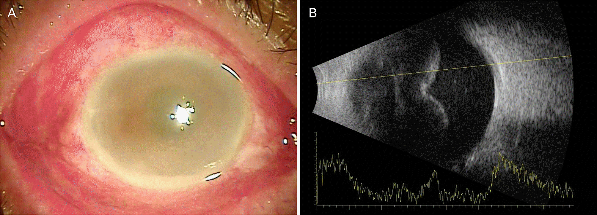
 | Figure 1.Anterior segment photograph and ultrasonogram at a day after cataract surgery. (A) Conjunctival injection, corneal edema and hypopyon were identified. (B) Thick vitreous opacity was observed. |
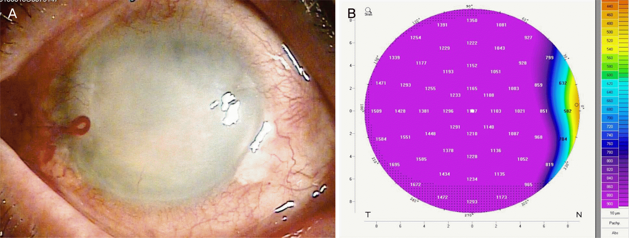
 | Figure 2.Anterior segment photograph and topography of the affected cornea at 3 months after cataract surgery. (A) Anterior seg-ment photograph shows severe corneal edema and opacity. (B) Topography revealed corneal thickness over 1,000 μ m due to corneal edema. |
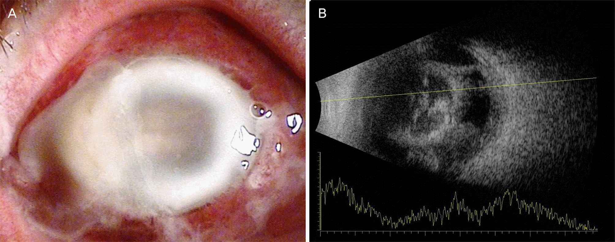
 | Figure 3.Anterior segment photograph and ultrasonogram of case 2 at 3 days after cataract surgery. (A) Severe conjunctival injection and discharge, corneal edema and ring-shaped stromal infiltration were observed. (B) Ultrasonogram shows heterogeneous vitreous opacity in the vitreous cavity. |
 | Figure 4.T2 enhanced image of orbital magnetic resonance imaging (MRI) at a week (A) and 2 months (B) after cataract surgery. (A) It shows proptosis of the right eye and heterogeneous enhancement of the retrobulbar area. (B) MRI revealed relieved proptosis and decrease of enhancement in the retrobulbar area. ASL = anterior, superior, left; ASR = anterior, superior, right; PIR = poste-rior, inferior, right; PIL = posterior, inferior, left. |
Table 1.
Clinical characteristics of postoperative Streptococcus dysgalactiae subspecies equisimilis associated endophthalmitis
F = female; DM = diabetes mellitus; NLP = no light perception; M = male; PPV = pars plana vitrectomy; IVAI = intravitreal antibiotics injection; PKP = penetrating keratoplasty; AIDS = acquired immunodeficiency syndrome; LP = light percepsion; n.d. = not described; VA = visual acuity; IVR = intravitreal ranibizumab injection; CVA = cerebrovascular accident.


 PDF
PDF ePub
ePub Citation
 Print
Print


 XML Download