Abstract
Purpose
We evaluated the short-term clinical outcomes of patients who underwent modified scleral fixation of an intraocular lens (IOL) using a scleral tunnel and groove.
Methods
From June 2016 to May 2017, 34 eyes of 34 patients who underwent modified scleral fixation of an IOL using a scleral tunnel and groove were retrospectively studied. We evaluated the best-corrected visual acuity (BCVA), corneal endothelial cell density, intraocular pressure (IOP), spherical equivalent, and postoperative complications at 1 week, 1 month, 3 months, and 6 months after surgery.
Results
The BCVA was 0.85 ± 0.83 logarithm of the minimal angle of resolution (logMAR) before surgery and 0.38 ± 0.61 logMAR at 6 months (p = 0.001). The corneal endothelial cell count was 1,955.12 ± 217/mm2 and 1,852.59 ± 190/mm2, before and after surgery, respectively, which was not significantly different (p = 0.186). Postoperative complications occurred in eight eyes (23.5%); IOP elevation in one eye (2.9%), IOL tilt or decentration in two eyes (5.7%), optic capture in four eyes (11.4%), and cystic macular edema in one eye (2.9%). The spherical equivalent showed myopic changes after surgery and decreased significantly over time (p = 0.001).
Conclusions
Modified scleral fixation of the IOL using a scleral tunnel and groove improved the BCVA, but did not significantly affect corneal endothelial cell loss. This procedure can be a good alternative to conventional scleral fixation of an IOL, which has advantages in shortened surgical time and easy surgical manipulation.
Go to : 
References
1. Engren AL, Behndig A. Anterior chamber depth, intraocular lens position, and refractive outcomes after cataract surgery. J Cataract Refract Surg. 2013; 39:572–7.

2. Sasaki K, Sakamoto Y, Shibata T, et al. Measurement of abdominal intraocular lens tilting and decentration using Scheimpflug images. J Cataract Refract Surg. 1989; 15:454–7.
3. Lee Y, Kim MH, Park YL, et al. Comparison of short-term clinical outcomes between scleral fixation vs. iris fixation of dislocated IOL. J Korean Ophthalmol Soc. 2017; 58:1131–7.

4. Oh SY, Kim SS. Astigmatic changes and clinical outcomes after scleral fixation of IOL. J Korean Ophthalmol Soc. 2014; 55:1452–9.

6. Scharioth GB, Prasad S, Georgalas I, et al. Intermediate results of sutureless intrascleral posterior chamber intraocular lens fixation. J Cataract Refract Surg. 2010; 36:254–9.

7. Agarwal A, Kumar DA, Jacob S, et al. Fibrin glue–assisted abdominal posterior chamber intraocular lens implantation in eyes with deficient posterior capsules. J Cataract Refract Surg. 2008; 34:1433–8.
8. Yamane S, Inoue M, Arakawa A, Kadonosono K. Sutureless 27-gauge needle guided intrascleral intraocular lens implantation with lamellar scleral dissection. Ophthalmology. 2014; 121:61–6.
9. Gabor SG, Pavlidis MM. Sutureless intrascleral posterior chamber intraocular lens fixation. J Cataract Refract Surg. 2007; 33:1851–4.

10. Rodríguez-Agirretxe I, Acera-Osa A, Ubeda-Erviti M. Needleguided intrascleral fixation of posterior chamber intraocular lens for aphakia correction. J Cataract Refract Surg. 2009; 35:2051–3.

11. Verbruggen KH, Rozema JJ, Gobin L, et al. Intraocular lens centration and visual outcomes after bag-in-the-lens implantation. J Cataract Refract Surg. 2007; 33:1267–72.

12. Zeh WG, Price FW Jr. Iris fixation of posterior chamber intraocular lenses. J Cataract Refract Surg. 2000; 26:1028–34.

13. Por YM, Lavin MJ. Techniques of intraocular lens suspension in the absence of capsular/zonular support. Surv Ophthalmol. 2005; 50:429–62.

14. Azar DT, Wiley WF. Double-knot transscleral suture fixation abdominal for displaced intraocular lenses. Am J Ophthalmol. 1999; 128:644–6.
15. Bloom SM, Wyszynski RE, Brucker AJ. Scleral fixation suture for dislocated posterior chamber intraocular lens. Ophthalmic Surg. 1990; 21:851–4.

16. Chan CK. An improved technique for management of dislocated posterior chamber implants. Ophthalmology. 1992; 99:51–7.

17. McAllister AS, Hirst LW. Visual outcomes and complications of scleral fixated posterior chamber intraocular lenses. J Cataract Refract Surg. 2011; 37:1263–9.
18. Ohta T, Toshida H, Murakami A. Simplified and safe method of abdominalless intrascleral posterior chamber intraocular lens fixation: Y-fixation technique. J Cataract Refract Surg. 2014; 40:2–7.
19. Wilgucki JD, Wheatley HM, Feiner L, et al. One-year outcomes of eyes treated with a sutureless scleral fixation technique for abdominal lens placement or rescue. Retina. 2015; 35:1036–40.
Go to : 
 | Figure 1.The surgical procedure of scleral fixation of intraocular lens (IOL) through scleral tunnel and groove. (A) The 8-lines corneal marker is used to mark 1 and 7 o'clock directions from corneal center. (B) Six points on sclera are marked at 1.5 mm posterior to the limbus (point 1: at 7 o'clock direction, point 2: at 1.0 mm away from point 1 counterclockwise, point 3: 3.0 mm away from point 2 counterclockwise, point 4: at 1 o'clock direction, point 5: at 1.0 mm away from point 4 counterclockwise, point 6: 3.0 mm away from point 5 counterclockwise). (C) Two limbal parallel scleral tunnels (3.0 mm length, 1/2 scleral thickness) are made using a 23-gauge needle (one: line between point 2 and 3, the other: line between point 5 and 6). (D) Two scleral grooves 1.0 mm long are made using a stab knife (one: line between point 1 and 2, the other: line between point 4 and 5). (E) Two full-thickness scleral incisions are made using a stab knife above point 1 and 4. (F) The leading haptic is held and then pulled out of the eye through the scleral incisions using 23-gauge microforceps. (G) The both haptics are inserted into the scleral tunnel. Then, the IOL is placed into position. (H) The conjunctiva is closed with 8–0 vicryl. |
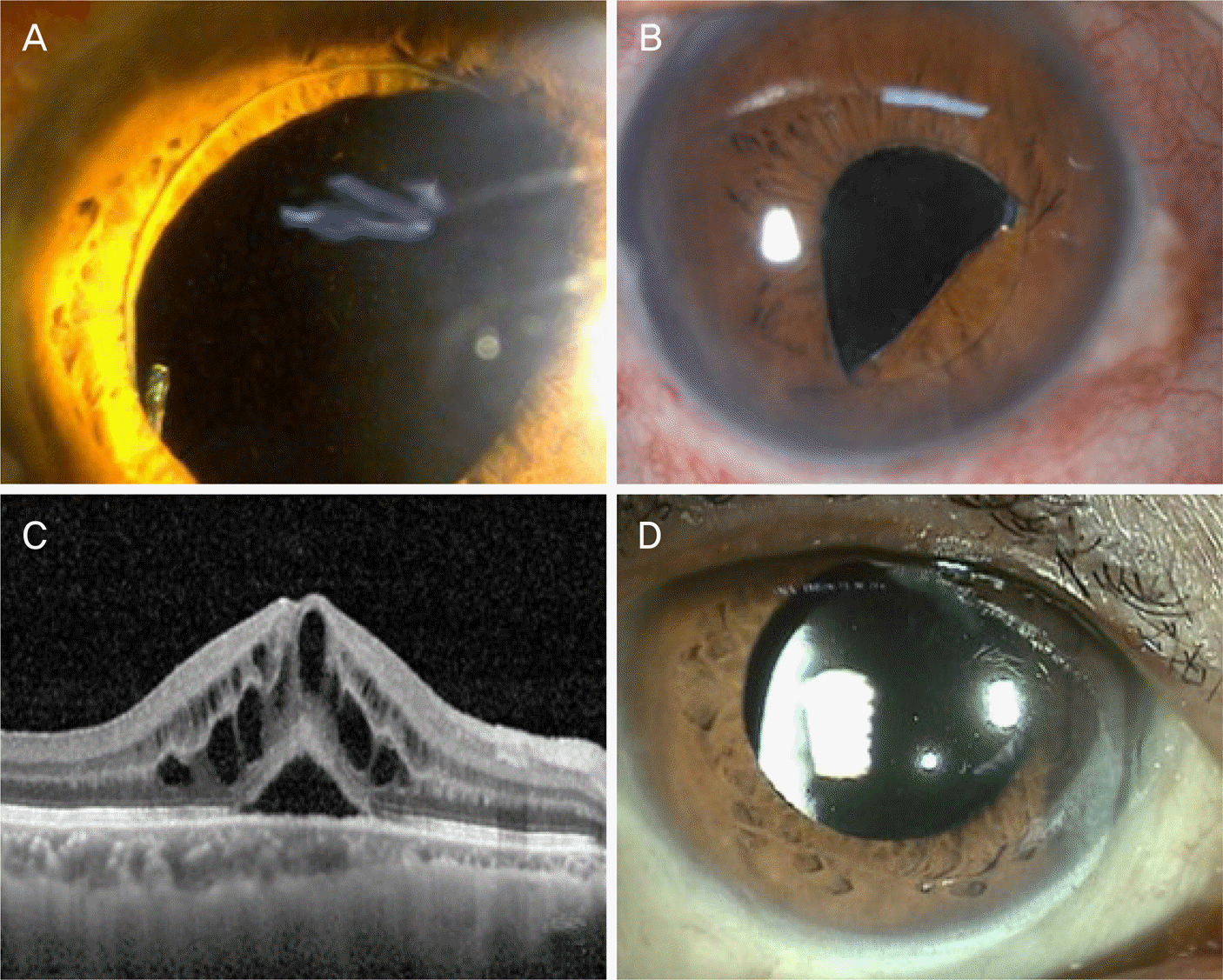
 | Figure 2.Complications of scleral fixation of intraocular lens (IOL) through sclera tunnel and groove. (A, B) Optic capture. (C) Cystoid macular edema. (D) IOL decentration. |
 | Figure 3Post-operative anterior segment optical coherence tomography (OCT). (A) A typical OCT image in the patient at 3 months after surgery. (B) OCT shows the scleral tunnel with the incarcerated haptic of the intraocular lens at 3 months. There are no signs of leakage or inflammation. |
Table 1.
Characteristics of 34 patients who underwent scleral fixation of IOL with scleral tunnel and groove
Table 2.
Mean BCVA and ECD at the time of preoperation and final follow-up after surgery
| Variable | Value |
|---|---|
| Baseline BCVA (logMAR) | 0.85 ± 0.83 |
| Final follow-up BCVA (logMAR) | 0.38 ± 0.61 |
| p-value* | 0.001 |
| Baseline ECD | 1,955.12 ± 217.64 |
| Final follow-up ECD | 1,852.59 ± 190.78 |
| p-value* | 0.186 |
Table 3.
Changes of IOP and mean spherical equivalent
| Variable | Value | p-value* |
|---|---|---|
| IOP | ||
| Pre-operation | 15.68 ± 3.13 | |
| Post 1 week | 17.38 ± 6.35 | 0.617 |
| Post 1 month | 16.47 ± 3.58 | 0.679 |
| Post 3 months | 15.24 ± 4.30 | 0.726 |
| Post 6 months | 14.94 ± 3.15 | 0.702 |
| Mean spherical equivalent (diopter) | ||
| Pre-operation | 4.32 ± 7.30 | |
| Post 1 week | −1.26 ± 1.96 | 0.001 |
| Post 1 month | −0.71 ± 1.56 | 0.001 |
| Post 3 months | −0.32 ± 1.60 | 0.001 |
| Post 6 months | −0.26 ± 1.30 | 0.001 |


 PDF
PDF ePub
ePub Citation
 Print
Print


 XML Download