

 PDF
PDF ePub
ePub Citation
 Print
Print


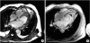
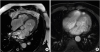
A 65-year-old man was admitted to the cardiology department with increasing dyspnea, orthopnea, and fatigue over the last month. Transthoracic echocardiography (TTE) revealed an ejection fraction of 60%, immobile thrombus in left ventricular (LV) apex, E/A ratio >2, biatrial dilatation, moderate mitral and tricuspid regurgitation, systolic pulmonary arterial pressure of 70 mmHg, and dilated inferior vena cava without respiratory variation (Supplementary Video 1). Blood tests were remarkable only for mild eosinophilia (1,000 cells/microliter). Similar to TTE findings, cardiac magnetic resonance imaging (MRI) showed a hypointense thrombus adjacent to LV apex with bilateral pleural effusion (Figure 1). Although only mild peripheral eosinophilia was present, bone marrow biopsy showed 25% infiltration of bone marrow with eosinophils. Based on these clinical, imaging and laboratory findings, the patient was diagnosed as having idiopathic hypereosinophilic syndrome (HES) and Loeffler endocarditis. He was administered 1 mg/kg/day methylprednisolon for three months, warfarin with a target international normalized ratio of 2 to 3, and 20 mg/day furosemide. At follow-up, his symptoms improved significantly. Control TTE performed 4 months after diagnosis showed complete resolution of thrombus with hypokinesia of LV apex (Supplementary Video 2). In addition, there was a decrease in systolic pulmonary arterial pressure by 15 mmHg without any change in E/A ratio. Control cardiac MRI confirmed TTE findings (Figure 2).
HES is characterized by excessive production of eosinophils that subsequently involve and damage multiple organs.1) HES can involve almost any organ system, but cardiac involvement is a serious cause of morbidity and mortality.2) This rare Loeffler endocarditis case in an idiopathic HES patient is interesting in a way that typical cardiac imaging findings and bone marrow hypereosinophilia were present in the presence of mild peripheral eosinophilia. There are very few cases of Loeffler endocarditis without prominent peripheral eosinophilia in the literature.3) Furthermore, complete resolution of LV apical thrombus was provided by medical treatment in this rare case.
 XML Download