

 PDF
PDF ePub
ePub Citation
 Print
Print


INTRODUCTION
With an aging population, the number of intertrochanteric femoral fractures treated each year continues to rise. In 2014, 16,000 people in the Netherlands were registered with a hip fracture12). Of these, roughly 14,000 were caused by a fall and three quarters of the patients were older than 80 years. The distribution between men and women was 1:3. Twenty-five percent of patients with a hip fracture are left permanently disabled and 25% die within the first year. Approximately 50% of the patients with a hip fracture can return to their own home environment after hospitalization13). The total annual costs of all hip fractures in the Netherlands are estimated to be €300 million134).
Operative treatment of a hip fracture remains the best option as it helps create an anatomical reduction of the fracture, stable fixation, and maintenance of blood supply, thereby providing an opportunity for early and full weight bearing with mobilization. As a result, the postoperative risks from immobilization—e.g., deep vein thrombosis and airway problems-sharply decrease15).
The helical blade received regulatory approval in 2005 and was designed to provide stronger fixation through impaction of the cancellous bone which should increase resistance to cut-out67). However, several randomized controlled trials comparing helical blade with the lag screw in intramedullary fixations have demonstrated greatly different outcomes compared with published data and theoretical concepts789101112).
The choice of implant used for surgical treatment of intertrochanteric femoral fractures depends on the surgeons' preference, not on empirical data or indication. Within the Department of Trauma Surgery, no consensus exists regarding the type of collum implant to be used in when surgically treating patients with an intertrochanteric femoral fracture.
This study aimed to assess if there were any differences in outcomes (i.e., cut-out and/or interventional variables) between a the Trochanteric Fixation Nail (TFN®; Synthes, Raynham, MA, USA) with helical blade and a TFN® with femoral neck screw when applied to surgical treatment of intertrochanteric femoral fractures.
Go to : 
MATERIALS AND METHODS
1. Inclusion/Exclusion Criteria
Within the Trauma Department at our institution, it was decided that surgeons would use TFN® for osteosyntheses of intertrochanteric femoral fractures. The surgeon could choose between a helical blade and femoral neck screw (“lag screw”) for the collum implant. Patients were included if they: i) were older than eighteen years of age, ii) had pre- and postoperative X-ray diagnostics available, and iii) were treated by/under direct supervision of a trauma surgeon. Candidates were excluded if they had a pathologic fracture, prior hip surgery on the ipsilateral side, additional fractures or an incorrectly registered operation code.
The cohort included those identified using the operation code 38535 (femur # intertrochanteric, TFN) from the digital registration system. The study group was comprised of 685 patients with an intertrochanteric femoral fracture treated with a TFN® with helical blade or a TFN® with femoral neck screw between January 1,2012 and December 31, 2016. A total of 631 patients where included in the study; 54 were excluded on the basis of a wrongly registered operation code after reassessment of the preoperative X-ray images (Fig. 1).

The study was approved by the local ethics committee of the Albert Schweitzer Hospital. Signed informed consent forms allowing use of medical records for research purposes were obtained from all patients.
2. Outcome Measures
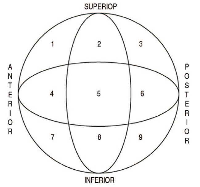
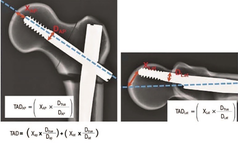
Mechanical complications (e.g., axial migration, axial cutout, lateral cut-out, non-union, periprothestic fracture, unacceptable position, mechanical complications with a tip-apex distance [TAD] <15 mm or >25 mm)413) were obtained from available post-operative X-ray examinations. Axial migration was considered a normal phenomenon due to collapsing of the fracture until sufficient stability was achieved41314). Intervention variables (i.e., TAD, surgery time, position of the collum implant [according to the Cleveland index as shown in Fig. 2]15), weight bearing prescriptions, length of stay, discharge destination after the hospital, postsurgical complications), were obtained from the medical records of electronic patient files. The TAD was calculated (Fig. 3) on the first post-operative X-ray for each patient as described by Baumgaertner et al.14). All measurements were performed by a single investigator, and 10% of his findings were double-checked by a trauma surgeon.
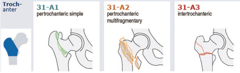
Fractures were classified using the AO Foundation/Orthopaedic Trauma Association (AO/OTA) classification and classified as stable (31-A1) or unstable (31-A2) (Appendix 1)16).
4. Statistical Analysis
Statistical analysis was performed using Statistic Package for Social Sciences (SPSS) version 24.0 (IBM Corp., Armonk, NY, USA). Differences were considered to be statistically signiflcant when P<0.05. The chi-square test was used to compare differences between the two groups for each variable, and the Student's t-test was used for quantitative variables.
Go to :
RESULTS
In total, 631 patients were treated surgically for an intertrochanteric femoral fracture. Of these, 239 (37.9%) were treated with a TFN® with helical blade and 392 (62.1%) with a TFN® with femoral neck screw (Fig. 1). The baseline characteristics demonstrated no statistically significant differences between the groups (Table 1); patients had a mean follow up of one year. The average age in each group was 81 years old and 76.1% were female. According to the AO/OTA classification 56.4% were classified as unstable fractures. There was no statistically significant difference (P=0.98) in the proportion of fractures that were unstable between the helical blade and femoral neck screw, helical blade 135 (56.5%), femoral neck screw 221 (56.4%), respectively.
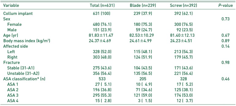
Table 1
Baseline Characteristics and Demographic Data
Values are presented as number (%), mean±stadard deviation, or number only.
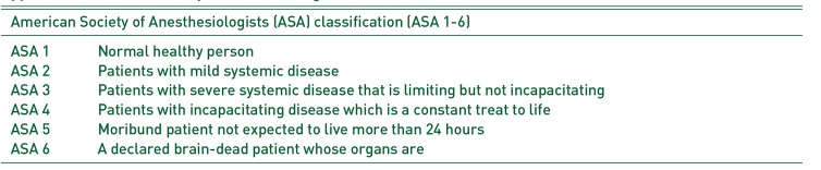
ASA: American Society of Anesthesiologists.
*Refer to Appendix 2.
![]()
An assessment of the primary outcome measure (i.e., cut-out) revealed that a total of 42 (6.7%) mechanical complications were recorded (axial cut-out, lateral cutout, non-union, periprothestic fracture or unacceptable position). A total of 17 (2.7%) cut-outs were recorded in both groups combined (helical blade 9 cut-outs, 3.8%; femoral neck screw 8 cut-outs, 2.0%; P=0.19) (Table 2). There was no statistically significant difference between the two groups when assessing the primary outcome (P=0.19). Also, no statistically significant difference existed in TAD (<15 mm or >25 mm) related to mechanical complications (helical blade 8, 3.3%; femoral neck screw 8, 2.0%; P=0.454). A total of 221 patients (35.0%) experienced axial migration of their collum implant (helical blade 78, 32.6%; femoral neck screw, 143; 36.5%; P=0.296).
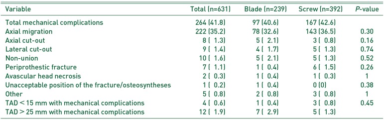
Table 2
Mechanical Complications
![]()
There were also no statistically significant differences among the secondary outcome measures between the two groups (Table 3, 4). The positions of the collum implant within the femoral head were evenly divided between the helical blade and screw groups; as shown in Fig. 4, 74.5% were placed in the optimal position (i.e., central in the femoral head, corresponding with position 5 in the Cleveland index) in each group. Post-operative complications were experienced by 605 out of 631 patients. The three most common postoperative complications were low hemoglobin for which transfusion was required (169/605 patients, 27.9%), delirium (129/605 patients, 21.3%) and urinary tract infection (72/605 patients, 11.9%).
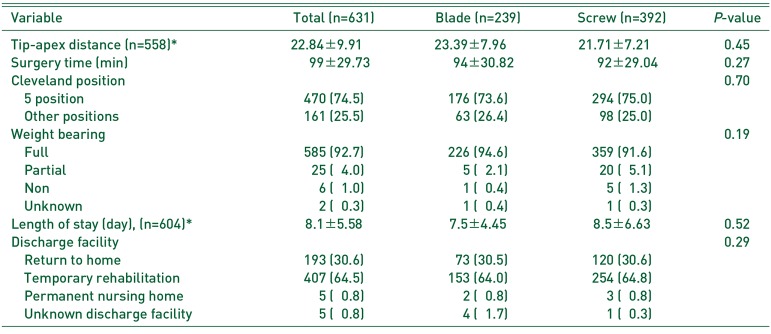
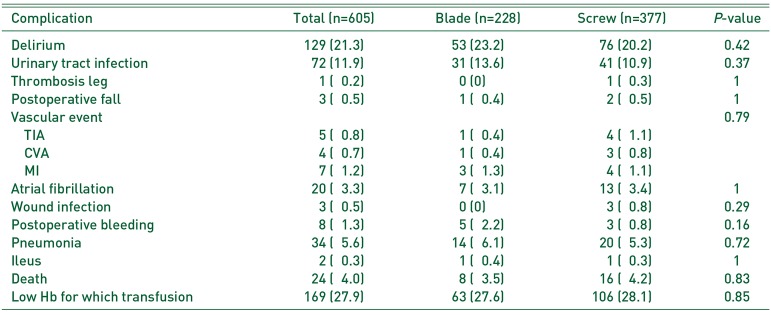
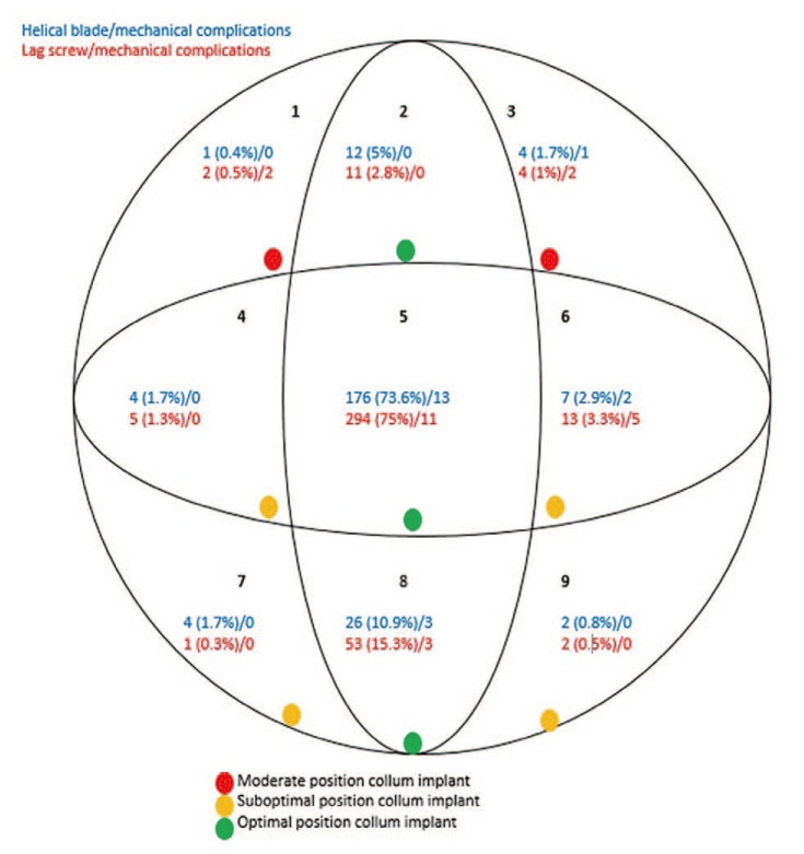
Table 3
Secondary Outcome Measures
![]()
Table 4
Postoperative Complications
![]()
Go to :
DISCUSSION
1. Mechanical Complications
When comparing outcomes associated with the use of helical blades and femoral neck screws for the surgical treatment of intertrochanteric femoral fractures, we observed no statistically significant differences in the frequency of cut-out or other mechanical complications. Axial migration may be an early symptom of cut-out, and in this study, the high number of axial migration events was considered a functional phenomenon as part of the physiological collapse of the fracture to obtain (sufficient) stability and a prerequisite for optimal bone healing. All figures from this study were in accordance with the figures found in recent literature713171819). In our study, the total percentage of mechanical complications (i.e., 6.7%) was lower than what is reported (i.e., 16–23%) in different studies20). One reason for this could be the differentiation within the surgical department. The trauma surgeons operate on greater than 75.3% of the intertrochanteric femoral fractures, thus developing a high level of competence in this type of operation.
2. Intervention Variables
For all intervention variables tested, we observed no statistically significant differences among the two treatment groups. An increased risk of mechanical complications according to the literature occurs at a TAD of <15 mm or >25 mm413). The disadvantage of TAD measurements is that they are made using the first postoperative X-ray images. In patients with a TAD of <15 mm or >25 mm, an expectative policy will be maintained with regard to re-operation of the patient and the clinical condition of the patient will lead treatment decisions. The increased risk of mechanical complications described in the literature with a TAD of <15 mm or >25 mm was not observed in this study11421). For the time being, the value of the TAD used in research and how these values are reflective of clinical outcomes remain open for discussion.
This study revealed no difference in postoperative complications between helical blade and femoral neck screws when used to surgically treat intertrochanteric femoral fractures. The actual number of postoperative complications will most likely be much higher since postoperative complications were only identified using data relating to the clinical admission of the patient. Also, postoperative complications are typically underreported and a subset of patients will develop a postoperative complication (e.g., delirium) after discharge from the hospital. Postoperative complications that were treated and resolved during admission and were no longer topical at discharge were often not mentioned in the discharge letter and therefore not reported.
3. Strengths and Weaknesses of the Research
The strengths of this research were that it evaluated patients treated over a five year time frame; all patients who received surgical treatment for an intertrochanteric femoral fracture using a TFN® with helical blade or femoral neck screw were included.
Weaknesses of this research include: i) underreporting of variables that could not be included in the analyses (e.g., blood loss), ii) underreporting of analyzed data, which led to missing data (especially in the registration of postoperative complications), and iii) surgeries were not all performed by the same trauma surgeon. All operators had the skills to place a TFN® with femoralneck screw, however, only three had the skills to place a TFN® with a helical blade; this factor may have possibly influenced the results.
Go to :
CONCLUSION
This study demonstrated no statistically significant differences in outcomes between the use of a TFN® with helical blade and a TFN® with femoral neck screw for the surgical treatment of intertrochanteric femur fractures. These findings suggest no outcomes-based rationale for choosing between the two collum implants tested in the surgical treatment of intertrochanteric femur fractures.
To generate empirically sound recommendations for the type of collum implant to use for intertrochanteric femur fractures, further research appears necessary.
Go to :
 XML Download