Journal List > Hip Pelvis > v.31(1) > 1117574

Kamo, Kido, and Kido: Comparison of the Incidence of Intra-operative Fractures in Hip Hemi-arthroplasty Performed in Supine and Lateral Positions

Abstract

Purpose

The difficulty of femoral preparation with supine-position hip hemi-arthroplasty (HA) often leads to intra-operative fractures (IOFs). We aimed to clarify the incidence and types of IOFs in HA for hip fractures performed in the supine and lateral positions.

Materials and Methods

We retrospectively investigated cases of HA for acute femoral neck fractures from June 2013 to May 2018. We examined the incidence and types of IOFs according to different approaches. We defined supine-position in HA as the supine and hip-hyperextended (over-range) femoral preparation position, and lateral position as the lateral and hip-flexed femoral preparation position. We used a short tapered wedged stem.

Results

Supine-position HA was used in 46 patients (23.7%) and lateral-position HA in 148 patients (76.3%). IOFs in supine-position HA occurred in 8 patients (17.4%) and included five Vancouver AGT and three Vancouver B2 fractures. IOFs in lateral-position HA occurred in 3 patients (2.0%) and included one Vancouver AGT and two Vancouver B fractures. Supine-position HA was a risk factor for IOFs (adjusted odds ratio, 9.71; 95% confidence interval, 2.37–39.8; P<0.01)

Conclusion

Supine-position in HA is an IOF risk factor and significantly increases the incidence of great trochanter fractures of Vancouver type A.

INTRODUCTION

In clinical practice, post-operative dislocations after hip hemi-arthroplasty (HA) receive much attention, but intra-operative fractures (IOFs) are often overlooked. The risk factors of IOFs include female sex, increasing age, osteoporosis, rheumatoid arthritis, bone morphology, and cementless stem123456). Supine-position HA with the anterolateral approach in the supine position (ALS) or direct anterior approach (DAA) has become prevalent. The difficulty of femoral preparations when using ALS or DAA often leads to IOFs78910). We also experienced some IOFs and stem mal-alignment in supine-position HA for hip fractures. Before using the supine-position approach, we had performed HA with the conventional posterior approach (PA) and experienced IOFs in 3.1% of the patients (4/128) and dislocations in 2.3% (3/128) over 5 years. All four IOFs were periprosthetic fractures requiring cerclage wiring. In contrast, supine-position HA caused IOFs of another type (great trochanter fractures) that we had never experienced when using PA. Based on this experience, we had returned to lateral-position HA (PA and superior approach [SA]11)), until we took measures to reduce the incidence of IOFs the supine-position HA. No studies have focused on the incidence of IOFs in supine-position and lateral-position HA. We aimed to clarify the incidence and types of IOFs in HA with the supine and lateral positions.

MATERIALS AND METHODS

We retrospectively investigated cases of HA for acute femoral neck fractures from June 2013 to May 2018. Since June 2013, we had used mainly tapered-wedge stem implants. We collated data regarding the approaches used, operation duration, intra-operative blood loss, and the following complications: IOFs (we used Vancouver classification6)), treatment for IOFs, post-operative dislocations, deep infections, symptomatic deep venous thrombosis, cerebrovascular infarction, and death. In addition, data on patient characteristics (age at operation, sex, body height, body weight, and body mass index [BMI]) and radiographic indices (limb length discrepancy [LLD] and varus angle), healthy (contralateral) center-edge angle, and canal flare index (CFI). This research was approved by the ethics review board of the authors' affiliated institution (H27-11) and written informed consents from the patients were obtained.

1. Surgery

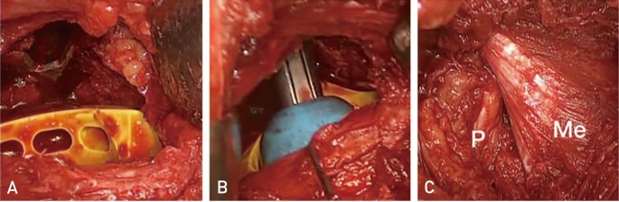
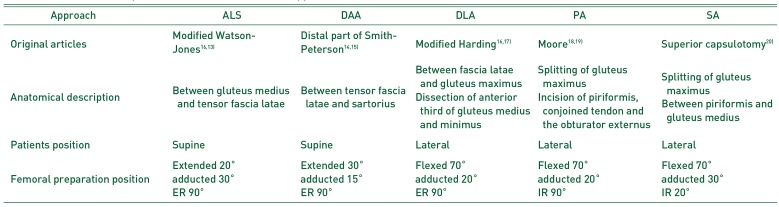
We categorized ALS and DAA as supine-position HA because of the supine position and hip hyperextended position in femoral preparation (Table 1121314151617181920)). Lateral-position HA was defined as HA using a direct lateral approach (DLA), PA, or SA because of the lateral-position and hip-flexed position in femoral preparation (Table 1). In procedures using DAA, we used a standard surgical table. In procedures using DLA, we used a pull-out technique to repair the attachments of the gluteus medius and minimus. In procedures using PA, the short rotators and posterior capsule were repaired. In procedures using SA, we preserved the short rotator muscles and used a Dual cup (Kyocera, Kyoto, Japan). This cup makes it possible to attach the outer and inner heads to the acetabular fossa. In addition, we used a trial outer head with a groove to facilitate trial reduction (Fig. 1). Flat broaches enabled neck osteotomy leaving the broach in place untill implantation.

2. Implants

We used a short tapered wedged stem (J-Taper Total Hip System) and a bipolar outer head (PHYSIO-HIP SYSTEM ceramic or metal; both from Kyocera). This system does not require reaming of the femur. Since September 2015, we used a snap-in type outer head (K-MAX Dual cup CLDCS; Kyocera) for all cases.

3. Choice of Approach

Our education system of orthopedic surgeons uses an internship of 6 years. We defined young surgeons as those whose career as orthopedic surgeons was under 6 years. They performed HA with PA as the first approach. Therefore, the surgeons had first mastered PA and then employed other approaches according to their preference.

4. Statistics

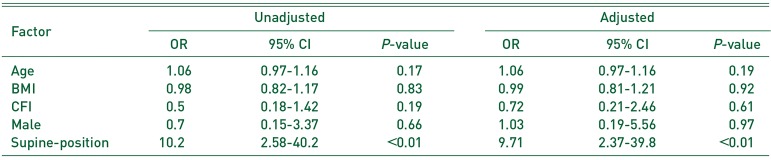
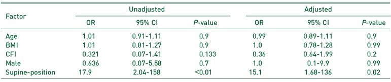
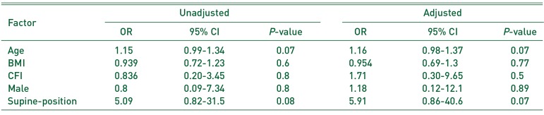
One-way ANOVA (Bonferroni correction) and the t-test were used to compare the operation duration, intra-operative blood loss, and radiographic evaluations using R 3.0.3 (R Foundation for Statistical Computing, Vienna, Austria). Fisher test was used to analyze categorical data. Multivariable logistic regression was performed to test the correlation between IOFs, Vancouver AGT type fractures (VAGT), Vancouver B type fractures (VB), the factors investigated in the previous study12345) (age, BMI, gender, and CFI), and the variables that showed significant correlation with patient's position (P<0.05).

RESULTS

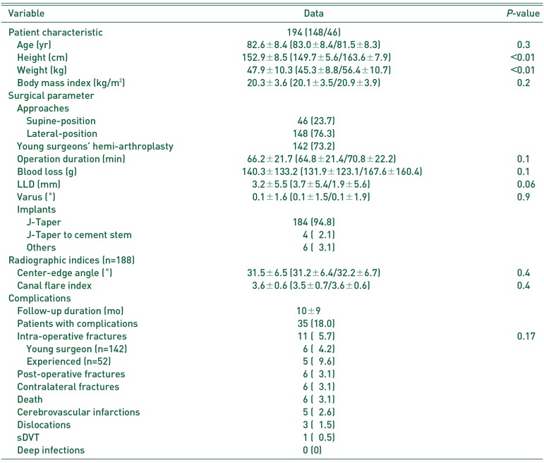
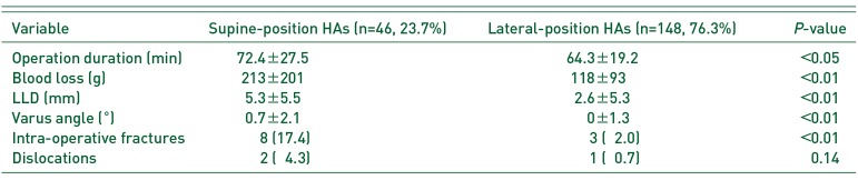
This study included 194 patients. The mean age at HA was 82.6±8.4 years (Table 2). The number of female patients was 148 (76.3%). The mean BMI was 20.3±3.6 kg/m2. ALS was used in 25 (12.9%) patients, DAA in 21 (10.8%), DLA in 9 (4.6%), PA in 50 (25.8%), and SA in 89 (45.9%) patients. Young surgeons performed 142 HAs (73.2%). The mean operation duration was 66.2±21.7 minutes and the mean operative blood loss was 140.3±133.2 g. IOFs occurred in 11 (5.7%) patients: six VAGT fractures, one VB1, and four VB2. IOFs occurred in 4.2% (6/142) of young surgeons' HAs and 9.6% (5/52) of experienced surgeons' HAs (P=0.17).
In the radiographic analysis, we excluded four IOFs (conversion to a cement stem), five straight stems, and one planned cement stem. The 184 patients for whom a tapered-wedge stem was used were evaluated by post-operative radiographs. The mean LLD was 3.2±5.5 mm and the mean varus angle was 0.1±1.6°. We excluded three hip arthroplasties and three multiple pinnings. The radiographic indices on the healthy side for the patients (n=188) were as follows: mean center-edge angle was 31.5±6.5°, mean CFI was 3.6±0.6.

1. Comparison between Approaches

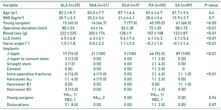
IOFs occurred in supine-position HAs in 8 (17.4%) patients and in lateral-position HAs in 3 (2.0%) patients (Table 3, 4). With ALS, IOFs occurred in 4 (16.0%) patients; three IOFs were VB2 fractures and required conversion from a taper wedged stem to a cement stem, and another IOF was a VAGT fracture, which required a claw plate. With DAA, IOFs occurred in 4 (19.0%) patients; all four cases were VAGT fracture and required no specific treatment. With PA, IOFs occurred in 2 (4.0%) patients: a VB2 fracture required cerclage wiring and a VAGT fracture required tension band wiring and conversion to a cement stem. With SA, IOFs occurred in 1 (1.1%) patient: VB1 was diagnosed on a postoperative radiograph and required non-weight bearing for 6 weeks.

2. The Risk Factor of IOFs

Multivariable logistic regression analysis revealed that supine-position HA was a risk factor for IOFs (adjusted odds ratio [OR], 9.71; 95% confidence interval [CI], 2.37-39.8; P<0.01) and VAGT fractures (adjusted OR, 15.1; 95% CI, 1.68-136; P=0.02) (Table 5,6,7).

DISCUSSION

In our study, IOFs occurred in 5.7% of the patients, which was higher than the frequency of post-operative dislocations (1.6%). IOFs were frequently (in 17.4% of the cases) encountered with the use of supine-position HA, this rate was higher than that with lateral-position HA (2.0%). IOFs were not related to surgical experience, in line with the previous studies18).
In ALS, the difficulty of implant insertion causes femoral shaft penetration (0.3%). In our patients, three VB2 fractures occurred during rasping or implantation. One VAGT occurred during elevation of the femur. In DAA, fractures of the great trochanter occur at a frequency of 1.4% to 4.5%92122). An insufficient release of the capsule9) and elevation of the femur with powerful levers10) result in great trochanter fractures. Additionally, the reduction maneuver results in periprosthetic fractures4). In our patients, all four VAGT fractures were diagnosed on post-operative radiographs. In DLA, VAGT occurs at a frequency of 0.7% to 3%, VALT at 2.8% to 8.1%, and VB fractures at 6.4%123). These two reports did not mention whether some particular procedures of DLA cause these fractures. Generally, VB fractures occur because of a mismatch between the stem and canal during rasping in an osteoporotic femur24). In PA, periprosthetic fractures occur at a frequency of 1.8% to 2.6%2122). In our cases, one VB2 fracture occurred during final stem insertion and one VAGT occurred during removal of the rasp. In SA, periprosthetic fractures occur at a frequency of 5.8%14). In our cases, one VB1 fracture was diagnosed on a radiograph at 1 week post-operatively. Theoretically, rasping before the neck cut would reduce hoop stress and prevent VB fractures14).
We thought that the hyperextended (over-range) position of the femoral rasping and soft tissue sparing would overload the tip of the great trochanter and the proximal femur in the supine position. Insufficient release of the hip capsule would result in excessive leverage momentum of the retractor behind the greater trochanter during hip extension, which may lead to VAGT. It would also lead to an oblique fracture of the proximal femur due to excessive external rotation torque applied to the distal femur to maintain the hip at the desired position to insert the stem. Lastly, an insufficient release of the capsule would lead to penetration of the femur by the femoral stem. In ALS and DAA, a release of the capsule and short external rotators as necessary would prevent IOFs and facilitate femoral preparation. The femoral preparation positions over a normal range of motion would require a release of muscles and the hip capsule. Fewer complications and correct implantation should precede muscle sparing.
Several limitations of this study should be acknowledged. First, this study was a retrospective case series. Second, we did not evaluate bone quality and disease-specific osteoporosis-like rheumatoid arthritis and further studies should be required. In our hospital (500-bed, secondary-level), lateral-position HAs decreased the incidence of IOFs. Meanwhile, the incidence of post-operative dislocations remained low. The post-operative LLD and varus angle of lateral-position HAs were more favorable than those of supine-position HAs. These results may be useful for young and experienced surgeons to employ new or unused approaches.

CONCLUSION

The supine position in HA surgery is an IOF risk factor and significantly increases the incidence of great trochanter fractures of Vancouver type A.

Notes

CONFLICT OF INTEREST: Kenta Kamo has received consulting fees from Kyocera. The authors declare that there is no potential conflict of interest relevant to this article.

References

1. Aslam-Pervez N, Riaz O, Gopal S, Hossain F. Predictors of intraoperative fractures during hemiarthroplasty for the treatment of fragility hip fractures. Clin Orthop Surg. 2018; 10:14–19. PMID: 29564042.
crossref
2. Duncan CP, Masri BA. Fractures of the femur after hip replacement. Instr Course Lect. 1995; 44:293–304. PMID: 7797866.
3. Davidson D, Pike J, Garbuz D, Duncan CP, Masri BA. Intraoperative periprosthetic fractures during total hip arthroplasty. Evaluation and management. J Bone Joint Surg Am. 2008; 90:2000–2012. PMID: 18762663.
4. Cooper HJ, Rodriguez JA. Early post-operative periprosthetic femur fracture in the presence of a non-cemented tapered wedge femoral stem. HSS J. 2010; 6:150–154. PMID: 21886528.
crossref
5. Miettinen SS, Mäkinen TJ, Kostensalo I, et al. Risk factors for intraoperative calcar fracture in cementless total hip arthroplasty. Acta Orthop. 2016; 87:113–119. PMID: 26541230.
crossref
6. van der Sijp MPL, van Delft D, Krijnen P, Niggebrugge AHP, Schipper IB. Surgical approaches and hemiarthroplasty outcomes for femoral neck fractures: a meta-analysis. J Arthroplasty. 2018; 33:1617–1627.e9. PMID: 29398259.
crossref
7. van den Bekerom MP, Sierevelt IN, Bonke H, Raaymakers EL. The natural history of the hemiarthroplasty for displaced intracapsular femoral neck fractures. Acta Orthop. 2013; 84:555–560. PMID: 24286565.
8. Schneider K, Audigé L, Kuehnel SP, Helmy N. The direct anterior approach in hemiarthroplasty for displaced femoral neck fractures. Int Orthop. 2012; 36:1773–1781. PMID: 22527335.
crossref
9. Baba T, Shitoto K, Kaneko K. Bipolar hemiarthroplasty for femoral neck fracture using the direct anterior approach. World J Orthop. 2013; 4:85–89. PMID: 23610757.
crossref
10. Unger AC, Dirksen B, Renken FG, Wilde E, Willkomm M, Schulz AP. Treatment of femoral neck fracture with a minimal invasive surgical approach for hemiarthroplasty - clinical and radiological results in 180 geriatric patients. Open Orthop J. 2014; 8:225–231. PMID: 25136389.
crossref
11. Kamo K, Kido H, Kido S, et al. [The concept of the superior approach for hip arthroplasty]. Jpn J Joint Dis. 2017; 36:457–460. Japanese.
12. Higuchi F, Gotoh M, Yamaguchi N, et al. Minimally invasive uncemented total hip arthroplasty through an anterolateral approach with a shorter skin incision. J Orthop Sci. 2003; 8:812–817. PMID: 14648270.
crossref
13. Moore AT. The self-locking metal hip prosthesis. J Bone Joint Surg Am. 1957; 39-A:811–827. PMID: 13438939.
crossref
14. Nakamura S, Matsuda K, Arai N, Wakimoto N, Matsushita T. Mini-incision posterior approach for total hip arthroplasty. Int Orthop. 2004; 28:214–217. PMID: 15168084.
crossref
15. Murphy SB, Ecker TM, Tannast M. THA performed using conventional and navigated tissue-preserving techniques. Clin Orthop Relat Res. 2006; 453:160–167. PMID: 17312591.
crossref
16. Tsukada S, Wakui M. Minimally invasive intermuscular approach does not improve outcomes in bipolar hemiarthroplasty for femoral neck fracture. J Orthop Sci. 2010; 15:753–757. PMID: 21116892.
crossref
17. Pala E, Trono M, Bitonti A, Lucidi G. Hip hemiarthroplasty for femur neck fractures: minimally invasive direct anterior approach versus postero-lateral approach. Eur J Orthop Surg Traumatol. 2016; 26:423–427. PMID: 27026091.
crossref
18. Bodrogi AW, Sciortino R, Fitch DA, Gofton W. Use of the supercapsular percutaneously assisted total hip approach for femoral neck fractures: surgical technique and case series. J Orthop Surg Res. 2016; 11:113. PMID: 27733183.
crossref
19. Chana R, Mansouri R, Jack C, et al. The suitability of an uncemented hydroxyapatite coated (HAC) hip hemiarthroplasty stem for intra-capsular femoral neck fractures in osteoporotic elderly patients: the Metaphyseal-Diaphyseal Index, a solution to preventing intra-operative periprosthetic fracture. J Orthop Surg Res. 2011; 6:59. PMID: 22099169.
crossref
20. Bertin KC, Röttinger H. Anterolateral mini-incision hip replacement surgery: a modified Watson-Jones approach. Clin Orthop Relat Res. 2004; (429):248–255. PMID: 15577495.
21. Pflüger G, Junk-Jantsch S, Schöll V. Minimally invasive total hip replacement via the anterolateral approach in the supine position. Int Orthop. 2007; 31(Suppl 1):S7–S11. PMID: 17668206.
crossref
22. Smith-Petersen MN. Approach to and exposure of the hip joint for mold arthroplasty. J Bone Joint Surg Am. 1949; 31A:40–46. PMID: 18122877.
crossref
23. Siguier T, Siguier M, Brumpt B. Mini-incision anterior approach does not increase dislocation rate: a study of 1037 total hip replacements. Clin Orthop Relat Res. 2004; (426):164–173.
24. Hardinge K. The direct lateral approach to the hip. J Bone Joint Surg Br. 1982; 64:17–19. PMID: 7068713.
crossref
Fig. 1

(A) The trial outer head with groove to minimize difficulty in trial reduction. The flat broaches made the neck osteotomy possible leaving the broach in place. (B) Trial reduction on the left hip hemi-arthroplasty. The piriformis was retracted posteriorly and the gluteus medius was retracted anteriorly. (C) The piriformis (P) and the gluteus medius (Me) were preserved after implantation.

hp-31-33-g001
Table 1

Anatomical Description and Patients Position of the Approaches

hp-31-33-i001
Approach ALS DAA DLA PA SA
Original articles Modified Watson-Jones1613) Distal part of Smith-Peterson1415) Modified Harding1617) Moore1819) Superior capsulotomy20)
Anatomical description Between gluteus medius and tensor fascia latae Between tensor fascia latae and sartorius Between fascia latae and gluteus maximus Splitting of gluteus maximus Splitting of gluteus maximus
Dissection of anterior third of gluteus medius and minimus Incision of piriformis, conjoined tendon and the obturator externus Between piriformis and gluteus medius
Patients position Supine Supine Lateral Lateral Lateral
Femoral preparation position Extended 20° Extended 30° Flexed 70° Flexed 70° Flexed 70°
adducted 30° adducted 15° adducted 20° adducted 20° adducted 30°
ER 90° ER 90° ER 90° IR 90° IR 20°

ALS: anterolateral supine approach, DAA: direct anterior approach, DLA: direct lateral approach, PA: posterior approach, SA: superior approach, ER: external rotation, IR: internal rotation.

Table 2

Patient Characteristics, Surgical Parameters, Radiographic Indices, and Complications

hp-31-33-i002
Variable Data P-value
Patient characteristic 194 (148/46)
 Age (yr) 82.6±8.4 (83.0±8.4/81.5±8.3) 0.3
 Height (cm) 152.9±8.5 (149.7±5.6/163.6±7.9) <0.01
 Weight (kg) 47.9±10.3 (45.3±8.8/56.4±10.7) <0.01
 Body mass index (kg/m2) 20.3±3.6 (20.1±3.5/20.9±3.9) 0.2
Surgical parameter
 Approaches
  Supine-position 46 (23.7)
  Lateral-position 148 (76.3)
 Young surgeons' hemi-arthroplasty 142 (73.2)
 Operation duration (min) 66.2±21.7 (64.8±21.4/70.8±22.2) 0.1
 Blood loss (g) 140.3±133.2 (131.9±123.1/167.6±160.4) 0.1
 LLD (mm) 3.2±5.5 (3.7±5.4/1.9±5.6) 0.06
 Varus (°) 0.1±1.6 (0.1±1.5/0.1±1.9) 0.9
 Implants
  J-Taper 184 (94.8)
  J-Taper to cement stem 4 (2.1)
  Others 6 (3.1)
Radiographic indices (n=188)
 Center-edge angle (°) 31.5±6.5 (31.2±6.4/32.2±6.7) 0.4
 Canal flare index 3.6±0.6 (3.5±0.7/3.6±0.6) 0.4
Complications
 Follow-up duration (mo) 10±9
 Patients with complications 35 (18.0)
 Intra-operative fractures 11 (5.7) 0.17
  Young surgeon (n=142) 6 (4.2)
  Experienced (n=52) 5 (9.6)
 Post-operative fractures 6 (3.1)
 Contralateral fractures 6 (3.1)
 Death 6 (3.1)
 Cerebrovascular infarctions 5 (2.6)
 Dislocations 3 (1.5)
 sDVT 1 (0.5)
 Deep infections 0 (0)

Values are presented as number only (female/male), mean±standard deviation (female/male), or number (%).

LLD: limb length discrepancy, sDVT: symptomatic deep venous thrombosis.

Table 3

Comparison between the Approaches

hp-31-33-i003
Variable ALS (n=25) DAA (n=21) DLA (n=9) PA (n=50) SA (n=89) P-value
Age (yr) 82.2±8.7 83.0±7.7 87.1±6.6 83.6±6.7 81.7±9.4 0.4
BMI (kg/m2) 20.7±3.3 20.2±3.6 21.4±4.1 20.6±3.4 19.9±3.7 0.7
Young surgeon 15 (60.0) 14 (66.7) 7 (77.8) 45 (90.0) 61 (68.5) <0.05
Operation duration (min) 80±33 63±14 82±28 72±20 58±15 <0.01
Blood loss (g) 222±225 202±174 128±9 107±108 123±87 <0.01
LLD (mm) 6.5±4.8 4.3±6.1 0.4±7.6 4.1±4.3 2.1±5.4 <0.01
Varus angle (°) 1.5±1.8 0.0±2.2 1.1±2.0 −0.2±1.0 −0.1±1.4 <0.01
Implants
 J-taper 19 (76.0) 21 (100) 9 (100) 46 (92.0) 89 (100) <0.01
 J-taper to cement stem 3 (12.0) 0 (0) 0 (0) 1 (2.0) 0 (0)
 Straight stem 3 (12) 0 (0) 0 (0) 2 (4.0) 0 (0)
 Cement 0 (0) 0 (0) 0 (0) 1 (2.0) 0
 Intra-operative fractures 4 (16.0) 4 (19.0) 0 (0) 2 (4.0) 1 (1.0) <0.01
 Vancouver AGT 1 (4.0) 4 (19.0) 0 (0) 1 (2.0) 0 (0)
 Vancouver B1 0 (0) 0 (0) 0 (0) 0 (0) 1 (1.0)
 Vancouver B2 3 (12.0) 0 (0) 0 (0) 1 (4.0) 0 (0)
 Young surgeon VAGT, 1/VB2, 1 VAGT, 2 0 (0) VAGT, 1/VB2, 1 0 (0) 0.56
 Dislocations 2 (8.0) 0 (0) 0 (0) 1 (2.0) 0 (0) 0.07

Values are presented as mean±standard deviation or number (%).

ALS: anterolateral supine approach, DAA: direct anterior approach, DLA: direct lateral approach, PA: posterior approach, SA: superior approach, BMI: body mass index, LLD: limb length discrepancy, HA: hemi-arthroplasty.

Table 4

Comparison between the Supine and Lateral-position Approach

hp-31-33-i004
Variable Supine-position HAs (n=46, 23.7%) Lateral-position HAs (n=148, 76.3%) P-value
Operation duration (min) 72.4±27.5 64.3±19.2 <0.05
Blood loss (g) 213±201 118±93 <0.01
LLD (mm) 5.3±5.5 2.6±5.3 <0.01
Varus angle (°) 0.7±2.1 0±1.3 <0.01
Intra-operative fractures 8 (17.4) 3 ( 2.0) <0.01
Dislocations 2 ( 4.3) 1 ( 0.7) 0.14

Values are presented as mean±standard deviation or number (%).

HA: hemi-arthroplasty, LLD: limb length discrepancy.

Table 5

The Risk Factors of Intra-operative Fracture

hp-31-33-i005
Factor Unadjusted Adjusted
OR 95% CI P-value OR 95% CI P-value
Age 1.06 0.97-1.16 0.17 1.06 0.97-1.16 0.19
BMI 0.98 0.82-1.17 0.83 0.99 0.81-1.21 0.92
CFI 0.5 0.18-1.42 0.19 0.72 0.21-2.46 0.61
Male 0.7 0.15-3.37 0.66 1.03 0.19-5.56 0.97
Supine-position 10.2 2.58-40.2 <0.01 9.71 2.37-39.8 <0.01

OR: odds ratio, CI: confidence interval, BMI: body mass index, CFI: canal flare index.

Table 6

The Risk Factors of Intra-operative Vancouver AGT Fracture

hp-31-33-i006
Factor Unadjusted Adjusted
OR 95% CI P-value OR 95% CI P-value
Age 1.01 0.91-1.11 0.9 0.99 0.89-1.11 0.9
BMI 1.01 0.81-1.27 0.9 1.0 0.78-1.28 0.99
CFI 0.321 0.07-1.41 0.133 0.36 0.64-1.99 0.2
Male 0.636 0.07-5.58 0.7 1.0 0.1-9.9 0.99
Supine-position 17.9 2.04-158 <0.01 15.1 1.68-136 0.02

OR: odds ratio, CI: confidence interval, BMI: body mass index, CFI: canal flare index.

Table 7

The Risk Factors of Intra-operative Vancouver B Fracture

hp-31-33-i007
Factor Unadjusted Adjusted
OR 95% CI P-value OR 95% CI P-value
Age 1.15 0.99-1.34 0.07 1.16 0.98-1.37 0.07
BMI 0.939 0.72-1.23 0.6 0.954 0.69-1.3 0.77
CFI 0.836 0.20-3.45 0.8 1.71 0.30-9.65 0.5
Male 0.8 0.09-7.34 0.8 1.18 0.12-12.1 0.89
Supine-position 5.09 0.82-31.5 0.08 5.91 0.86-40.6 0.07

OR: odds ratio, CI: confidence interval, BMI: body mass index, CFI: canal flare index.

TOOLS
Similar articles