

 PDF
PDF ePub
ePub Citation
 Print
Print


INTRODUCTION
Nails are hard keratinized structures on the top surfaces at the end of fingers and toes. It is important that nails are not just shields for protection. They are active structures: finger nails grow 1.8~4.5 mm per month and toe nails about of half of that1. Nails often reflect pathologic features of adjacent tissue and various nail diseases are found in about 10% of patients visiting our dermatologic clinic and approximately 6.8% of infants had nail alterations23. Nail disorders are caused by various conditions. Infections such as periungual wart or herpetic whitlow, or onychomycosis and acute traumatic damages are obvious reasons. Some drugs (like chemotherapeutic agents) are associated with nail dystrophy because they act on rapidly proliferating organs. Systemic diseases such as anemia, Raynaud's disease, hepatic failure, chronic heart failure, renal failure, thyroiditis, inflammatory bowel disease, and chronic bronchitis also affect nail growth4. In addition, many dermatologic diseases including psoriasis, lichen planus, lichen nitidus, pityriasis rubra pilaris, alopecia areata, and Darier's disease are known to affect nails56.
Atopic dermatitis (AD) is a common skin disease characterized by chronic, relapsing forms of skin inflammation from a disturbance of epidermal-barrier function that culminates in dry skin7. It is known to be one of the main predisposing factors for chronic hand and foot eczema. According to a previous study on dermatologic diseases associated with AD in Korean, 6% of patients had hand or foot eczema. The prevalence was increased by age until 30, showing 3% at ages 0~10 and 7.9% at 21~308. Another study reported approximately 58.9% of active AD patients had hand eczema and 16% of them have nail change9.
However, no large-scale studies have been reported here, focusing on nail problems associated with AD. Even so, some related cases have been reported: osteomyelitis on the distal phalanges of AD patients10 and onycholysis on atopic eczema treated with PUVA photochemotherapy11. A study involving a small number of childhood AD subjects showed higher prevalence of nail dystrophy (4 of 48 patients, 8.3%) than did the control (1 of 89 patients, 1.1%), although this was not statistically significant12.
The aims of our study were to determine the incidence and types of nail abnormalities associated with AD and to determine their relation to the severity of AD.
MATERIALS AND METHODS
Subjects
This cross-sectional study was conducted at Department of Dermatology, Hallym University Kangnam Sacred Heart Hospital, Seoul, Korea. Eligible participants included all AD patients ages 2~19 who visited the outpatient clinic of during January 2014 to April 2016. AD was diagnosed by dermatologists clinically according to the criteria of Hanifin and Rajka13. Patients were excluded who had been diagnosed for other diseases known to affect abnormal nails: 1) Systemic conditions such as anemia, Raynaud's disease, hepatic failure, chronic heart failure, renal failure, thyroiditis, inflammatory bowel disease, and chronic bronchitis; 2) Infectious disease such as onychomycosis, viral warts, and herpes infections; 3) Dermatologic diseases, including psoriasis, lichen striatus, lichen planus, lichen nitidus, pityriasis rubra pilaris, alopecia areata, and Darier's disease. One subject with a history of traumatic nail effects (from biting them) was also excluded.
This study was approved by the institutional review board of the Hallym University Kangnam Sacred Heart Hospital (IRB no. 2013-08-68) and was conducted in accordance with the Declaration of Helsinki. Written informed consent was obtained from all patients or their guardians.
Evaluation of nail features
In order to evaluate the nail abnormalities, patients had physical exams of all finger and toe nails by dermatologists who checked their shape, color, and hardness. Clinical pictures were taken if any nail abnormalities were detected. Nail abnormalities were classified as: transverse groove (Beau's line), trachyonychia, leukonychia, koilonychia, onycholysis, nail pitting, onychoschizia, melanonychia, brachyonychia, and onychomadesis. Epidemiologic features such as age and gender were also collected.
Clinical assessment
The severity of the AD was assessed using the eczema area and severity index (EASI) score, which evaluates four clinical parameters: erythema, induration/papulation, excoriation, and lichenification on a 0~3 scale within four defined body regions (head/neck, upper limbs, trunk, and lower limbs). The scoring was done by dermatologists14.
Statistical analysis
The results were expressed as ‘mean±standard deviation.’ The chi-square test for nominal variables and the Student's t-test for continuous variables were used to determine the significance of differences. The relative frequency of risk factors was analyzed by logistic regression using the backward model. Significance levels for all analyses were set at p=0.<0.05. All statistical analyses were conducted using PASW Statistics 18.0 (IBM Co., Armonk, NY, USA).
RESULTS
Demographic features of patients
A total of 235 patients were recruited, of whom the average age was 7.43±4.92. The average EASI score for all patients was 9.81±8.75. The study population consisted of 123 male and 112 females (male to female ratio, 1:0.91). There was no difference on average age between genders but males (11.82±11.03) had higher EASI scores than did the females (7.68±4.55).
The EASI score and age showed a weak correlation in this study group, with total correlation coefficient (CC=0.297, p=0.006), upper limb (CC=0.239, p=0.029), and lower limb (CC, 0.325, p=0.003) all showing statistical significance.
The prevalence and types of nail dystrophy
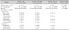
The detailed data on the prevalence and types of the nail dystrophy are provided in Table 1. Of the 235 AD patients, 24 (10.2%) patients had nail abnormalities. Males showed more nail abnormalities on fingers and toes than female, with an odds ratio (OR) of 2.4 (not statistically significant). There were 15 patients with finger nail abnormalities, 9 males and 6 females. The average age was greater in patients with finger nail abnormalities (10.13±4.36) than in those without (7.21±4.91), and this was a statistically significant difference (p=0.026). Males had more finger nail abnormalities than did females but this was not statistically significant (OR, 1.395; 95% confidence interval [CI], 0.480~4.051; p=0.539). There were 14 patients with toe nail abnormalities, 12 males and 2 females. The average age was higher in patients with toe nail abnormalities (9.21±5.67) than in those without (7.29±4.86), but the difference was not statistically significant (p=0.155). Males had far more toe nail abnormalities than did females (OR, 5.634; 95% CI, 1.051~25.166; p=0.029).
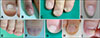
The nail abnormalities observed were classified as transverse groove (Beau's line) (Fig. 1A; 6 patients: 25.0%), nail pitting (Fig. 1B; 4 patients: 16.7%), koilonychia (Fig. 1C; 4 patients: 16.7%), trachyonychia (Fig. 1C; 3 patients: 12.5%), leukonychia (Fig. 1D; 3 patients: 12.5%), brachyonychia (Fig. 1E; 2 patients: 8.3%), melanonychia (Fig. 1F; 2 patients: 8.3%), onychomadesis (Fig. 1G; 2 patients: 8.3%), onychoschizia (Fig. 1H; 2 patients: 8.3%), onycholysis (Fig. 1I; 2 patients: 8.3%). There were 4 patients who had multiple types of nail changes; three of them had Beau's line.
Relationships of nail abnormality with EASI score and hand/foot eczema
The detailed data on the relation of nail abnormalities with EASI score and hand or foot eczema are provided in Table 2. The average EASI score of patients with total nail abnormalities was higher than patients without (11.80±8.31 vs. 9.30±9.50, p=0.236). The EASI score confined to upper extremity was 2.78±2.65 for the finger nail change group and 2.24±1.72 for the normal finger nail group (not statistically significant, p=0.346). The EASI score limited to lower extremities was higher in the toe nail change group (7.23±5.64) than in those without them (4.15±4.29), and this was statistically significant (p=0.032). The logistic regression analysis, controlling for age and sex, showed statistical significance for toe nail changes in relation to EASI score of lower limbs (OR, 1.115; 95% CI, 1.014~1.316; p=0.030).
The prevalence of hand or foot involvement of AD patients with nail abnormality was 54.2% (OR, 2.364; 95% CI, 0.923~6.055, p=0.091) and that of patients without nail abnormality was 33.2% (OR, 0.423; 95% CI, 0.165~ 1.083; p=0.091). There was hand involvement of AD in 40% of patients with finger nail abnormalities (OR, 1.778; 95% CI, 0.607~5.208, p=0.294), and 42.9% (OR, 6.156; 95% CI, 1.969~19.252; p=0.002) of patients with toe nail abnormalities had AD foot lesions. That is, nail change patients had more hand and foot eczema than those without nail change. Moreover, patients with foot eczema had higher incidence of toe nail changes (statistically significant).
The prevalence of finger or toe nail abnormalities in hand or foot eczema was 15.7% (13/83; OR, 2.364; p=0.091) and that of patients without hand or foot eczema was 7.2% (11/152; OR, 0.236, p=0.091). Finger nail changes were seen in 9.1% (6/66; OR, 1.778; p=0.294) of hand eczema patients, and toe nail changes were seen in 20.0%(6/30; OR, 6.156; p=0.032) in foot eczema patients.
There was no statistically significant difference in EASI scores according to types of nail abnormalities.
DISCUSSION
This study was conducted to determine the prevalence and types of nail abnormalities in AD, and to evaluate the relation between nail abnormalities and the severity of AD. As it is well known, patients with AD have a high proportion of children and adolescents. In year 2014, the national prevalence of AD in children (under age 19) was 5.8% and that of adults was 0.9%. Actually, most of the patients are under the age of 20 in our outpatient clinic. Therefore, we targeted patients under 20 years of age and excluded onychomycosis because onychomycosis accounts for about 50% of nail disease, but it is very rare in children1516.
Of the 235 AD patients, 24 patients had nail abnormalities. There was no statistically significant difference in the total EASI score in relation to the presence of nail abnormalities. However, when the EASI score was confined to lower extremities, a relationship was shown with the prevalence of toe nail dystrophy.
Among the numerous diseases affecting nails, dermatologic ones make up the majority. Nail psoriasis accompanies about 10%~55% in psoriasis patients and nail pitting is the most common feature1718. Other nail changes that accompanied psoriasis included Beau's line, onycholysis, leukonychia, subungual hyperkeratosis, splinter hemorrhage, crumbling, onychomadesis, and red spot lunula19. In psoriasis, nail involvement increases with age, duration, and extent of the disease. Moreover, the presence of psoriatic arthritis also caused diminished quality of life due to its high visibility20. Nail lichen planus occurs in 10% of patients with systemic lichen planus and may also appear in isolated nail lesions with absence of other cutaneous symptoms. Each of the nails can be affected separately or together. Thinning, ridging, and distal splitting of the nail plate are most common findings of nail lichen planus, and nail pterygium can be seen21. Nail changes in pityriasis rubra pilaris can be seen about 21% of patients, and is characterized by yellow-brown discoloration, subungual hyperkeratosis, nail thickening, and splinter hemorrhage22. Darier's disease is an autosomal dominantly inherited disease characterized by greasy hyperkeratotic papules in seborrheic regions, nail abnormalities, and mucous membrane changes. Nail involvement occurred in 85%~100% of the subjects and included: longitudinal red and/or white lines extending from the base of the nail across the lunula to the free margin of the nail, nail fragility, V-shaped notches at the free edge of the nail, longitudinal ridging of the nail, painful splits, and subungual hyperkeratotic fragments2324. Onychomycosis can present onycholysis, subungual hyperkeratosis, nail-plate thickening, and leukonychia. AD patients are known to have higher susceptibility to viral and fungal infection due to altered immune responses and decreased T lymphocytes825. In this study, the above diseases were excluded, except for AD itself. Nail change may accompany AD but there is little information about the types of nail changes associated with AD. Compared to a reported prevalence of 8.3%, in this study, 10.2% of AD patients appeared to have nail changes12.
Different nail dystrophies occur depending on the nail area in which the problem occurs. Eczematous change infiltrating the nail matrix can cause anonychia, nail pitting, trachyonychia, longitudinal ridges, transverse groove (Beau's line), onychomadesis, and onychoschizia26. Damage to the nail bed can cause brachyonychia, chromonychia, clubbing, koilonychia, subungual exostosis, yellow nail syndrome, onycholysis, and splinter hemorrhage27. In this study, the most common nail symptom was Beau's line which formed in 26% of patients with nail change. It is caused by decreased function of the proximal nail matrix and if the dysfunction persists over a few weeks, onychomadesis also appears (which formed 8.3% of nail abnormalities in this study). Three of four patients with multiple types of nail changes had Beau's line in common, in each case accompanied by koilonychia, nail pitting, and onychoschizia (Table 1). Along with Beau's line, a large portion of the nail dystrophies observed in this study, such as trachyonychia, pitting nail, and onychoschizia; have their origin in problems in the nail matrix. Regarding the results of this study, and the fact that other diseases causing nail abnormalities have biopsy-proven inflammatory effects on the nail matrix, it is reasonable to think that regional eczematous changes around nails cause the nail dystrophies observed in AD6212829. This might be caused by non-specific inflammation of the nail matrix, which then interrupts proper synthesis of the nails.
The average EASI score was higher in the nail abnormality group (11.80±8.31) than in those with normal nails (9.30±9.50), but the difference was not statistically significant. Because the EASI score is calculated based on erythema, induration/papulation, excoriation, and lichenification over the whole body area, finger and toe lesions contribute little to the score due to their small surface area. Moreover, because nails reflect their past like tree rings do, there may be no distinctive eczema involving the nail matrix when lesions are present. And since this study was cross-sectional study, only the lesion shown at the time of visit was able to be checked. Although it was not statistically significant, the EASI score of the upper extremities was higher in the finger nail abnormality group. More than that, the EASI score confined to lower extremities was higher in patients with toe nail abnormalities (statistically significant). Because older age and male gender contributed to higher EASI scores in the study population, logistic regression analysis was performed and the EASI scores of lower extremities and toe nail changes revealed a positive relation (OR, 1.115; 95% CI, 1.014~1.316; p=0.030). Patients with hand or foot eczema had higher rates of nail changes. Especially in foot involvement of AD, there was a significant correlation with toe nail abnormalities (OR, 6.257; 95% CI, 1.748~22.394, p=0.002).
There are some limitations in this study. There was no control group from the general population to compare the prevalence of nail dystrophies. And skin biopsies were not performed to confirm histological features of nail apparatus due to patient's relatively young age and the possibility of permanent nail dystrophy. However, we were able to know which position of nail was abnormal through clinical appearance of nail. Nevertheless, there is significance as the first study to examine the type of nail dystrophy in AD and the association of nail change with the severity of AD. In this study, both EASI scores confined to lower limbs and AD involvement of the feet showed positive relations to toe nail changes in this study. Toe nail changes might be predicted by the EASI score in the lower limbs in AD patients.

 XML Download