

 PDF
PDF ePub
ePub Citation
 Print
Print


Dear Editor:
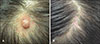
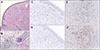
Perinevoid alopecia is one of the atypical hair loss disorders1. We describe a rare case of perinevoid alopecia. A 33-year-old woman presented with a solitary patch of alopecia with a central skin-colored papule on her vertex for 2 months (Fig. 1A). The match-head-sized skin-colored papule was observed when she was 10 years old, although the patch of alopecia was observed 2 months prior to presentation. Broken hairs were seen at the site of the patch of alopecia. There was no history of local irritation resulting in hair breakage. She reported an unremarkable past history and family history. Her laboratory findings were within the reference range. The central skin-colored papule was completely removed, and histopathologically this papule showed numerous nevocytes and a sparse inflammatory cell infiltrate (Fig. 2A). The periphery of the papule showed sparse hair follicles with perifollicular inflammatory cell infiltrates (Fig. 2B). The inflammatory cell infiltrates within the papule and at the periphery primarily comprised CD3- and CD8-positive cells (Fig. 2C~F). Incidentally, follicular rupture with inflammatory infiltrate was shown under the nevus cell nests. She was diagnosed with perinevoid alopecia, and surgical removal of the central papule was followed by regrowth of hair at the affected site (Fig. 1B).
Perinevoid alopecia is an extremely rare disorder with a clinically distinctive feature of alopecic patch with central pigmented nevus and a histologically specific finding of inflammatory cell infiltration in nevus cell nests and perifollicular areas. Since Quiroga and Pecoraro2 reported a case of perinevoid alopecia in 1958, few cases have been published in the literature1. Previous reports have described the development of perinevoid alopecia on the scalp and the chin in young adults in whom this rare condition is commonly observed. Although pathogenesis of perinevoid alopecia is unclear, it may be similar to that as sociated with the development of a halo nevus. Histopathological examination of a halo nevus shows dense inflammatory cell infiltrates invading the nevus cell nests in the upper dermis and degeneration of peripheral melanocytes3. The inflammatory cells are observed to be CD3- or CD8-positive lymphocytes. In this patient who presented with perinevoid alopecia, histopathological examination also showed nevus and inflammatory cells at the site of the central nevus lesion and sparse hair follicles and perifollicular inflammatory cell infiltrates in the perinevoid area. Immunohistochemistry examination showed the inflammatory cells noted in this patient were CD8-positive lymphocytes. Gilhar et al.4 have described that the association between nevi and alopecia is attributable to an immunological reaction in that melanocyte-associated T-cell epitopes act as auto-antigens to induce an autoimmune reaction against hair follicles and nevus cells. We think incidental development of minute follicular rupture in this case was not associated with perinevoid alopecia. Although pseudocyst of scalp is a much more severe inflammatory disease of scalp, it does not develop perilesional alopecia5. Perinevoid alopecia can be effectively managed with surgical removal of the nevus.
 XML Download