

 PDF
PDF ePub
ePub Citation
 Print
Print


INTRODUCTION
Dermal melanocytosis is a common pigmented skin disease, characterized by an increased number of ectopic melanocytes in the dermis1. It is usually divided into four main groups: nevus of Ota, nevus of Ito, blue nevus, and Mongolian spots. Rare variants of dermal melanocytosis that do not belong to these four typical groups are called dermal melanocyte hamartoma, also referred as congenital dermal melanocytosis (CDM) because it mostly appears from birth2345. Here, we report a case of CDM on the foot with a literature review of previously reported cases of CDM.
CASE REPORT
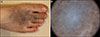
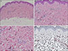
A 22-year-old woman presented with an asymptomatic, confluent, and pigmented skin lesion on the right foot dorsum since her birth. No specific changes occurred, including color and texture, except that the lesion enlarged in proportion to the growth of the body. She denied any significant medical problems and family history of pigmented disorders. On physical examination, mottled, confluent, and blue-gray macules and patches were observed on the right foot dorsum (Fig. 1A). We received the patient's consent form about publishing all photographic materials. Dermoscopic examination revealed a steel blue or gray structureless area surrounded by mottled brown globules (Fig. 1B). Skin biopsy demonstrated increased scattered melanocytes in the dermis without nest formation. Spindle and epithelioid melanocytes with melanin pigment were randomly oriented and dispersed among the collagen bundles. Immunohistochemical staining for Melan A showed positive staining for the melanocytes (Fig. 2). Based on the clinical and pathologic findings, the patient was diagnosed with CDM. She has been treated with three sessions of Q-switched Nd- YAG 1,064 mm laser without remarkable improvement.
DISCUSSION
Embryologically, melanocytes are derived from neural crest cells originating from the ectoderm5. Dermal melanocytosis has been suggested to occur due to the arrest of melanocyte migration, resulting in increased number of melanocytes in the dermis5. Except for blue nevus, the other types of dermal melanocytosis share similar histopathologic findings and differ only in the concentration and location of melanocytes. The histopathologic finding shows elongated dermal melanocytes scattering between collagens1. Clinically, the types of dermal melanocytosis, such as nevus of Ota or Ito, are distinguished by their unique location–trigeminal or acromioclavicular distribution5. Dermal melanocyte hamartoma is a rare dermal melanocytosis pattern that does not fit into the typical classification of dermal melanocytosis25. In the present case, the pigmented lesion distribution differed from the nevus of Ota, nevus of Ito, and Mongolian spots. Additionally, the lesion was not matched to blue nevus, which shows a high concentration of dermal melanocytes in the dermis1.
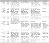
Several cases of CDM have been previously reported (Table 1)1234678910. Among the 10 cases including ours, 6 were reported at adults. Female predominance (male:female=2:8) was observed similar to other dermal melanocytosis, such as the nevus of Ota. Segmental or dermatomal distribution was observed in five cases, localized pattern in four cases, and generalized form in one case. Among the cases with a localized pattern, the lesions were confined to the trunk or upper extremities3910. CDM developing as an isolated patch on the foot similar to our case, has not been described. Clinically, CDM usually appears as uniform or mottled gray-blue patches with or without macules. Histopathologic findings revealed scattered melanocytes without nest formation throughout the dermis, which were subtly different from the nevus of Ota or Ito (melanocytes detected mainly in the upper dermis), and Mongolian spot (mostly in the lower dermis)1. In most cases, the pigmentation did not disappear with age. However, in one case, a 4-day-old neonate had shown generalized blue-gray pigmentation that gradually faded until 15 months of age. Although the case was reported as a generalized dermal melanocytosis, we speculate that the case might be difficult to be differentiated from extrasacral Mongolian spot because it tends to disappear or fade during childhood7.
In conclusion, we report a rare case of CDM on the foot with relevant literature review. Further studies are necessary to elucidate the clinical and pathogenetic characteristics of CDM.
 XML Download