

 PDF
PDF ePub
ePub Citation
 Print
Print


INTRODUCTION
Symmetrical giant facial plaque-type juvenile xanthogranuloma (SGFP-JXG) is an uncommon type of xanthogranuloma, which was first described by Gunson et al.1 in 2008. Cutaneous manifestation of JXG typically presents as a solitary, well-separated yellow or red-brownish papule or nodule, measuring up to 1 cm in most cases2. A large or diffuse plaque-type lesion as shown in this case is notably rare. Histologically, JXG consists of a well demarcated mixed infiltrates of mononuclear histiocytes with vacuolated cytoplasm and multinuclear giant cells, mostly Touton type giant cells, on a background of scattered lymphocytes, neutrophils, and mast cells3. Immunohistochemical staining confirms the diagnosis of JXG, with the histiocytes being stained in CD68. JXG is negative for protein S-100 and CD1a, therefore differentiating it from Langerhans cell histiocytosis4.
Plaque types of JXG at extrafacial sites have been occasionally reported in the literature25. SGFP-JXG, however, is reported only in two previous cases6. Herein, we report another rare case of SGFP-JXG on a 3-year-old boy, which persisted for 2 years without any signs of regression. This is the first case report of a SGFP-JXG on which a single fractional ablative laser therapy was performed with a promising outcome.
CASE REPORT
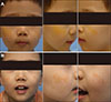
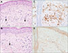
A 3-year-old Korean boy was referred to our clinic with bilateral yellowish indurated plaques on both cheeks since 1 year after birth (Fig. 1A). Physical examinations revealed no ophthalmic or mucosal involvements, and palpable lymph nodes were not observed. Laboratory findings including lipid profiles such as total cholesterol, triglyceride, high density lipoprotein-, and low density lipoprotein-cholesterol were within normal limits. A skin biopsy was performed, and the histologic findings showed numerous foam cells with several Touton giant cells consisting of a lipid cytoplasm with a ring of nuclei, and occasional foreign body giant cells, throughout the upper dermis (Fig. 2A, B). The immunohistochemical staining result was positive for CD68 and negative for S-100 (Fig. 2C, D). Based on the clinicopathologic features, the patient was diagnosed as a SGFP-JXG. As the lesion did not show any signs of spontaneous regression, we performed a single session of fractional ablative CO2 laser, and a significant reduction of the size was observed throughout the next 6 months of follow-up period without signs of recurrence (Fig. 1B).
DISCUSSION
SGFP-JXG is reported in only two case studies since 200816. In both cases, the lesions persisted for longer than 6 years, and had not shown a sign for spontaneous resolution. In one case, the 10-year-old patient simultaneously had JXG on the arms, and the extrafacial site disappeared spontaneously6. Furthermore, none of the cases observed internal organ involvements.
A confounding factor to our final diagnosis was its differential diagnosis with diffuse normolipemic plane xanthoma (DNPX). DNPX was first described by Altman and Winkelmann in 1962, and the features of this entity are: (a) xanthelasma palpebrarum, (b) diffuse plane xanthomas of the head, neck, trunk, or extremities, and (c) plasma lipid values that are within normal limits78. Its skin biopsy shows an accumulation of foam cells and Touton giant cells, along the infiltrated histiocytes that are CD68 positive, and CD1a and S-100 negative. DNPX is a rare xanthoma variant that has been associated with hematologic disorders, and only 45 cases are reported in the English literature between 1962 and 20139. Although histologically challenging to distinguish with JXG, DNPX typically shows characteristic xanthelasma appearing first in most cases, and tends to occur more frequently in adults81011. Therefore, the clinical presentation of our case without preceding appearance of xanthelasma, the patient's age, and its greater degree of similarity with the previously reported cases of SGFP-JXG, favors the current diagnosis.
In conclusion, we report another rare case of SGFP-JXG occurred on the face of a 3-year-old boy. As shown in the previous reports, a subset of the lesion can persist for several years without resolution; hence persistent lesions may require an attempt for a treatment. A few previous DNPX cases, which have similar histologic features as our case, reported successful treatment outcomes with ablative lasers and dermabrasion111213. Ablative lasers such as CO2 and Er:YAG lasers are safe and effective devices for the removal of epidermal or superficial dermal lesions. The current report attempted a single session of fractional ablative CO2 laser, which resulted in a significant regression in size of the lesion. To our knowledge, there is only one other case that attempted ablative CO2 laser on xanthogranulomas of the skin, and it reported some scarring left after the laser therapy14. The use of fractional laser in our case might have contributed to the avoidance of post-laser scarring, since limited ablation was induced at each microthermal zone, thus augmenting collagen remodeling with a decreased risk of scarring compared to fully ablative technology15. A long term follow-up after the treatment showed a continuous effect of the treatment. Hence, this is the first case report of a persistent SGFP-JXG on which a single fractional ablative laser therapy was performed with a promising outcome.
 XML Download