

 PDF
PDF ePub
ePub Citation
 Print
Print


INTRODUCTION
Riehl's melanosis is a form of contact dermatitis that is characterized by a brown to gray pigmentation over the face1. Although the pathogenesis of Riehl's melanosis remains largely unknown, recent studies suggest that Riehl's melanosis is associated with Riehl's melanosis caused by ingredients of cosmetics12. Facial pigmentation can be a devastating psychological burden in patients with this condition. However, conventional treatments are often unsatisfactory. The mainstay of treatment of Riehl's melanosis is as follows: avoidance of identified contact allergens, sun protection, and topical application of bleaching agents.
Favorable results using low-fluence Q-switched neodymium-doped yttrium aluminum garnet (Nd:YAG) laser in the treatment of facial pigmentary disorders, including melasma, nevus of Ota, and postinflammatory hyperpigmentation, have been reported34567. Although the specific mechanism of low-fluence Nd:YAG laser for pigmentary disorders was unknown, ultrastructural changes in melanosomes were found after irradiation with low-fluence Nd:YAG laser8. Recently, studies have reported the use of low-fluence Nd:YAG laser in the treatment of Riehl's melanosis29.
In this study, we investigated 10 patients with clinicopathologically confirmed Riehl's melanosis and assessed the effectiveness of low-fluence Nd:YAG laser in the treatment of the disease. In addition, we analyzed clinical parameters in terms of treatment response to low-fluence Nd:YAG laser to identify prognostic factors in treating Riehl's melanosis.
MATERIALS AND METHODS
Ten patients with Riehl's melanosis were enrolled in this study. The patients visited Seoul National University Hospital (SNUH) for the management of hyperpigmentation of their facial region. The diagnosis of Riehl's melanosis was established based on clinical and histopathologic analyses of patients. To distinguish from simple postinflammatory hyperpigmentation, the absence or presence of preceding symptoms for overt inflammation at the site of pigmentation was thoroughly investigated. Skin biopsies were performed to rule out other pigmentary disorders such as melasma, nevus of Ota, acquired bilateral nevus of Ota-like macules, and ochronosis. To screen possible causative allergens, a patch test was performed in all except three patients. After confirming the causative allergens, the patients were educated to avoid the use of any cosmetics that contained the causative allergens.
To treat Riehl's melanosis, the patients received a treatment using a 1,064-nm Q-switched Nd:YAG laser (Pastelle®; WONTECH, Seoul, Korea) at a three-week interval. The parameters of the laser treatment were as follows: fluence started from 0.9 J/cm2 with a 7-mm-sized spot and frequency of 10 Hz. The fluence of the laser was adjusted up to 2.0 J/cm2 based on the patient's sensitivity to pain and response to the previous treatment. The laser treatment was applied to the pigmented lesion with appropriate overlap and repeated with three to four passes until the pigmented lesions showed subtle erythema without petechiae. Each treatment session took about 20 minutes.
At each visit before the treatment session, we asked the patient about the degree of improvement and the development of adverse events and took standardized clinical photographs under the same lighting condition using a digital camera (550D; Canon, Tokyo, Japan) to record the progress. In addition, the degree of improvement and the development of adverse events were further confirmed by using wood light. The additional session of laser treatment was determined by the patient's response to previous treatments, satisfaction, and need for further treatment. During the laser treatment, 4% hydroquinone cream (Eldoquin Forte®; ICN Pharmaceuticals Inc., Costa Mesa, CA, USA) was concurrently used on the hyperpigmented lesions in eight patients.
Objective assessments of improvement were performed by two investigators using standardized photographs taken before each treatment session. They compared the standardized photographs with the pretreatment photographs and evaluated the improvement of each treatment session based on the following grading scale (1~5): grade 0 (worsening or stable disease), grade 1 (minimal improvement, 0%~25% improvement), grade 2 (moderate improvement, 26%~50% improvement), grade 3 (marked improvement, 51%~75% improvement), and grade 4 (near total improvement, more than 75% improvement).
The effectiveness of low-fluence Q-switched Nd:YAG laser in the treatment of Riehl's melanosis was investigated by evaluating the grade of improvement and number of treatment sessions required to achieve each grade of improvement. To identify the prognostic factors in the treatment of Riehl's melanosis, we compared the improvement grade and number of treatment sessions required for improvement according to the clinical characteristics or demographic factors, using the Mann-Whitney U and Fisher's exact tests. SPSS 22.0 (IBM Co., Armonk, NY, USA) was used to perform statistical analysis. Data are shown as a mean±standard deviation. A p-value of <0.05 was considered statistically significant.
This study was approved by the Institutional Review Board of SNUH (IRB no. H-1705-097-856). We received the patient's consent form about publishing all photographic materials.
RESULTS
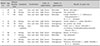
The demographic data and clinical information of the 10 patients are summarized in Table 1. All patients were females of Korean descent, and the mean age of the enrolled patients was 60.3±11.9 years (ranged from 46 to 81 years). All patients had pigmented lesions on their face and nine patients on their neck. Before the development of pigmented lesions, seven patients experienced symptoms of mild itching or erythema. The color of the pigmented lesion was dark brown in six patients and light brown in the remaining four patients. The pattern of pigmentation was homogenous in six and reticulated in four patients. The histopathologic findings included a variable degree of pigmentary incontinence, lymphocytic infiltration in the upper dermis, and focal basal vacuolar alteration of the epidermis in all patients.
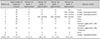
The response to low-fluence Q-switched Nd:YAG laser treatment is summarized in Table 2. The mean number of performed treatment was 17.9±5.9 (ranged from 10 to 28). All but one patient achieved grade 3 improvement. Specifically, seven patients reached grade 4 while two patients showed grade 3 improvement. However, one patient failed to reach grade 2 improvement even after 10 sessions of treatment. The mean number of laser treatment sessions to reach grade 1, 2, 3, and 4 improvement was 4.5±2.9 (range, 2~11), 8.8±3.8 (4~15), 12.1±4.0 (6~17), and 14.6±4.4 (9~20), respectively. Among the patients, three complained of guttate hypopigmentation; however, the hypopigmented lesions spontaneously improved after interruption of the laser treatment at the site of hypopigmentation. In addition, two patients experienced a transient aggravation of pigmentation after laser treatment. However, lowering the fluence improved the aggravated pigmentation.
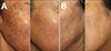
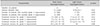
To identify prognostic factors determining the treatment response for Riehl's melanosis, we compared the results of laser treatment according to the clinical characteristics or demographic factors. We found that the color of pigmentation can be a determining factor for the response to the treatment: patients with dark brown hyperpigmentation showed more favorable responses than those with light brown hyperpigmentation (Fig. 1). The subgroup analysis between the groups revealed the proportion of patients who reached grade 4 improvement was higher (100% in dark brown and 25% in light brown, p-value=0.03) and the mean needed numbers of laser treatment sessions to reach grade 1 improvement were less in patients with dark brown pigmentation (2.7±0.8 in dark brown and 7.3±2.6 in light brown, p-value=0.01) (Table 3). In our data, the disease duration was shorter in patients with dark brown pigmentation (11.2±8.6 months) than in those with light brown pigmentation (57.0±43.1 months). Therefore, we performed a subgroup analysis on the effect of disease duration in comparing the outcome measures of patients who have had the disease for less than or more than 12 and 24 months, respectively. Results showed no statistically significant association between disease duration and treatment outcome (data not shown). We could not find a significant association in other variables such as age, total number of treatment sessions, and the proportion of patients experiencing symptoms or adverse events.
DISCUSSION
The pathophysiology of pigmentations in Riehl's melanosis remains unknown. Previous studies suggested that Riehl's melanosis is a form of pigmentary disorder caused by ingredients of cosmetics10. In our study, seven patients underwent patch tests, and six patients (85.7%) showed positive response to at least one allergen (Table 1). Considering that the pigmentation of Riehl's melanosis is caused by inflammation10, it can be inferred that the treatment modalities used for postinflammatory hyperpigmentation will be also useful for Riehl's melanosis. Recent studies have shown that low-fluence 1,064-nm Nd:YAG laser treatment is effective for postinflammatory hyperpigmentation511. Therefore, we attempted low-fluence 1,064-nm Nd:YAG laser treatment for Riehl's melanosis. In the present study, we found that low-fluence Nd:YAG laser treatment for Riehl's melanosis showed satisfactory results with a steady improvement: 90.0% of patients showed grade 2 improvement and 70.0% achieved grade 4 improvement. Since Riehl's melanosis is thought to be a form of postinflammatory hyperpigmentation12, treatments with a strong stimulus can exacerbate the preexisting pigmentation. In the present study, we experienced cases of exacerbation of existing pigmentation in two patients, although we used a laser modality with low energy. However, the rebound hyperpigmentation improved after additional low-fluence Nd:YAG laser treatment with the parameters adjusted. Based on our results, we suggest that low-fluence Nd:YAG laser can be a suitable treatment for Riehl's melanosis.
Since the wide use of the low-fluence Nd:YAG laser for pigmentary lesions, there have been increasing reports of potential adverse events after the treatment. Among them, a mottled hypopigmentation after multiple sessions of low-fluence Nd:YAG laser treatment has been reported.212 Previous studies suggested that mottled hypopigmentation was caused by the impairment of melanogenesis rather than apoptosis of melanocytes1213. Because there is no established treatment for mottled hypopigmentation and it can cause serious emotional distress to patients, it is necessary to detect it at an early stage and prevent its progression. In this study, to prevent the development of mottled hypopigmentation, the patient was treated in an interval of at least three weeks. Moreover, we used wood light to identify the development of hypopigmentation at each visit. In this study, three patients experienced the development of guttate hypopigmentation. However, because of the early interruption of the laser treatment at the site of hypopigmentation, further pigmentary change was prevented. Eventually, the hypopigmentation improved on its own.
In this study, we further analyzed factors affecting the response to low-fluence Nd:YAG laser treatment and found that patients with dark brown hyperpigmentation showed more favorable responses than those with light brown hyperpigmentation. The proportion of patients who reached near total improvement was higher and the response to low-fluence Nd:YAG laser treatment was quicker in patients with dark brown hyperpigmentation. Specifically, our data showed that significant difference in the proportion of patients and the mean number of laser treatment sessions for grade 1 and 4 exist between light brown and dark brown pigmentation. The results can be meaningful because grade 1 is the first notable treatment improvement and grade 4 represents a significant improvement. In addition, although statistically nonsignificant, the number of treatment sessions for moderate and marked improvement was less in patients with dark brown hyperpigmentation. We assumed the amount of melanin pigment in dark brown hyperpigmentation can be more abundant, and the response may be conspicuous during the same treatment. Furthermore, considering that the color of pigmentation is related to the position of the pigment in the skin along with the amount of melanin pigment14, the difference in response to the treatment may be associated with the location as well as the amount of the melanin pigment in Riehl's melanosis. Additionally, although analysis of our data did not determine a statistically significant effect of disease duration on the treatment outcome, a further study with large samples is necessary to exclude any confounding effects of disease duration.
In conclusion, we found that low-fluence Nd:YAG laser is an effective and safe treatment modality for Riehl's melanosis. Multiple sessions of low-fluence Nd:YAG laser treatment resulted in marked or near total improvement in the majority of patients. However, it should be noted that adverse effects, such as temporary aggravation or mottled hypopigmentation, can occur, although they can be managed successfully. A further prospective study with a large number of enrolled subjects is necessary to validate the results of our study.
 XML Download