

 PDF
PDF ePub
ePub Citation
 Print
Print


Min Kyu Noh1, SangHan Lee2
Abstract
Wischnewski spots (WS) are multiple black spots observed in the gastric mucosa at autopsy that are considered a reliable and important feature of hypothermia. Nonetheless, the frequency of WS varies widely. WS were discovered in 20 cases out of 3,493 autopsies (0.57%) conducted between 2001 and 2017 in the Department of Forensic Medicine of the School of Medicine, Kyungpook National University in Korea. This study aimed to investigate the distribution and size of WS in these cases and analyze the respective causes of death. Nine cases that occurred in winter were the same as the nine cases with hypothermia as the cause of death or contributory cause. The post-mortem blood alcohol test was positive in eight cases, with acute or chronic alcoholism determined as the cause of death in two of these cases. There were two cases of acute poisoning by pesticides. Putrefaction was noted in six cases (30%). WS presented in various sizes ranging from pinpoint to more than 5 mm in diameter, and the number of WS varied from 5 to 100. WS distribution was diffuse in four cases (20%) and localized in 13 cases (65%). Microscopic examination showed brown to black pigmentation but no neutrophil infiltration or vital reactions in the WS. Thus, WS are associated with hypothermia and are considered post-mortem alterations with variable appearance, size, and distribution. Hypothermia is an exclusive diagnosis at autopsy that should result from a combined assessment of toxicological tests, circumstance of death, and autopsy findings.
Figures and Tables
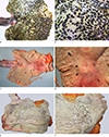 | Fig. 1Wischnewski spots (WS) of gastric mucosa. (A, B) Gross appearance of the stomach of case No. 6 (large sized WS). (C, D) Gross appearance of the stomach of case No. 2 (medium sized WS). (E, F) Gross appearance of the stomach of case No. 1 (pinpoint to small sized WS).
|
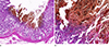 | Fig. 2H&E stain of gastric mucosa of case No. 2. The brown to black pigmentation is shown, but there is no neutrophil infiltration (A, ×40; B, ×200).
|
References
1. Birchmeyer MS, Mitchell EK. Wischnewski revisited: the diagnostic value of gastric mucosal ulcers in hypothermic deaths. Am J Forensic Med Pathol. 1989; 10:28–30.

2. Sperhake JP, Rothschild M, Risse M, et al. Histomorphology of Wischnewsky spots: contribution to the forensic histopathology of fatal hypothermia. In : Oehmichen M, editor. Hypothermia: clinical, pathomorphological and forensic features. Lubeck: Schmidt-Romhild;2004. p. 211–220.
3. Turk EE. Hypothermia. Forensic Sci Med Pathol. 2010; 6:106–115.

4. Madea B, Tsokos M, Preuss J. Death due to hypothermia: morphological findings, their pathogenesis and diagnostic value. In : Tsokos M, editor. Forensic pathology review. . Totowa: Humana Press;2008. Vol. 5:p. 3–21.
5. Dettmeyer RB. Hypothermia. Forensic histopathology: fundamentals and perspectives. New York: Springer;2011. p. 65–171.
6. Hirvonen J. Necropsy findings in fatal hypothermia cases. Forensic Sci. 1976; 8:155–164.
7. Wolf DA, Aronson JF, Rajaraman S, et al. Wischnewski ulcers and acute pancreatitis in two hospitalized patients with cirrhosis, portal vein thrombosis, and hypothermia. J Forensic Sci. 1999; 44:1082–1085.
8. Tsokos M, Rothschild MA, Madea B, et al. Histological and immunohistochemical study of Wischnewsky spots in fatal hypothermia. Am J Forensic Med Pathol. 2006; 27:70–74.
9. Hirvonen J, Elfving R. Histamine and serotonin in the gastric erosions of rats dead from exposure to cold: a histochemical and quantitative study. Z Rechtsmed. 1974; 74:273–281.
10. Bright F, Gilbert JD, Winskog C, et al. Additional risk factors for lethal hypothermia. J Forensic Leg Med. 2013; 20:595–597.
11. Zhou C, Byard RW. Armanni-Ebstein phenomenon and hypothermia. Forensic Sci Int. 2011; 206:e82–e84.
12. Knight B. Simpson's forensic medicine. 11th ed. London: Arnold;1997. p. 146–147.
13. DiMaio DJ, DiMaio VJ. Forensic pathology. Boca Raton: CRC Press;1993. p. 384–387.
14. Hirvonen J. Systemic and local effects of hypothermia. In : Tedeschi CG, Eckert WG, Tedeschi LG, editors. Forensic medicine. Philadelphia: W.B. Saunders Company;1977. Vol. 1:p. 758–774.
15. Nixdorf-Miller A, Hunsaker DM, Hunsaker JC 3rd. Hypothermia and hyperthermia medicolegal investigation of morbidity and mortality from exposure to environmental temperature extremes. Arch Pathol Lab Med. 2006; 130:1297–1304.
16. Coe JI. Hypothermia: autopsy findings and vitreous glucose. J Forensic Sci. 1984; 29:389–395.
17. Dolinak D, Matshes EW, Lew EO. Environmental injury. In : Dolinak D, Matshes EW, Lew EO, editors. Forensic pathology: principles and practice. Amsterdam: Elsevier Academic Press;2005. p. 239–258.
18. Mant AK. Autopsy diagnosis of accidental hypothermia. J Forensic Med. 1969; 16:126–129.
19. Clark KH, Stoppacher R. Gastric mucosal petechial hemorrhages (Wischnewsky lesions), hypothermia, and diabetic ketoacidosis. Am J Forensic Med Pathol. 2016; 37:165–169.
 XML Download