

 PDF
PDF ePub
ePub Citation
 Print
Print


INTRODUCTION
The role of laparoscopic surgery for the management of gallbladder cancer remains controversial. The current standard of care for preoperatively suspected gallbladder cancer is still an open radical cholecystectomy.1 In laparoscopic radical cholecystectomy, overcoming the innate complexity in the performance of adequate lymphadenectomy, liver resection, and possible common bile duct resection remains a challenge to surgeons.2
Recently, however, several high volume centers have demonstrated the efficacy and safety of laparoscopic radical cholecystectomy, which includes extended cholecystectomy and hepatoduodenal ligament lymphadenectomy in gallbladder cancer.3 Nevertheless, only a few reports on the feasibility of laparoscopic radical cholecystectomy with a common bile resection.4
With our expanding experience in minimally invasive surgery, our center has begun to employ the laparoscopic radical cholecystectomy in select patients with a preoperatively suspected early gallbladder cancer. We believe that as long as oncologic principles of open surgery have been carefully followed, laparoscopic surgery has a potential role in the management of early gallbladder cancer. Thus, we aim to report and describe our first successfully performed laparoscopic radical cholecystectomy with combined segmental resection of the bile duct in a patient with preoperatively suspected T2 gallbladder cancer near the cystic duct.
CASE
Patient's clinical profile
A 74-year-old woman with background history of hypertension presented in the outpatient department of Yonsei University Health System complaining of 2 weeks history of fever, abdominal pain and vomiting. She does not smoke or drink alcoholic beverages. Laboratory examinations revealed the following values: White blood count of 6.10 103/µl, alkaline phosphatase at 133 IU/L (47–147), aspartate aminotransferase 26 IU/L (13–34), alanine aminotransferase 9 IU/L (5–46), gamma-glutamyl transferase at 123 (7.0–35.0), total bilirubin at 0.8 mg/dL (0.4–1.5), fasting blood glucose at 113 mg/dL (70–110), carcinoembryonic antigen at 2.73 ng/ml, and cancer antigen 19–9 at 140 U/ml (0.0–5.0).
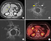
The imaging from the computed tomography (CT) scan of the abdomen showed irregular wall thickening on the liver and peritoneal side of the gallbladder body with no definite liver invasion. The tumor also extended to the cystic duct. An enlarged lymph node at portocaval space, and a borderline sized lymph node at the hepatoduodenal ligament were also noted (Fig. 1A). Likewise, the endoscopic ultrasound (EUS) confirmed a liver side tumor in the body of the gallbladder with cystic duct involvement without serosal and liver invasion (Fig. 1B, C). On positron emission tomography (PET) scan, an intense fluorodeoxyglucose (FDG) uptake on the body of the gallbladder and portocaval lymph node was noted, most likely a lymph node metastasis (Fig. 1D). Our preoperative impression was T2 gallbladder carcinoma with lymph node metastasis. Patient subsequently underwent laparoscopic radical cholecystectomy with common bile duct resection and Roux-en Y hepaticojejunostomy on January 12, 2018.
Operative technique
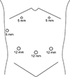
Laparoscopic radical cholecystectomy was performed using 6 abdominal ports (Fig. 2). A 12-mm trocar was inserted at the umbilicus, and pneumoperitoneum was established. The patient was then placed in a reverse Trendelenburg position and tilted to the lateral left. Staging laparoscopy was performed. Since there was no sign of distant metastasis, the additional 5 trocars were inserted. Lymphadenectomy was started at the junction of the common hepatic artery and gastroduodenal artery, removing all the lymph nodes and connective tissues surrounding the common hepatic artery and superior margin of the pancreas. Dissection continued along the proper hepatic artery towards the left and right hepatic artery. Since the patient had a tumor involving the cystic duct, the common bile duct resection was performed. The common bile duct was dissected and transected at its most distal extrapancreatic extension. Two 10 mm hemoclips were applied to secure distal stump. The common bile duct was retracted laterally and superiorly to clearly expose the portal vein. All remaining connective tissues and lymph nodes around the portal vein were dissected up to the confluence of the right and left hepatic ducts. The gallbladder was carefully dissected in a retrograde manner using an electrocautery and was sent for frozen section. It was noted that the tumor invaded the perimuscular connective tissue of the gallbladder and there was no tumor invasion on the liver bed by frozen section. As a result, the liver resection was not performed. The common hepatic duct was resected below the confluence of right and left hepatic duct and the proximal margin was sent for frozen section. There were no tumor cells identified by frozen section.
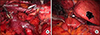
Next, a full Kocher's maneuver was performed to expose the inferior vena cava and posterior surface of the pancreas. All lymph nodes and connective tissues anterior to the inferior vena cava, retropancreatic, and aortocaval area were removed. Fig. 3A showed the extent of laparoscopic lymphadenectomy. All specimens were collected and placed inside an endopouch.
The hepaticojejunostomy was constructed laparoscopically (Fig. 3B). The jejunum was transected using an endoscopic gastrointestinal anastomosis (GIA) stapling device approximately 50 cm from the ligament of Treitz. Approximately 50 cm of retrocolic roux limb was brought up to the common hepatic duct stump and an end-to-side anastomosis was created using a 4-0 vicryl single layer suture (posterior side, continuous running suture; anterior side, simple interrupted suture). Hemostasis was carefully observed and any bleeding vessels were clipped using the endoclip. Two hemovac drains were placed; one in right hepatic area posterior to the hepaticojejunostomy anastomosis, and the other, anterior to the anastomosis.
Finally, the 4 cm umbilical incision was extended enough for specimen extraction and extracorporeal jejuno-jejunal anastomosis. A hand sewed side-to-side, double-layer anastomosis was created using vicryl 3-0 sutures.
Pathological findings
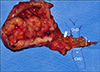
Final pathology report revealed a 4.1×3.0 cm, moderately differentiated adenocarcinoma (Fig. 4). The tumor invades the perimuscular connective tissue on the hepatic side, with no extension into the liver (T2b; American Joint Committee on Cancer 8th Ed.). There were no lymphovascular and perineural invasions. The presence of adenoma, low grade dysplasia, in the cystic duct was noted. A metastatic carcinoma in 1 out of 9 lymph nodes without perinodal soft tissue extension was also reported. The resection margin of the common bile duct was free of tumor.
Post-operative recovery and follow-up
Patient was started on liquid diet 24 hours postoperatively. Full return of bowel activity was noted on the 3rd postoperative day so patient was placed on full diet. The patient was discharged on the 7th hospital stay with drains removed. Adjuvant chemotherapy with gemcitabine was started at 14 days after surgery and had a regular follow-up check-up in the outpatient department.
DISCUSSION
The current recommended surgical approach for preoperative suspected T2 gallbladder cancer is open radical cholecystectomy, which includes en bloc liver resection (wedge resection or segment 4b/5 resection) and regional lymphadenectomy. Yet, until recently, there is still no consensus as to whether an open approach is superior to laparoscopic surgery.5 However, in high volume centers with more experience in minimally invasive surgery, laparoscopic radical cholecystectomy is currently being employed with an oncologic safety and long-term outcome comparable to open approach.678
In this report, a laparoscopic radical cholecystectomy for preoperatively suspected T2 gallbladder cancer with suspected invasion of the cystic duct was performed. In this approach, an en bloc wedge or segment 4b/5 liver resection was not done. For T2 gallbladder cancer, we believe that a simple cholecystectomy with lymph node dissection is adequate as long as intraoperative histologic evaluation confirms a tumor-free liver margin. However, a proper preoperative imaging, including CT scan and EUS, and an intraoperative histopathological evaluation to assess the extent of the tumor invasion is of paramount importance in the decision making whether or not to do extended cholecystectomy. Moreover, in case of liver sided tumor, we usually employed preoperative EUS or intraoperative ultrasound to identify the potential hepatic invasion. As previously reported by Kim et al.,9 a simple cholecystectomy and lymph node dissection without liver resection had a similar overall survival and recurrence patterns compared with those of radical cholecystectomy in patients with T2 gallbladder cancer. In addition, although hepatic side tumor had a worse long-term outcome compared to peritoneal side tumor, liver resection was not associated with long-term survival.10 Yet, even simple cholecystectomy alone has an acceptable oncologic outcome in a properly selected patient with T2 gallbladder cancer.11
Secondly, the extent of lymphadenectomy included the aortocaval lymph nodes (Station 16). Our patient had enlarged lymph nodes at the hepatoduodenal ligament and portocaval space, thus an aortocaval lymph node dissection was performed. The previous study had demonstrated that aortocaval lymph nodes were the common site of tumor recurrence in T2 gallbladder cancer after radical cholecystectomy.12 Moreover, although long-term outcome with aortocaval lymph nodes metastasis is poor and does not benefit from radical resection,13 we believe that a proper prognostication can be achieved through extended lymphadenectomy in aortocaval area.
Finally, we employed common bile duct resection to have an adequate tumor-free margin because of the clinically evident tumor invasion to the cystic duct. Our experience in laparoscopic pancreaticoduodenectomy which also includes common bile duct resection and hepaticojejunostomy reconstruction14 is one our consideration of adopting the feasibility of minimally invasive approach in gallbladder cancer even with common bile duct invasion. Technically, we employed bile duct to mucosa, end to side hepaticojejunostomy anastomosis with continues running suture on the posterior side and simple interrupted suture on the anterior side, using a vicryl 4-0 suture. With the help of a magnified view during laparoscopy, a healthy and viable tissue of the bile duct can be carefully identified and included for the anastomosis. Currently, however, common bile duct resection in gallbladder carcinoma is indicated only if there is evidence of tumor invasion in the cystic duct to achieve an R0 resection of the primary tumor and if the performance of a complete lymph node dissection that will not be achieved without common bile duct resection.1516
Therefore, with our increasing experience in minimally invasive surgery and mastery in the hepatobiliary anatomy, we are confident that common bile duct resection is not a contraindication for the laparoscopic approach. Nevertheless, this case report is relevant in that it suggests the potential of extending the indication of minimally invasive surgery for gallbladder cancer. Further, extensive lymphadenectomy can be safely and effectively performed laparoscopically especially in high volume center.
It is noteworthy to emphasize the undeniable advantages of the laparoscopic approach over open surgery such as a shorter length of hospital stay, less pain, and early return to work. More importantly, an adjuvant therapy could be started early after the surgery. In certain circumstances when effective systemic chemotherapeutic agents are clinically available in near future, these will be the fundamental advantages of minimally invasive surgery over open surgery.
In summary, this case shows laparoscopic radical cholecystectomy with combined common bile duct resection is technically feasible. This approach should be re-evaluated in terms of reproducibility, and oncologic outcomes based on large number of the patients with long-term follow up data.
 XML Download