

 PDF
PDF ePub
ePub Citation
 Print
Print


INTRODUCTION
In living donor liver transplantation (LDLT), every anatomical structure of the recipient liver must be meticulously dissected for reuse during graft implantation. Such surgical procedures in LDLT often lead to excessive bleeding during mobilization of the recipient native liver, especially in patients with portal hypertension or a past history of previous liver surgery.12
Many surgeons performing liver transplantation (LT) and hepatobiliary surgery are well acquainted with the Pringle maneuver because it effectively reduces bleeding during hepatic parenchymal transection.3 In the Pringle maneuver, repeated intermittent occlusion of the hepatoduodenal ligament for up to 15 to 30 minutes has been attempted to minimize detrimental hepatic ischemia. Unlike liver resection, hepatic ischemia of the recipient native liver does not become a matter of concern because it will be removed. We hypothesized that such a hepatic inflow occlusion could be applied during liver mobilization for LDLT when difficult dissection is anticipated. Since there is no need to shorten to the duration of inflow occlusion, it can be prolonged over a few hours.
We herein introduce our technique for prolonged occlusion of the hepatoduodenal ligament applied to recipient hepatectomy for LDLT.
PATIENTS AND METHODS
This study consisted of two parts. The first part was a simulated assessment of splanchnic hemodynamics on prolonged occlusion of the hepatoduodenal ligament in patients with cirrhotic livers. The second part was its clinical application with technical refinement.
Simulative hemodynamic assessment on occlusion of the hepatoduodenal ligament in patients with cirrhotic liver
The native liver of a recipient requiring LT is often cirrhotic, and development of portal hypertension and extrahepatic portal collaterals are often seen. Because of portal hypertension, it is vulnerable to induction of brisk bleeding during perihilar mobilization and dissection of the hepatoduodenal ligament. If the blood flow within the hepatoduodenal ligament is abruptly occluded, what changes happen in the splanchnic blood flow system?
In patients with normal or non-cirrhotic liver, sudden occlusion of the main portal vein results in congestion of the splanchnic flow system, by which bowel congestion can develop since there are no sizable venous collaterals. However, the portal vein pressure in such patients is not highly elevated, thus such splanchnic congestion is often well-tolerated and easily resolved after restoration of portal flow. Occlusion of the hepatic artery does not induce any noticeable systemic effect.
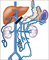
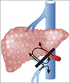
In patients with liver cirrhosis and portal hypertension, sudden occlusion of the main portal vein results in pressure increase in the splanchnic flow system, but the pressure increase is often not great because venous collaterals have already developed. Since the liver is perfused with high-pressure portal flow, any damage to the portal vein branches at the liver surface and hepatoduodenal ligament results in brisk bleeding. Complete occlusion of the hepatoduodenal ligament can cease portal vein-associated bleeding (Fig. 1). Hepatic arterial flow is also increased due to the compensatory effect on portal hypertension, thus such arterial hyperperfusion can induce excessive bleeding during perihilar mobilization and dissection of the hepatoduodenal ligament. Occlusion of the hepatic artery also prevents such risk of hepatic artery-associated bleeding (Fig. 2).
Optimized technique for prolonged hepatoduodenal ligament occlusion
For the Pringle maneuver, several methods have been used, such as vascular tourniquets or vascular clamps with cover shoes, to occlude the hepatic inflow securely. Such instruments are regarded as atraumatic, but can induce tearing of the swollen or weakened hepatoduodenal ligament in the LT recipients.
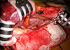
We accumulated experience on the Pringle maneuver with curved intestinal clamps, thus we decided to use the curved intestinal clamps in our study (Fig. 3). The inflow occlusion with curved intestinal clamps has the following two merits: its application and release are simple, thus it enables us to control occlusion easily; and the strength of clamping can be adjusted intuitively, thus clamping per se becomes truly atraumatic. We learned that clamping power to the first 2–3 jaw steps was enough to occlude the portal flow.
RESULTS
Between early 2014 and the end of 2018, prolonged occlusion of the hepatoduodenal ligament using curved intestinal clamps has been applied to more than 60 cases of adult LDLT.
Until the end of 2016, this technique was limited to patients showing heavy bleeding during perihilar mobilization. Application of this technique effectively reduced bleeding during mobilization of the right liver and detachment from the retro-hepatic vena cava. After a learning period, this technique was applied at the start of mobilization and stopped after complete mobilization of the retro-hepatic vena cava (Fig. 3).
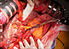
Since early 2017, this technique has also been applied during dissection of the hepatoduodenal ligament. The clamps were applied at the distal part of the hepatoduodenal ligament, and then dissection of the proximal hilum continued (Fig. 4). The clamps were temporarily released to palpate the running course of the hepatic arteries. Such a prolonged occlusion enabled us to interrupt the hepatic arterial flow effectively, by which the risk of tumor spread could be reduced theoretically. Thus, LT for advanced HCC became an eligible indication of this technique.
Analysis of the first 60 cases showed that the mean total occlusion duration was 67±13 minutes. One-third of patients (20 cases) underwent two sessions of occlusion, the first during mobilization of the right liver and detachment from the retro-hepatic vena cava, and the second for hilar dissection. No patient suffered from major serosal peritoneal tearing-associated bleeding or hepatic artery problems. Intentional prolonged occlusion over two hours was applied to five patients who had advanced multiple hepatocellular carcinoma (HCC), and none of them showed tumor recurrence within six months after LDLT.
DISCUSSION
Prevention of excessive bleeding during LT operation presents a major challenge. We have performed more than 6000 LT operations so far, but a considerable proportion of patients still required high transfusion amounts during LT operation. In deceased donor LT, mobilization of the recipient native liver and hilar dissection can be performed promptly. In contrast, LDLT requires additional procedures for preservation of the retro-hepatic inferior vena cava and small hepatic artery branches, thus risk of bleeding inevitably increases.145
Here, we have share our surgical experiences learned from non-transplant hepatobiliary surgeries and LTs. The Pringle maneuver is one of the important techniques of liver surgery. James Hogarth Pringle reported his notes on the arrest of hepatic hemorrhage due to trauma in 1908.6 Thereafter, the Pringle maneuver was settled upon as the standard procedure for liver surgery. However, as far as we know, its application was confined to liver transection alone.
We learned that the Pringle maneuver was also helpful to dissect livers heavily-adhered to the diaphragm, primarily due to previous hepatectomy. We also know that complete inflow occlusion with tight squeeze is not necessary for the Pringle maneuver. Simple application of a loose clamp may satisfy the purpose of the Pringle maneuver for reduced-bleeding hepatectomy.
In a novel move, we introduced the concept of the Pringle maneuver to the field of LT, where hepatic parenchymal transection is not necessary. Many LT surgeons intend to perform transfusion-less LT, but that goal is difficult to achieve. Various surgical techniques, including pinch-burn-cut techniques, high hilar dissection, and dissection with energy devices have been developed to reduce intraoperative bleeding during LT operations.789 Considering impaired coagulation profiles and concurrent portal hypertension, hemostasis in LT recipients is quite different from that of non-transplant liver surgery patients.
Our simulated hemodynamic analysis revealed that occlusion of the hepatoduodenal ligament in LT recipients can work as a stop-gap to weaken the bleeding-prone effect from portal hypertension. When a brisk bleeding occurs after sudden tearing of the venous collaterals around the liver, it is often difficult to quickly achieve local bleeding control. If the venous collateral is formed proximal to the main portal vein, hepatoduodenal ligament occlusion will work effectively. Based on our experience in more than 60 cases, we think that prolonged occlusion of the hepatoduodenal ligament is a simple and effective method to reduce intraoperative bleeding during LT operation.
Prevention of intraoperative tumor spread is another potential benefit of prolonged occlusion of the hepatoduodenal ligament. Excessive handling of the liver containing HCC can lead to hematogenous spread of the tumor into the blood stream. The surgical concept of anterior approach for large HCC was introduced clinically to reduce the risk of intraoperative tumor spread.10 Similarly, hepatoduodenal ligament occlusion can be applied during right liver mobilization with intention to minimize hematogenous spread of HCC cells in LT. This beneficial effect has not been objectively proven yet, thus we are performing a prospective study. We think that its primary indication is presence of HCC ≥5 cm because this size limit is an important prognostic factor in the LDLT setting.
In conclusion, we believe that prolonged occlusion of the hepatoduodenal ligament is a simple effective method to reduce intraoperative bleeding, and has potential benefit to reduce the risk of intraoperative tumor spread during LDLT operations.
 XML Download