

 PDF
PDF ePub
ePub Citation
 Print
Print


I. Introduction
Osteoradionecrosis (ORN) is one of the most severe complications resulting from radiotherapy (RT) in patients with head and neck cancer (HNC) and can occur between four to twenty-four months after RT1. In the literature, there are several definitions for ORN that are based mainly on clinical findings when, after head and neck radiation, the bone becomes exposed and devitalized, persisting without proper repair for a 3 to 6 month period, resulting in ORN234. Firstly described by Regaud5 in 1922, ORN involves a combination of four factors: radiation, hypoxia, hypovascularization, and hypocellularization resulting in bone necrosis. The mandible is the most affected bone in head and neck radiation patients678.
ORN can occur spontaneously or due to local factors including tumor localization, dose and/or type of RT9, trauma due to dental prosthetics, dental surgeries before, during, or after RT, deficient oral hygiene, and local infections including periodontal disease and dentoalveolar abscesses9101112. Arteriopathies, diabetes, alcoholism, and malnutrition are systemic factors that contribute to ORN13. The diagnosis of this complication is based on clinical characteristics including bone exposure, infection, halitosis, fistula, pathological fracture, and local pain14. In asymptomatic cases, ORN can be presumptively diagnosed with radiographic exams15.
Once the cause of ORN is determined, various types of treatment can be employed, including irrigation with chlorhexidine 0.12%, use of antibiotics, and surgical intervention1,1617. Surgical resection and hyperbaric oxygen therapy (HOT) have been reported to be therapies of choice18, although perhaps the maximum benefit is obtained through a combination of various therapy strategies11617. Currently there is no gold standard treatment for ORN nor widely accepted guidelines. Early diagnosis and oral condition monitoring are crucial for the prevention and successful treatment of ORN16.
Considering how few studies have evaluated the conduct of clinical protocols for ORN, the present study aimed to describe the profile and dental management of ORN in patients undergoing head and neck RT in an oncological clinical research center.
II. Materials and Methods
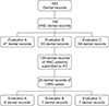
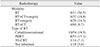
A retrospective descriptive study was performed using dental record data of patients with ORN treated in an oncological clinical research center. At first, a search was performed in the electronic registration system containing 583 dental records of all the treated patients. Afterwards, HNC patients were selected, totaling 158 records which were distributed to three examiners for manual assessment. Each examiner was then responsible for selecting dental records that contained a description of ORN, resulting in a final total of 20 dental records.(Fig. 1)
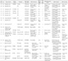
From the 20 dental records of patients with ORN, information was collected regarding their age, sex, type and/or HNC localization, cancer stage, type of treatment, dose, and RT type. Regarding ORN, the localization, time between the last dental appointment and first description of ORN, predisposing factors for ORN, type of treatments administered, and outcomes between the first and last description of ORN were considered. In addition, ORN cases were classified according to the Støre and Boysen18 score based on clinical characteristics described in the dental records. Following this score, isolated mucosal defects were classified as stage 0 (zero); cases with radiological evidence of necrotic bone with intact mucosa were classified as stage I; cases with exposed bone and radiological evidence were classified as stage II; and cases with exposed bone, radiological evidence, extraoral fistula, and infection were classified as stage III. All patients signed the informed consent form in accordance with the Declaration of Helsinki.
III. Results
In a 4 year period (2013–2017), 158 individuals with HNC were treated at the Clinical Research Center of Bauru School of Dentistry, University of São Paulo (Bauru, Brazil). One hundred thirty-nine patients (88.0%) received RT in the head and neck region and 20 of them were diagnosed with ORN. Sixteen patients (80.0%) with ORN were male and 4 patients (20.0%) were female. The mean age was 60.3 years with a range from 29 to 85 years.(Table 1)
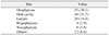
The most common types of cancer in patients who underwent head and neck RT were oropharyngeal (38.1%) and oral (31.7%) cancers.(Table 2) There was a higher incidence of ORN in individuals with squamous cell carcinoma (SCC) on the floor of the mouth at 70.0% followed by the alveolar ridge SCC with 50.0% of cases in patients with oral cancer. Patients with oral and oropharyngeal cancers developed ORN 50.0% of the time.(Table 3)
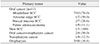
Related to antineoplastic treatment modalities, of the 11 patients who underwent only RT, 6 developed ORN; of the 32 patients who underwent RT associated with chemotherapy and surgery, 6 developed ORN; of the 28 patients who underwent RT associated with surgery, 4 developed ORN; and of the 60 patients who underwent RT and chemotherapy, 4 developed ORN. The majority of ORN cases occurred in individuals who underwent conventional/cobalt RT (10 cases). The type of RT was not reported in 18 patients who underwent RT in the head and neck region, and only one of these cases presented with ORN.(Table 4) The dose of RT varied between 5,000 cGy and 7,020 cGy.(Table 1)
The most common ORN site was the posterior region of the mandible, with 12 individuals (60.0%), followed by the anterior region of the mandible with 4 cases (20.0%), posterior maxilla with 2 cases (10.0%), 1 case (5.0%) in the anterior to posterior maxilla, and 1 case (5.0%) that affected the anterior and posterior regions of the mandible. Regarding ORN stage, 14 out of the 20 (70.0%) cases were classified as stage 2 and 6 out of the 20 cases (30.0%) were classified as stage 3.(Table 1)
Among the ORN predisposing factors, 14 cases (70.0%) were related to dental conditions (dental extractions, dentoalveolar abscesses, periodontal disease, dental prosthetics, and residual roots), 2 cases (10.0%) were associated with systemic factors (tobacco and alcohol use), 1 case (5.0%) was associated with surgery for tumor removal, and it was not possible to find descriptions of associated predisposing factors in 3 cases (15.0%). Dental extractions and tobacco use were present in 8 cases (40.0%), followed by badly adapted prosthetics and alcohol use in 3 cases (15.0%), and residual roots, dentoalveolar abscesses, and dehiscences following tumor removal in 1 case (5.0%) each. ORN minimal clinical presentation time was immediate in 2 cases (10.0%), and the larger clinical presentation time after RT totaled 49 months with an average of 6.5 months. Half of the ORN cases (50.0%) were diagnosed 6 months after RT and after 2 months for 4 cases (20.0%).(Table 1)
Many different approaches were used for ORN treatment with the most frequent ones involving curettage and 0.12% chlorhexidine irrigation in 9 cases (45.0%), 2 cases (10.0%) of sequestrectomy associated with curettage and 0.12% chlorhexidine irrigation, and 2 cases (10.0%) of curettage associated with 0.12% chlorhexidine irrigation and antibiotic therapy. The other associated treatment modalities (1 per case) are presented in Table 1.
ORN outcomes included 10 cases (50.0%) that resulted in closure of the exposed bone and in 1 case (5.0%) presenting in the anterior and posterior regions of the mandible, there was only posterior region closure. In 4 cases (36.0%) where there was bone exposure closure/epithelialization, the treatment modality used was curettage associated with 0.12% chlorhexidine irrigation.(Table 1) The other closure cases encompassed several treatment types aforementioned.
IV. Discussion
In the present study, ORN was found more often in elderly (61–85 years) males (80.0%) with an average age of 60.6 years; a little older than findings from previous studies1920 (54.021, 55.222, and 57.819 years). ORN was commonly found in patients with SCC on the floor of the mouth (70.0%) in our study922.
Complications resulting from RT are more frequent in irradiated regions, resulting in ORN risk factors based on RT type and radiation dose. Therapy with intensive modulated radiotherapy (IMRT) presents an advantage in ORN prevention, since the ORN rate with IMRT (4%) is lower compared to three-dimensional (3D) conformal radiation therapy (3D-CRT) (19%)2223242526. In this study, most individuals with ORN underwent conventional RT (18.5%) and IMRT (15.1%) with only 1 case (7.1%) receiving 3D-CRT.(Table 4) Doses higher than 5,000 cGy also raise the risk of ORN, with greater risk seen in doses higher than 6,000 cGy2728. ORN occurred in irradiated fields with doses ranging from 5,000 to 7,020 cGy with more frequent total doses of 6,000 cGy (4/20) and 7,000 cGy (4/20). ORN occurred independent of the treatment modality (RT and/or CT and/or surgery) and conveyed the fact that higher radiation doses contributed significantly to ORN.
When ORN occurs in the context of dental conditions associated with systemic factors (e.g., tobacco and alcohol use), including dental extractions, infections or abscesses, and trauma caused by dental prosthetics in the irradiated field, there is an increased risk for ORN2429303132 even though ORN can occur spontaneously. In this study, ORN was found more frequently in the mandible (80.0%), which is the region adjacent to the location of most tumors (oral floor, tongue, and retromolar trigone region), but was also present in the maxilla (10.0%), localized next to tumors. These regions supposedly received direct radiation in high doses, seemingly the determinant factor for ORN in this study. The concept of bone density differences and blood supply between the maxilla and mandible becomes secondary.
Most ORN cases in this study were related to dental conditions (70.0%) with only 2 cases (10.0%) not being related to local predisposing factors, where ORN could have possibly occurred spontaneously which would be contrary to previous studies in which 79%23 and 82%33 of the ORN cases occurred spontaneously. Among the dental conditions, dental extractions and tobacco use were the predisposing factors more often associated (40.0%) with ORN, followed by trauma and alcohol use (15.0%). In another study, a low ORN rate (1.7%) related to dental extractions was found23. However, the association with tobacco and alcohol use increased ORN risk and severity2722. The time for ORN development after the end of RT was usually within 3 years, and varied from 0 months to 192 months in cases of chronic trauma934. In this study, half (50.0%) of the ORN cases occurred 6 months after the end of RT and ranged between 0 to 49 months with an average (6.5 years) below the one found in previous studies (8 months)2122.
Challenges for daily clinical assessments include case identification, pain source, and differentiation of ORN clinical signs when it is still in its initial stages from other side effects resulting from SCC treatment that can affect the oral mucosa, including oral mucositis23. Furthermore, it is difficult to identify radiographic alteration signs for ORN since they are only detectable when 30% to 40% of the bone density is compromised1835. These facts can explain the lack of description in the medical records of this study as well as signs of ORN initial stages (0 or 1), thus making it impossible to perform ORN retrospective ratings for stages other than stages 2 (70.0%) and 3 (30.0%) which had clinical sign descriptions.
ORN treatment is difficult, involves a combination of therapies, depends on available therapeutic resources, and relies on patient compliance with instructions and their individual biological response. For many researchers, conservative treatment is performed only in small ORN areas since for more advanced conditions, surgical resection is considered more efficient1. The conservative and surgical approach associated with HOT is well documented11936. A less invasive option for ORN control and healing involves 0.12% chlorhexidine which, when administered topically, acts as a bactericide against gram-positive and gram-negative microorganisms and some yeasts. Despite exhibiting good results when associated with superficial necrotic bone curettage, there is still no protocol for the use of chlorhexidine for ORN treatment117.
Less invasive treatment options were the first choice for ORN cases in this study. Even for more advanced cases, 0.12% chlorhexidine irrigation was administered. For all stage 3 ORN cases, the use of an associated systemic antimicrobial was introduced based on the presence of local suppuration. There was epithelialization in 8 (57%) stage 2 ORN cases and in 2 (33%) stage 3 ORN cases. In one case, there was total ORN resolution due to surgical resection. Bone exposition closure was observed in most cases (11/20) in this study which were treated with less invasive therapies.
 XML Download