

 PDF
PDF Citation
 Print
Print


INTRODUCTION
Monkeys are used significantly in various studies of veterinary medicine and human medicine, owing to their similarities.12345 Therefore, veterinarians and researchers in these fields should understand the anatomy of monkeys as fundamental knowledge of the subject. However, materials for studying monkey anatomy are insufficient.67 Another serious problem is that it is difficult to obtain monkeys for dissection due to required legal and ethical permissions.8
Recently, computed tomographs (CTs) and magnetic resonance images (MRIs) of monkeys are commonly used to verify results of neuroscience studies.910111213 To interpret CT and MRI accurately, veterinarians and researchers need to learn sectional anatomy.141516 Nevertheless, materials to study sectional anatomy of monkeys are insufficient. Existing sectional specimens of monkeys cannot show whole body and detailed structures together.17 In the case of CT and MRI, it is almost impossible to produce three-dimensional (3D) models of minute structures due to low resolution and gray color.1819
In the Visible Human Project, cross-sectional images of a cadaver were produced. It was a milestone, enabling observations of human body structures with high resolution and vivid colors.2021 Voxel-man, a 3D model made from Visible Human Project data, reached another level of realistic display of the human body.22 After the Visible Human Project, succeeding projects to obtain improved sectioned images of cadavers were launched in Korea and China.232425 Using these data, numerous two-dimensional (2D) applications and 3D models of humans were developed to aid education and research of anatomy and radiology.26272829303132 Once cross-sectional images in real color and 3D models of a monkey can be produced like those of humans, difficulties of real monkey dissection and shortage of materials for monkey sectional anatomy will be overcome.
To enhance the understanding of monkey anatomy, the objective of this study was to provide a Visible Monkey data set containing high quality and real color sectioned images of a rhesus monkey with corresponding CT and MRI. To achieve this purpose, the whole body of a rhesus monkey was serially ground and photographed to make sectioned images.
METHODS
A female rhesus monkey (head-to-feet length, 758 mm; weight, 4.3 kg; age at sacrifice, 93 months) was donated from the National Primate Research Center of Korea. In the state of living, the head of the monkey was 3 Tesla (3T) MR scanned using both T1 and T2 methods (repetition time, shortest; echo time, shortest). The monkey was sacrificed by an intramuscular injection of ketamine (10 mg/kg) followed by an intravenous injection of potassium chloride (100 mg/kg). Neither formalin nor dye was injected.
The sacrificed monkey was fixed on a wooden plate. Its palms were fixed to face forward to yield the anatomical position of a human. Its soles were fixed to make the ankle joint to be at a 90-degree angle. The whole body of the sacrificed monkey was scanned with CT (voltage, 140 kVp; electric current time, 240 mAs) and 3T MRI (repetition time, 675 ms; echo time, 10.944 ms).
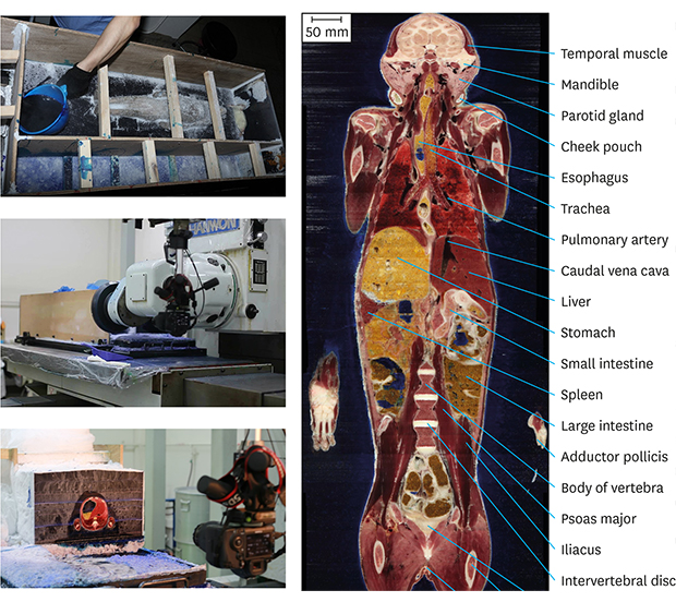
After CT and MR scanning of the whole body, the sacrificed monkey was frozen at −70 °C for one week. After placing the monkey in gelatin solution (water 1,000 mL, gelatin 30 g, methylene blue 0.5 g) in an embedding box, the box was frozen in a freezer for one week (Fig. 1).33
 | Fig. 1Sacrificed and frozen rhesus monkey being embedded (left), serially sectioned (middle), and photographed (right).
|
The frozen monkey was serially ground with a cryomacrotome at displacement accuracy of 0.001 mm.25. Serial sectioning was performed at 0.05 mm intervals for the head region and at 0.5 mm intervals for the remainder of the body. After grinding, frost was wiped away using 99% ethyl alcohol. Any protruding connective tissue on the sectioned surface was removed with a scalpel (Fig. 1).
A Canon™ EOS 5Ds R digital single-lens reflex (DSLR) camera (resolution, 8,688 × 5,792; color depth, 48 bit color) equipped with a Canon™ EF 100mm f/2.8L macro IS USM lens (Canon, Tokyo, Japan) was used. The distance from the digital camera to the sectioned surface was adjusted to provide a photograph area of 205 mm in horizontal length and 137 mm in vertical length on the sectioned surface, corresponding to a pixel size of 0.024 mm × 0.024 mm. Two strobes (Elinchrom™ digital S, Renens, Switzerland) with power pack (Elinchrom™ digital 2, Renens, Switzerland) were used to maintain constant brightness of the sectioned surface. A color control patch (Tiffen™, Hauppauge, New York, NY, USA) was placed on the sectioned surface and photographed every day for post processing of these images. The sectioned surface was photographed using a digital camera (ISO 100, shutter speed 1/250, aperture F/8, manual focus). The photograph was checked by anatomists on a computer monitor and saved in tagged image file format in Adobe Photoshop CS6 (Adobe Systems, Inc., San Jose, CA, USA). The procedure was repeated from the head to the feet (Fig. 1).
RESULTS
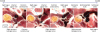
After obtaining horizontal 3T MRI of the head of the living monkey, 3T MRI and CT of the whole body of the sacrificed monkey were then produced. A total of 2,967 sectioned images of the horizontal plane were manufactured without missing cuts (resolution, 8,688 × 5,792; single file size, 288 mbyte; total file size, 834 gbyte). These sectioned images were produced at 0.05 mm intervals for the head region and at 0.5 mm intervals for other regions. The pixel size was 0.024 mm × 0.024 mm and the color depth was 48-bit color (Fig. 2 and Table 1). Reconstructed coronal and sagittal planes of these sectioned images revealed the whole extent of monkey anatomy (Fig. 3).
 | Fig. 2MRI, CT, and sectioned images of the rhesus monkey's whole body.MRI = magnetic resonance image, CT = computed tomography.
|
Table 1
Features of horizontal CT, MRI, and sectioned images of the rhesus monkey

3T = 3 Tesla, CT = computed tomography, MRI = magnetic resonance image.
aMonkey before death; bMonkey after death.
![]()
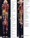 | Fig. 3Reconstructed coronal sectioned image viewed from anterior side (left) and sagittal sectioned image viewed from right side (right) of the monkey's whole body. These reconstructed images were resized proportionally regarding pixel size of the horizontal images (0.024 mm × 0.024 mm) and sectioning intervals (0.05 mm for head region and 0.5 mm for the rest).
|
Extracranial and intracranial structures were clearly visible in sectioned images of the head. Eye structures including extraocular muscles and nerves were clearly visible on these images (Fig. 4A). Connection from the retina to the lateral geniculate nucleus was found (Fig. 4A-C). Even the reticular nucleus and external medullary lamina of the thalamus were observed on these images. The stria medullaris of the thalamus and fornix were very close to each other without touching. Eventually, the stria medullaris went to the habenula, while the fornix went to the hippocampus (Fig. 4B-D). Moreover, minute blood vessels were observed on these images. The nerve to the caudal rectus muscle from the oculomotor nerve was visible (Fig. 4C and D). The paramedian pontine perforating artery was also observed on the image (Fig. 4E).
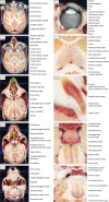 | Fig. 4Sectioned images of the head of a rhesus monkey. Images of full extent (left column) were cropped and magnified to show minute structures (right column). Levels of these images include lens (A), thalamus (B), caudal rectus muscle (C), midbrain (D), and pons (E).
|
In sectioned images of the thorax, the dorsal vagal trunk was found around the esophagus (Fig. 5A). The four chambers of the heart were identified in the middle mediastinum. In the heart, small structures such as the tendinous cord and septomarginal trabecula were visible. At the lateral side of the heart, the phrenic nerve passed the diaphragm and the coronal vessels could be observed. Moreover, the airway from the lobar bronchus to the bronchiole was visible (Fig. 5B). The thoracic sympathetic ganglion was thicker than the sympathetic trunk. The gray and white rami communicantes could be distinguished (Fig. 5C). From the left side to the right side, thoracic aorta, thoracic duct, and azygos vein were located (Fig. 5D). Through whole levels of the spinal cord, gray matter, white matter, spinal ganglion, ventral spinal artery, dorsal spinal artery, and even the radicular artery were all clearly identified (Fig. 5E).
 | Fig. 5Sectioned images of the thorax of a rhesus monkey. These images of full extent (left column) were cropped and magnified to show minute structures (right column). Levels of these images include the apex of the lung (A), upper heart (B), lower heart (C), base of the lung (D), and crus of the diaphragm (E).
|
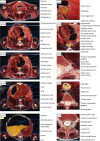
In sectioned images of the abdomen, the pancreatic duct, common bile duct, and minor duodenal papilla were identified around the duodenum. Even the gastroduodenal artery from the celiac trunk was found (Fig. 6A). In the kidney, minute structures including the cortical radiate vessel and minor calyx were observed (Fig. 6B). The central canal in the medullary cone and artery in the ventral ramus of the 5th lumbar nerve were also visible in the spinal canal (Fig. 6C). Regarding the pelvic structures, ovarian follicles at multiple stages were observed, owing to the specimen's young age at sacrifice (7 years old) (Fig. 6D). The pudendal nerve and artery were found in the pudendal canal. Cavernous structures inside the clitoris were also observed on these images (Fig. 6F).
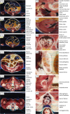 | Fig. 6Sectioned images of the abdomen and pelvis of a rhesus monkey. These images of full extent (left column) were cropped and magnified to show minute structures (right column). Levels of these images include the celiac trunk (A), kidney (B), medullary cone (C), ovary (D), pudendal canal (E), and clitoris (F).
|
Owing to the high resolution and narrow intervals of these sectioned images, whole courses of gracile structures could be traced. For example, the whole course of the sympathetic nerve (Fig. 5C), spinal cord with spinal nerves (Figs. 3, 5, and 6), and pancreatic ducts (Fig. 6A) were traced. Moreover, the right recurrent laryngeal nerve coming off from the right vagus nerve (Fig. 7) and the facial nerve from the stylomastoid foramen through the parotid gland to the facial muscles were traced (Fig. 8). Similarly, any continuous structure could be traced on these serially sectioned images.

DISCUSSION
The Visible Monkey data set of this study is the first trial to obtain high-quality and real-color sectioned images of a whole monkey body (intervals, 0.05 mm or 0.5 mm; pixel size, 0.024 mm × 0.024 mm; color depth, 48 bits color). The superb image quality enabled observations of detailed anatomical structures of a rhesus monkey (Figs. 3-8).
Through experience since the year 2000 in making sectioned images of humans14233337 and animals,3839 the authors have accumulated several skills to yield high quality sectioned images, as follows. The first is cadaver processing skill. When receiving a donation, an appropriate specimen without lesions or prosthetics is chosen. To preserve the normal anatomy as much as possible, neither formaldehyde fixative nor dye is injected. An ideal freezing environment is formed with a deep freezer. The second is photography skills. The digital camera and lighting device are set properly according to the environment. Every sectioned surface is amended delicately. Even small flaws invisible to the naked eye are removed by observing the high-resolution photograph. The third is the post processing skill. Consistent alignment, color, and brightness of images could be achieved through post processing of these images by skilled technicians (Fig. 3).3435
In addition to these skills, novel equipment of this study enabled high quality images. First, the cryomacrotome used in this study has high precision reliability.23 The machine installed for previous research is still being used in the current research since 2002. Second, a high performance DSLR camera has been continuously developed by engineers.4041 We always utilize the newest DSLR camera. In this study, the pixel size of sectioned images was 0.024 mm by using Canon EOS 5Ds R DSLR camera, while that of previous research was 0.2 mm with Kodak DSC 56023 and 0.1 mm with Canon EOS 5D14 and Canon EOS-1Ds mark III.3338 The improvement of resolution can be verified by comparing images of this study with those of the authors' previous study.14
Comparative anatomy of a human and rhesus monkey is a highly interesting topic because these two species are derived from a common ancestor, catarrhini species. These sectioned images of this study will be a good source for comparative biology research.42
The Visible Monkey data set of this study will be upgraded continuously with segmentation and 3D reconstruction, just like the Visible Korean data set of a human.434445 As an initial step, the skin of the head and brain have been segmented and made into 3D models. These 3D models made from sectioned images are superior in accuracy to those made by CT or MRI. By observing these 3D models and corresponding sectioned images together, the sectional anatomy of a monkey can be elucidated in a concrete way (Fig. 9).
 | Fig. 9Surface models of the monkey's skin and brain with embedded horizontal sectioned image and coronal sectioned image.
|
As for future steps, Visible Monkey can be used for educational purposes. By identifying numerous anatomical structures on these sectioned images, an atlas of the monkey anatomy and 3D models for a virtual dissection can be made.163233 These data sets can be also used for neuroscience research, including virtual experiment to determine effects of electromagnetic wave and radiation on an organism. The validity of the virtual experiment can be confirmed by determining if the result of the virtual experiment on a monkey corresponds to the result of an actual experiment on a monkey.464748495051
The Visible Monkey will function as a reference for monkey anatomy. It can be used as a mutual supplementation with existing MRI and CT (Fig. 2). Once researchers understand monkey anatomy by using images of Visible Monkey, they can conduct studies on monkey more accurately. These data sets are expected to enhance understanding of primate studies. These images can be shared with any researcher free of charge. Researchers may send an email to the corresponding author to obtain these images.
 XML Download