Abstract
Background
The purpose of this study was to compare the tibial tunnel aperture contact characteristics simulating an anteromedial and transtibial anterior cruciate ligament (ACL) tunnel preparation.
Methods
Seven matched pairs of cadaveric knees were tested. From each knee, a 10-mm quadriceps ACL graft was prepared. The native ACL was arthroscopically removed and tibial tunnels were drilled. In one knee, a transtibial technique was performed with femoral tunnel drilling approached through the tibial tunnel. For the anteromedial technique on the contralateral knee, the posterior tibial tunnel was chamfered with a rasp. The knees were then disarticulated and tibial tunnel aperture geometry was measured. A pressure sensor was placed between the graft and the posterior aspect of the tibial tunnel and the graft was secured with an interference screw. Contact force, contact area, contact pressure, peak contact pressure, hysteresis and stiffness were measured at cyclic loads of 50 N, 100 N, 150 N, and 200 N.
Results
Tibial tunnel aperture area, diameter and deviation from a circle were significantly larger with the transtibial technique (p < 0.05). There was no significant difference in hysteresis, stiffness, contact area, contact force and mean contact pressure. The peak contact pressure between the ACL graft and the tibial tunnel was significantly higher with the anteromedial technique for 100 N (p = 0.04), 150 N (p = 0.01), and 200 N (p = 0.002) cyclic loading.
Anterior cruciate ligament (ACL) reconstruction is a commonly performed procedure but debate still exists whether drilling the femoral tunnel using a transtibial approach or through an independent technique, such as through an anteromedial portal is a better approach. Multiple studies have established the biomechanical superiority of anatomically positioning the femoral tunnel.123) To achieve more anatomic reconstructions, there has been a focus on placing the grafts as close as possible to the native ACL footprints in order to restore normal knee kinematics and to recreate anatomic graft angles.34)
The transtibial technique has been shown to produce successful outcomes and was previously the most commonly used option for ACL reconstruction.56) However, the transtibial technique does not fully place the ACL at its anatomic footprints and tends to place the graft in a more vertical orientation due to anatomic constraints of the tibial tunnel.478910) In comparison, the anteromedial portal approach, where the femoral tunnel is drilled through a separate portal, has been increasing in popularity due to its ability to produce more anatomic footprints and graft angles.3411121314) Biomechanically, the anteromedial technique has been shown to be superior in terms of rotational stability and in restoring anterior-posterior translation during walking.41315) Despite the apparent advantages of the anteromedial technique, it has not necessarily led to improved clinical outcomes and could lead to difficulties in guide positioning, short femoral tunnels and risk of cartilage injury.131617) Furthermore, while the transtibial approach may increase the odds of ipsilateral leg surgery, the anteromedial approach has been associated with a higher ACL revision rate.1819)
Given the differences in approach, the transtibial technique and the anteromedial technique can create differing amounts of stress on the graft as it bends over both the tibial and femoral apertures. It is thought that the repetitive bending stresses at these tunnel apertures could be responsible for graft damage due to the bony tunnel edges. 2021) However, previous studies have mainly focused on the bending angles at the femoral tunnel interface rather than the tibial tunnel.222324)
Previous studies have compared the effects of the transtibial technique and the anteromedial technique on the shape and size of the tibial aperture.41625) They have shown that the intra-articular tibial aperture created using a transtibial approach after femoral drilling is generally larger and more elliptical than one created by the anteromedial technique.41725) This suggests that femoral drilling using a transtibial approach results in a more voluminous and funnel-shaped tibial tunnel due to the expansion from a circular to an elliptical aperture. In contrast, the anteromedial approach leaves a more cylindrical tunnel without an enlarged aperture.
The purpose of this study was to compare the anteromedial and transtibial ACL tunnel preparation techniques in terms of tibial tunnel aperture contact characteristics. We hypothesized that due to the varying tibial tunnel geometry created by the transtibial and anteromedial techniques, the anteromedial technique will result in greater pressures at the interface between the graft and the tibial tunnel aperture.
A total of 14 fresh-frozen human cadaveric knees (seven matched pairs, six males and one female; mean age, 68 years; standard error of the mean [SEM], 0.6) without gross evidence of tibial plateau defects were used for biomechanical testing. This was an institutional review board exempt study as this is a basic science cadaver study.
The native ACL was arthroscopically removed from each specimen. An arthroscopic surgical approach was used to drill the tibial tunnels. Specifically, the tibial tunnels were drilled using a guide at 55°, aimed at the center of the tibial footprint. A 50-mm tunnel was created by setting the bullet of the guide at 50 mm and the bullet tip was set at the tibial cortex without tension.26) The tunnels were overreamed with a 10-mm cannulated reamer (Arthrex, Naples, FL, USA). Each knee was then randomly assigned to the anteromedial or transtibial group. For the transtibial group, the standard transtibial technique with the knee in 90° flexion was performed, using an over-the-top guide to hit the ACL footprint on the femur to determine the femoral tunnel position. Without disarticulating the knee, a 10-mm acorn reamer (Arthrex) was then placed over the guide pin and drilled through the tibia to simulate drilling beyond the tibia into the femoral tunnel (Fig. 1). For the anteromedial technique, the posterior tibial tunnel was chamfered with a rasp. After all surgical procedures were completed, the femur was disarticulated and soft tissues were removed. The distal end of the tibia was then potted into a 6-cm-long PVC pipe using plaster of Paris, while ensuring that the tibial plateau was parallel to the floor.
A three-dimensional digitizer (MicroScribe 3DLX; Revware Inc., Raleigh, NC, USA) was used to record the aperture geometry using eight points around the tibial plateau aperture. The points were then reconstructed using Rhinoceros 3D Modeling software (McNeel North America, Seattle, WA, USA), and the shape and area of each aperture were determined (Fig. 2).
A 10-mm-wide and 8-cm-long quadriceps graft with a 2.5-cm-long patellar bone block was removed from one side of each of the matched pairs, tagged at each end using a Krackow stitch with #2 Fiberwire (Arthrex), and then tensioned at 40 N for 10 minutes (Fig. 3).27) A single graft was utilized for each matched pair to eliminate any effect of graft size, with randomization of the order of testing transtibial or anteromedial first. A pressure mapping sensor (Sensor 4041; Tekscan, South Boston, MA, USA) was placed through the tibial tunnel along with the graft, taking care to keep the sensor at the posterior aspect of the tunnel with a portion of the sensor pad outside of the tunnel to ensure capture of forces on the aperture (Fig. 4). The distal portion of the pressure sensor pad was stabilized with a SutureTak suture anchor (Arthrex) to the distal tibia. The graft was then stabilized with a 9-mm biointerference screw (Arthrex) and the distal stitch was tied over a cortical screw distally to ensure fixation and to prevent graft slippage during testing.
After the placement of the sensor and the graft, the specimen was placed into a custom jig that held the specimen so that the tibial plateau was tilted forward to create an inclination angle of 50° then rotated 20° in order to simulate anatomical orientation.2829) The jig was positioned using a two-axis translator directly under the crosshead of the Instron materials testing machine (Model #3365; Instron, Canton, MA, USA). The proximal end of the graft was cryoclamped 30 mm from the tibial plateau (Fig. 5). The geometric and contact characteristics of the tibial tunnel aperture were then quantified. Specifically, the location of the proximal tibial aperture was recorded, then the contact characteristics (contact force, surface contact area, mean contact pressure and peak contact pressure) between the graft and the tibia were collected for the entirety of the testing process using a Tekscan pressure sensor. Before testing, the sensor was calibrated to be within 5 N of the Instron load for the range between 20 N and 100 N. The Instron was set to complete cyclic axial loading along the length of the graft at 10 N (preload), 50 N, 100 N, 150 N, and 200 N for 5 cycles at each load at 20 mm/min.30) These loads and cycle numbers were selected to span a range of physiologic loads with the max load chosen to not damage the graft or the pressure sensor.31) This protocol was completed twice, with the first run to precondition the graft to minimize viscoelastic effects, and the second was run used for analysis.
To determine the aperture geometric characteristics, a digital reconstruction was created to determine the area of the aperture and a best-fit circle was defined. From this, the diameter and deviation from a circle were calculated. For the aperture contact characteristics, Tekscan sensor pad data were collected within a 132-mm2 area below the aperture, which was based on Tekscan capabilities, pilot testing and the available distance between the aperture and the bio-interference screw. For each cycle, dynamic data for contact area, force, pressure and peak pressure of the graft on the posterior tibial tunnel were recorded and matched to load and displacement data. Linear stiffness and hysteresis were also calculated. Based on pilot data, a sample size of seven matched pairs was estimated to be sufficient to detect a 100-kPa difference in peak contact pressure with 80% power (α = 0.05) and a standard deviation of 75 kPa. Statistical comparison of the matched pairs was completed using paired t-tests with p-values < 0.05 considered statistically significant.
The transtibial technique created a significantly larger and more elliptically shaped aperture in the tibial plateau than the anteromedial technique. In terms of aperture area, the transtibial technique created a significantly larger aperture mean that was 168.8 mm2 (SEM, 17.9 mm2) compared to a mean of 116.6 mm2 (SEM, 12.9 mm2) for the anteromedial technique (p = 0.002). Additionally, the anterior-posterior diameter of the transtibial aperture was significantly greater with a mean of 15.2 mm (SEM, 0.8 mm) than the anteromedial aperture mean of 12.3 mm (SEM, 0.7 mm; p = 0.001). The transtibial aperture also showed a significantly larger deviation from a circle at a mean of 2.3 mm (SEM, 0.5 mm) versus a mean of 1.0 mm (SEM, 0.4 mm) for the anteromedial technique (p = 0.002).
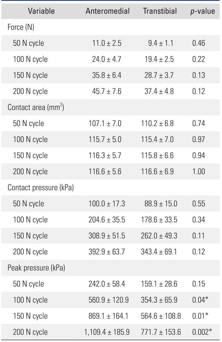
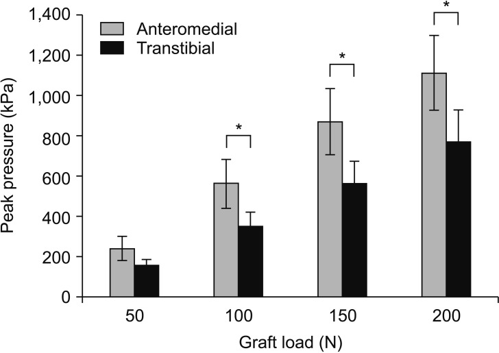
There were no statistically significant differences between the anteromedial and transtibial approaches for cyclic loading parameters of linear stiffness and hysteresis (Table 1). There were also no significant differences between the two techniques for contact area, contact force and mean contact pressure at any of the loading cycles (Table 2). There was also no statistically significant difference between the anteromedial and transtibial approaches at the 50 N loading cycle for peak contact pressure (p = 0.15). However, the peak contact pressure between the ACL graft and the tibial tunnel aperture was significantly greater using the anteromedial technique than the transtibial technique at 100 N (p = 0.04), 150 N (p = 0.01), and 200 N (p = 0.002) (Fig. 6).
This study evaluated the differences between the transtibial technique and the anteromedial portal technique in terms of geometry of the intra-articular tibial tunnel aperture and the contact characteristics between the graft and the tibial tunnel aperture. For the geometric characteristics of the tibial tunnel aperture, our study found that the transtibial technique created a funnel with an elliptical aperture and in contrast, the anteromedial technique created a tunnel with parallel walls as intended by the procedure. This is likely due to the approach for drilling the femoral tunnel in the transtibial technique where the drill must be slightly angulated relative to the tibial tunnel to achieve an appropriate angle, resulting in the expansion of the posterior tibial tunnel aperture. Additionally, while this study did not find any statistically significant difference in terms of linear stiffness, hysteresis, contact area, contact force or contact pressure, there were greater peak contact pressures when using the anteromedial technique compared to the transtibial technique.
Previous studies have shown this phenomenon of increased tunnel aperture size with the use of the transtibial technique. Miller et al.25) noted that transtibial femoral drilling made the aperture more elliptical in shape and increased the area of the intra-articular tibial tunnel aperture by 28.4% from 92.4 mm2 to 118.6 mm2. In comparison, the group that underwent the anteromedial technique had an area of 106.3 mm2. Our results showed similar trends and the tunnel aperture achieved with the anteromedial technique was comparable in size. However, our study showed a greater increase with transtibial drilling, with an increase in area of 45%. This could be because the authors25) started at different drilling positions for each method. Bedi et al.4) also found that the aperture created by the transtibial technique was elliptical, with the anteroposterior dimension being 38% larger than the medial-lateral dimension at 14.9 mm and 10.6 mm, respectively. In contrast, the anteromedial portal technique created a more circular aperture, with anteroposterior and medial-lateral dimensions of 10.8 mm and 10.4 mm. The authors4) found that the transtibial approach mainly expanded the tibial aperture in the anteroposterior direction. In another study, Youm et al.16) found using a modified version of transtibial drilling with varus and internal rotation of the tibia that the mean area of the tunnel orifice was significantly greater with transtibial approach drilling at 11.6 mm × 9.2 mm versus 10.3 mm × 9.1 mm with anteromedial approach drilling. The areas of these apertures were smaller than the apertures we obtained, possibly due to the use of smaller reamers. However, they still showed the effect of transtibial drilling creating a more elliptical orifice when compared to the anteromedial portal method.
The geometric differences between the two approaches likely contributed to the difference in peak contact pressures experienced by the graft at the tibial tunnel aperture. In any reconstruction, the graft must bend over two separate apertures when going from the tibia to the femur, creating areas of graft-bone contact.32) The transtibial approach creates a funnel-shaped tibial tunnel, leading to a less acute bend of the graft at the tibial aperture. This results in reduced peak contact pressures and lessens the possible abrasive effects. Previous studies that looked at the stresses experienced by the graft due to tunnel apertures focused mainly on the intra-articular aperture on the femoral side.
In a cadaveric study, Nishimoto et al.22) found that the graft bending angle at the femoral tunnel aperture was significantly more acute using the transtibial technique than the anteromedial portal technique, especially at low flexion angles. However, in contrast with the authors' findings,22) Wang et al.23) found that in an in vivo study comparing single bundle transtibial and double bundle anteromedial portal approaches, the femoral graft was more acutely bent in the anteromedial group. Despite this contradiction, both studies theorized that a greater bend in the graft over the intra-articular femoral aperture could lead to abrasion and future failure of the graft. In our study, instead of comparing the possible stresses on the graft at the femoral aperture, our focus was on the tibial aperture.
With any anteriorly directed force on the tibia, the graft will load on the posterior tibial tunnel and bend over the tibial aperture. In our study, the stiffness and hysteresis of the graft were not statistically significantly different, suggesting that the graft itself acted similarly despite the difference in drilling technique. Furthermore, the transtibial and the anteromedial techniques were not significantly different in terms of force, contact area and mean contact pressure, demonstrating that the general contact characteristics between the graft and posterior tibial tunnel were similar between the two techniques. However, the anteromedial technique showed a greater peak contact pressure under physiologic loads, which was seen at the aperture where the graft bends over the rim of the tibial tunnel. The greater peak contact pressure could possibly contribute to future graft failure when using an anteromedial approach due to the increased stress on the graft over time and should be considered as a factor when deciding the approach for ACL reconstruction. Therefore, a less acute angle when drilling the tibial tunnel should be considered when using the anteromedial technique to reduce the bending angle over the tibial aperture. Transtibial drilling may also lessen the chance of graft failure by reducing tibial-sided stresses on the graft itself. However, the larger tibial tunnel that is created by this technique could possibly be detrimental to graft-to-bone healing over time due to greater graft-tunnel motion before graft incorporation is complete and under-filling of the tunnel could lead to delays in graft incorporation and decreased graft fixation.33)
As with any cadaveric biomechanical study, one of the recognized limitations is that only time zero geometries, kinematics and stresses can be determined. The experimental setup was selected to isolate the effects of the tunnel drilling technique and has its limitations. This includes a static angle at which the load is applied, which does not fully replicate the changing bending angles of an ACL at the femoral and tibial apertures during flexion and extension.2534) While graft ruptures are more frequently found at the aperture of the femoral tunnel, any portion where the graft bends over a bony edge could be a source of graft failure. Furthermore, because our model includes only the tibial portion, it does not perfectly replicate post-ACL reconstruction knee kinematics nor the full range of movements and dynamic forces that the graft could experience. The difference in techniques may also cause varying placement of the femoral tunnel, which could affect the angles and forces experienced by the ACL reconstruction. The anteromedial technique generally places the graft lower on the femur, which would result in a more acute angle against the plateau aperture. In our testing setup, using the same angle for both specimens actually represents a better case scenario for the anteromedial technique. The same grafts were used for each technique to eliminate variance in graft size; however, it could also be a limitation if there was any creep in the graft. In order to minimize the effect of graft creep on the results, the testing order was randomized and a preconditioning protocol was performed prior to testing of each construct.
In conclusion, the contact characteristics between the anteromedial and transtibial techniques were generally similar. However, the anteromedial technique showed a smaller, more circular tibial tunnel and a greater peak contact pressure at the tibial tunnel aperture under physiologic loading conditions. Increased peak contact pressure on the graft at the tibial aperture may increase the stress on the graft and possibly lead to failure following ACL reconstruction with the anteromedial technique.
ACKNOWLEDGEMENTS
Partial funding was provided by Congress Medical Foundation, VA Rehabilitation Research and Development Merit Review and the John C. Griswold Foundation.
References
1. Lee MC, Seong SC, Lee S, et al. Vertical femoral tunnel placement results in rotational knee laxity after anterior cruciate ligament reconstruction. Arthroscopy. 2007; 23(7):771–778. PMID: 17637414.

2. Pearle AD, Shannon FJ, Granchi C, Wickiewicz TL, Warren RF. Comparison of 3-dimensional obliquity and anisometric characteristics of anterior cruciate ligament graft positions using surgical navigation. Am J Sports Med. 2008; 36(8):1534–1541. PMID: 18390491.

3. Gadikota HR, Sim JA, Hosseini A, Gill TJ, Li G. The relationship between femoral tunnels created by the transtibial, anteromedial portal, and outside-in techniques and the anterior cruciate ligament footprint. Am J Sports Med. 2012; 40(4):882–888. PMID: 22302206.

4. Bedi A, Musahl V, Steuber V, et al. Transtibial versus anteromedial portal reaming in anterior cruciate ligament reconstruction: an anatomic and biomechanical evaluation of surgical technique. Arthroscopy. 2011; 27(3):380–390. PMID: 21035990.

5. Maletis GB, Cameron SL, Tengan JJ, Burchette RJ. A prospective randomized study of anterior cruciate ligament reconstruction: a comparison of patellar tendon and quadruple-strand semitendinosus/gracilis tendons fixed with bioabsorbable interference screws. Am J Sports Med. 2007; 35(3):384–394. PMID: 17218661.
6. Buchner M, Schmeer T, Schmitt H. Anterior cruciate ligament reconstruction with quadrupled semitendinosus tendon: minimum 6 year clinical and radiological follow-up. Knee. 2007; 14(4):321–327. PMID: 17512735.
7. Strauss EJ, Barker JU, McGill K, Cole BJ, Bach BR Jr, Verma NN. Can anatomic femoral tunnel placement be achieved using a transtibial technique for hamstring anterior cruciate ligament reconstruction? Am J Sports Med. 2011; 39(6):1263–1269. PMID: 21335354.

8. Giron F, Cuomo P, Edwards A, Bull AM, Amis AA, Aglietti P. Double-bundle “anatomic” anterior cruciate ligament reconstruction: a cadaveric study of tunnel positioning with a transtibial technique. Arthroscopy. 2007; 23(1):7–13. PMID: 17210421.

9. Rue JP, Ghodadra N, Lewis PB, Bach BR Jr. Femoral and tibial tunnel position using a transtibial drilled anterior cruciate ligament reconstruction technique. J Knee Surg. 2008; 21(3):246–249. PMID: 18686488.

10. Steiner ME, Battaglia TC, Heming JF, Rand JD, Festa A, Baria M. Independent drilling outperforms conventional transtibial drilling in anterior cruciate ligament reconstruction. Am J Sports Med. 2009; 37(10):1912–1919. PMID: 19729364.

11. Bedi A, Raphael B, Maderazo A, Pavlov H, Williams RJ 3rd. Transtibial versus anteromedial portal drilling for anterior cruciate ligament reconstruction: a cadaveric study of femoral tunnel length and obliquity. Arthroscopy. 2010; 26(3):342–350. PMID: 20206044.

12. Franceschi F, Papalia R, Rizzello G, Del Buono A, Maffulli N, Denaro V. Anteromedial portal versus transtibial drilling techniques in anterior cruciate ligament reconstruction: any clinical relevance? A retrospective comparative study. Arthroscopy. 2013; 29(8):1330–1337. PMID: 23906273.

13. Silva A, Sampaio R, Pinto E. ACL reconstruction: comparison between transtibial and anteromedial portal techniques. Knee Surg Sports Traumatol Arthrosc. 2012; 20(5):896–903. PMID: 21850428.

14. Keller TC, Tompkins M, Economopoulos K, et al. Tibial tunnel placement accuracy during anterior cruciate ligament reconstruction: independent femoral versus transtibial femoral tunnel drilling techniques. Arthroscopy. 2014; 30(9):1116–1123. PMID: 24907026.

15. Wang H, Fleischli JE, Zheng NN. Transtibial versus anteromedial portal technique in single-bundle anterior cruciate ligament reconstruction: outcomes of knee joint kinematics during walking. Am J Sports Med. 2013; 41(8):1847–1856. PMID: 23752955.
16. Youm YS, Cho SD, Lee SH, Youn CH. Modified transtibial versus anteromedial portal technique in anatomic single-bundle anterior cruciate ligament reconstruction: comparison of femoral tunnel position and clinical results. Am J Sports Med. 2014; 42(12):2941–2947. PMID: 25269655.
17. Koutras G, Papadopoulos P, Terzidis IP, Gigis I, Pappas E. Short-term functional and clinical outcomes after ACL reconstruction with hamstrings autograft: transtibial versus anteromedial portal technique. Knee Surg Sports Traumatol Arthrosc. 2013; 21(8):1904–1909. PMID: 23203338.

18. Duffee A, Magnussen RA, Pedroza AD, Flanigan DC, Kaeding CC. MOON Group. Transtibial ACL femoral tunnel preparation increases odds of repeat ipsilateral knee surgery. J Bone Joint Surg Am. 2013; 95(22):2035–2042. PMID: 24257662.

19. Rahr-Wagner L, Thillemann TM, Pedersen AB, Lind MC. Increased risk of revision after anteromedial compared with transtibial drilling of the femoral tunnel during primary anterior cruciate ligament reconstruction: results from the Danish Knee Ligament Reconstruction Register. Arthroscopy. 2013; 29(1):98–105. PMID: 23276417.

20. Graf BK, Henry J, Rothenberg M, Vanderby R. Anterior cruciate ligament reconstruction with patellar tendon: an ex vivo study of wear-related damage and failure at the femoral tunnel. Am J Sports Med. 1994; 22(1):131–135. PMID: 8129096.
21. Toritsuka Y, Shino K, Horibe S, et al. Second-look arthroscopy of anterior cruciate ligament grafts with multistranded hamstring tendons. Arthroscopy. 2004; 20(3):287–293. PMID: 15007317.

22. Nishimoto K, Kuroda R, Mizuno K, et al. Analysis of the graft bending angle at the femoral tunnel aperture in anatomic double bundle anterior cruciate ligament reconstruction: a comparison of the transtibial and the far anteromedial portal technique. Knee Surg Sports Traumatol Arthrosc. 2009; 17(3):270–276. PMID: 19048229.

23. Wang JH, Kim JG, Lee DK, Lim HC, Ahn JH. Comparison of femoral graft bending angle and tunnel length between transtibial technique and transportal technique in anterior cruciate ligament reconstruction. Knee Surg Sports Traumatol Arthrosc. 2012; 20(8):1584–1593. PMID: 22120838.

24. Shin YS, Ro KH, Jeon JH, Lee DH. Graft-bending angle and femoral tunnel length after single-bundle anterior cruciate ligament reconstruction: comparison of the transtibial, anteromedial portal and outside-in techniques. Bone Joint J. 2014; 96-B(6):743–751. PMID: 24891573.
25. Miller MD, Gerdeman AC, Miller CD, et al. The effects of extra-articular starting point and transtibial femoral drilling on the intra-articular aperture of the tibial tunnel in ACL reconstruction. Am J Sports Med. 2010; 38(4):707–712. PMID: 20164306.

26. Denti M, Bigoni M, Randelli P, et al. Graft-tunnel mismatch in endoscopic anterior cruciate ligament reconstruction: intraoperative and cadaver measurement of the intra-articular graft length and the length of the patellar tendon. Knee Surg Sports Traumatol Arthrosc. 1998; 6(3):165–168. PMID: 9704323.
27. Chen CH, Chen WJ, Shih CH. Arthroscopic anterior cruciate ligament reconstruction with quadriceps tendon-patellar bone autograft. J Trauma. 1999; 46(4):678–682. PMID: 10217233.

28. Illingworth KD, Hensler D, Working ZM, Macalena JA, Tashman S, Fu FH. A simple evaluation of anterior cruciate ligament femoral tunnel position: the inclination angle and femoral tunnel angle. Am J Sports Med. 2011; 39(12):2611–2618. PMID: 21908719.
29. Ahn JH, Lee SH, Yoo JC, Ha HC. Measurement of the graft angles for the anterior cruciate ligament reconstruction with transtibial technique using postoperative magnetic resonance imaging in comparative study. Knee Surg Sports Traumatol Arthrosc. 2007; 15(11):1293–1300. PMID: 17721777.

30. Caborn DN, Urban WP Jr, Johnson DL, Nyland J, Pienkowski D. Biomechanical comparison between BioScrew and titanium alloy interference screws for bone-patellar tendon-bone graft fixation in anterior cruciate ligament reconstruction. Arthroscopy. 1997; 13(2):229–232. PMID: 9127082.

31. Sakane M, Fox RJ, Woo SL, Livesay GA, Li G, Fu FH. In situ forces in the anterior cruciate ligament and its bundles in response to anterior tibial loads. J Orthop Res. 1997; 15(2):285–293. PMID: 9167633.

32. Bae JY, Kim GH, Seon JK, Jeon I. Finite element study on the anatomic transtibial technique for single-bundle anterior cruciate ligament reconstruction. Med Biol Eng Comput. 2016; 54(5):811–820. PMID: 26296801.

33. Wilson TC, Kantaras A, Atay A, Johnson DL. Tunnel enlargement after anterior cruciate ligament surgery. Am J Sports Med. 2004; 32(2):543–549. PMID: 14977688.

34. Guenoun D, Vaccaro J, Le Corroller T, et al. A dynamic study of the anterior cruciate ligament of the knee using an open MRI. Surg Radiol Anat. 2017; 39(3):307–314. PMID: 27515305.

Fig. 1
Schematic of the different angles of approach in the anteromedial (A) and transtibial (B) tunnel drilling techniques. ACL: anterior cruciate ligament.
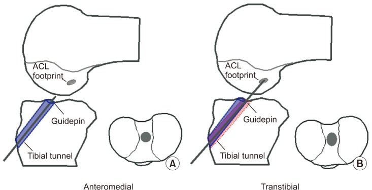
Fig. 2
The anteromedial versus transtibial aperture geometries were digitized, loaded into the Rhinoceros three-dimensional modeling software and the tibial tunnel aperture size was calculated.
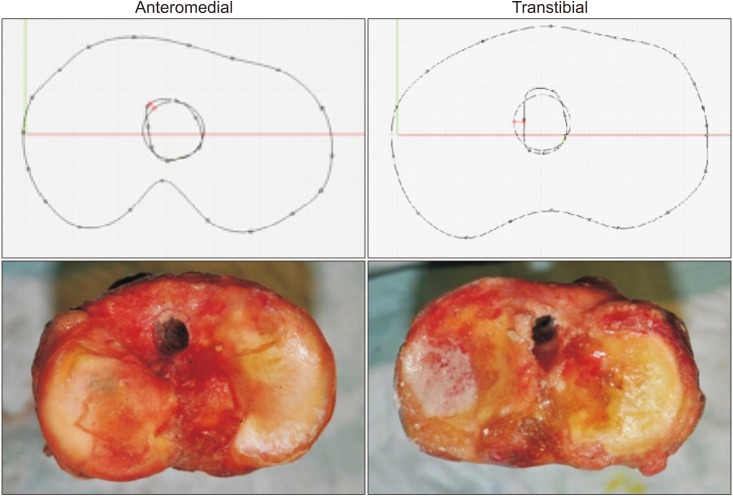
Fig. 4
A guidewire was used to ensure that the Tekscan sensor remained posterior to the graft when inserting the interference screw.
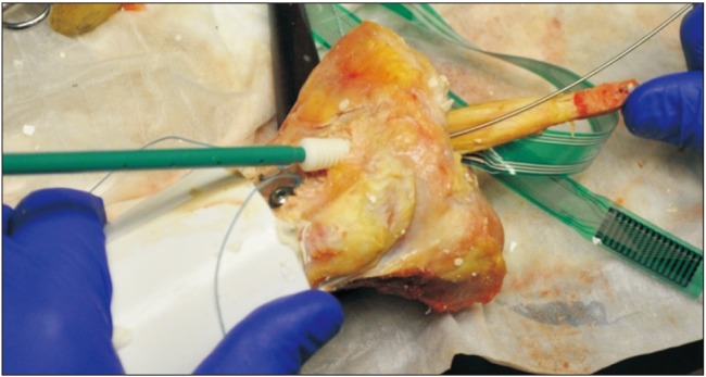
Fig. 5
The potted tibia was mounted to a custom testing cylinder and the bone block of the graft was cryo-clamped to the Instron material testing system.
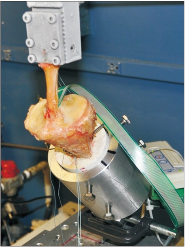
Table 1
Cyclic Loading Parameters
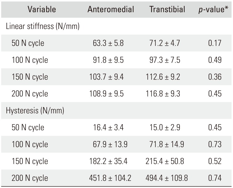
Table 2
Cyclic Loading Tibial Tunnel and Graft Contact Parameters


 PDF
PDF ePub
ePub Citation
 Print
Print


 XML Download