

 PDF
PDF Citation
 Print
Print


INTRODUCTION
Bacteria have been identified as the main cause of pulp infection and periapical disease [12]. It has been established that irrigants, coupled with mechanical instrumentation, are mandatory to ensure the elimination or greatest possible reduction of pathogens from the infected root canal. Irrigants have varying degrees of antimicrobial activity. The characteristics of an ideal endodontic irrigant includes good antimicrobial activity, non-cytotoxicity towards the surrounding periapical tissue, and the ability to inactivate endotoxins, to act as a lubricant, and to dissolve organic tissue remnants [3456]. The present study focused on the antimicrobial aspect of irrigants, as there are over 700 bacterial species in the oral cavity, with any individual harboring 100–200 species [7]. Furthermore, it has also been proven beyond doubt that the presence of bacteria is a major contributor to endodontic infections.
Sodium hypochlorite (NaOCl) has long been the gold-standard endodontic irrigant due to its good antimicrobial activity and tissue-dissolving capability [35]. However, it has been found to cause severe inflammatory reactions when placed in contact with vital tissues [8]. Chlorhexidine (CHX) is a widely used antimicrobial agent that has emerged as an endodontic irrigant. It has also been used as an inter-appointment medication during the endodontic treatment of teeth with apical periodontitis. As an irrigant, CHX has shown antimicrobial effectiveness comparable to NaOCl in several clinical and laboratory studies [910111213]. However, CHX has shown cytotoxic potential, although to a lesser extent than NaOCl. Therefore, adequate precautions should be taken when CHX is used in oral treatments at higher concentrations or for a longer duration [1415].
Octenidine dihydrochloride (OCT; N,N′-[1,10 decanediyldi-1[4H]-pyridinyl-4ylidene]bis[1-octanamine]dihydrochloride) is a bispyridine antimicrobial compound that carries 2 cationic active centers per molecule and demonstrates broad-spectrum antimicrobial effects, covering both gram-positive and gram-negative bacteria, fungi, and several viral species [16]. It exerts bactericidal/fungicidal effects by interfering with cell walls and membranes. OCT is currently widely used in the medical field for skin burns and decontaminating mucous membranes and open wounds [17]. It is also used in mouthwash formulations and other dental applications. Nonetheless, OCT is not currently popular as an endodontic irrigant because insufficient information is available about its properties in vivo. OCT is unique due to its relative non-cytotoxicity at the site of action [18] and good antimicrobial activity. These characteristics make OCT attractive as a potential alternative antimicrobial agent in the field of endodontics.
Enterococcus faecalis (E. faecalis) and Candida albicans (C. albicans) have repeatedly been found in root canals undergoing retreatment due to persistent infections [12]. Extensive research has been performed on E. faecalis and C. albicans. However, recent studies have identified Staphylococcus epidermidis (S. epidermidis) as one of the most prevalent species in cases of post-endodontic treatment disease with endodontic failure, along with E. faecalis [19]. S. epidermidis is known to be a facultative anaerobe that can grow through aerobic respiration or fermentation in anaerobic conditions [20]. The aim of the present study was to compare the antimicrobial effects of OCT with 3% NaOCl and 2% CHX as root canal irrigants against S. epidermidis.
MATERIALS AND METHODS
The present study was approved by the International Medical University Ethics Committee (project number: BDS I1-12(03)2015). The model proposed by Haapasalo and Ørstavik [21] was modified for the present study.
Growth and culture of S. epidermidis
Sterilized trypticase soy broth/agar (Hadry Diagnostics, Santa Maria, CA, USA) was prepared following the manufacturer's instructions, and the agar media was poured into a sterilized petri dish to a depth of 5 mm, under a laminar flow hood. The poured plates were allowed to solidify and were refrigerated until use. S. epidermidis was initially grown on a trypticase soy agar plate at 37°C for 24 hours. A single colony was then suspended in 5 mL of trypticase soy broth and grown overnight at 37°C. Its turbidity was then adjusted to a McFarland standard of 0.5.
Determination of the minimum inhibitory concentration
The minimum inhibitory concentration (MIC) was determined using serial dilutions of 0.1% OCT, 2% CHX, and 3% NaOCl. S. epidermidis was prepared to obtain the 0.5 McFarland turbidity. The 96-well plate was then incubated in a rotary incubator (180 rpm) at 37°C for 24 hours. The MIC was defined as the lowest concentration of each irrigant in which no visible turbidity was noted.
Determination of the zone of inhibition
The agar diffusion method was used to determine the zone of inhibition. S. epidermidis was subcultured onto trypticase soy agar plates and incubated at 37°C for 24 hours. The colonies were suspended in broth and the turbidity was adjusted, corresponding to a McFarland standard of 0.5. The bacterial suspension was then spread evenly over the surfaces of the trypticase soy agar plate and left for 5 minutes to dry. Pre-sterilized Whatman discs (6 mm in diameter) were soaked in 20 µL of different concentrations of each irrigant: 1%, 2% and 3% of NaOCl; 0.5%, 1% and 2% of CHX; 0.0125%, 0.025%, 0.05% and 0.1% of OCT; saline as a control. The discs were then placed on the trypticase soy agar plate with sufficient distance between them and incubated at 37°C for 24 hours. The zones of inhibition were then measured using a transparent ruler and recorded. Triplicate data for each concentration were obtained and the results were averaged.
Preparation of tooth samples
After obtaining informed consent from patients attending the Oral Health Clinic of International Medical University, 40 non-carious single-rooted human mandibular premolars extracted for orthodontic or periodontal reasons were collected. The calculus and tissue remnants on the exterior root surface were removed using a curette. The teeth were then embedded in cubical blocks of auto-polymerizing custom tray resin. The blocks were mounted and de-coronated using a high-speed precision diamond disc under water cooling. Then, 6 mm dentin blocks were cut from the cementoenamel junction. The samples were kept in a moist environment during all the procedures to prevent dehydration.
The inner diameter of the root canals of each sample was standardized using a Gates Glidden size 3 drill at 300 rpm with a lowspeed handpiece. The dentin blocks were cleaned in an ultrasonic water bath for 3 cycles of 20 minutes to remove the smear layer. The dentin blocks were then autoclaved, and the external surfaces of the samples were coated with nail varnish, placed in 70% alcohol for sterilization, and allowed to dry at room temperature for 1 hour in a biosafety cabinet under aseptic conditions. The tooth samples were pretreated with plain culture media before contamination. All procedures were done in a biosafety cabinet (biosafety level II) under aseptic conditions.
Sample inoculation
A McFarland standard of 0.5 of S. epidermidis was used for contaminating the dentin blocks. The dentin blocks were then inoculated by immersing them in the bacterial suspension in pre-sterilized containers at 37°C for 21 days, with a change of the bacterial suspension once every 3 days.
Irrigation of the dentin blocks
After incubation for 21 days, the dentin blocks were removed from the bacterial suspension using sterilized artery forceps. The excess culture was removed from the surface of the dentin samples using sterile paper. The samples were then randomly divided into 4 groups of 10 blocks: saline, 2% CHX, 3% NaOCl, and 0.1% OCT. Each group was then irrigated with 5 mL of its respective irrigant for a period of 3 minutes. The excess irrigant was then removed with sterile paper points from each dentin block.
Dentin sampling
Dentin chips were harvested from the root canal by circumferential removal using a lowspeed handpiece with a Gates Glidden size 4 drill. The dentin chips obtained were collected in Eppendorf tubes containing 0.5 mL of saline. The suspension was then homogenized by vigorous vortexing to release the adhered bacteria and left for 5 minutes to allow sedimentation of the dentin chips. The supernatant, which contained the bacterial suspension, was then used for microbiological analysis to determine the amount of colony-forming units (CFUs).
Statistical analysis
A Kruskal Wallis test was conducted on the data of zones of inhibitions and CFUs to evaluate differences among the groups studied. Post hoc tests were conducted to evaluate pairwise comparisons among the groups by using Duncan's test of multiple comparisons. The data was statistically analyzed with a level of significance set at 5% (IBM SPSS statistics software version 20.0, IBM, Armonk, NY, USA).
RESULTS
The MIC values of each tested irrigant were 0.05% for CHX, 0.25% for NaOCl, and 0.0125% for OCT. The inhibition zones of CHX, NaOCl, and OCT at each tested concentration are presented in the Figure 1. All tested irrigants showed concentration-dependent increase of zones of inhibition, and 3% NaOCl showed the largest zone of inhibition amongst all tested irrigants (p < 0.05). OCT in 0.0125% solution showed significantly larger zone of inhibition than 0.5% and 1% CHX (p < 0.05), and 0.025% OCT showed similar zone of inhibition with 2% CHX and 1% NaOCl.
Figure 1
Measurements of the inhibition zones of the irrigants (mm).
CHX, chlorhexidine; NaOCl, sodium hypochlorite; OCT, octenidine dihydrochloride.
Different lower superscript letters indicate statistically significant differences.

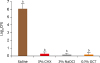
CFUs evaluated in dentin block model showed significant antibacterial effect of tested irrigants on S. epidermidis compared to saline (p < 0.05). However, there were no significant differences among the CFU measurements of 2% CHX, 3% NaOCl, and 0.1% OCT showing complete elimination of S. epidermidis in all samples (Figure 2).
DISCUSSION
Irrigation of the root canal is one of the key determinants imperative for successful endodontic treatment. In the present study, antimicrobial effects of OCT against S. epidermidis was investigated and found that 0.1% OCT showed comparable antimicrobial effect with 2% CHX and 3% NaOCl. The effective concentration of OCT was determined by MIC and agar diffusion technique, and validated the antimicrobial effect by quantitative analysis of CFUs.
Evidence from literature suggested that antimicrobial efficacy of OCT was sufficient in the complex environment of the root canal system. The results of present study also indicated the same. This bispyridine derivative was first developed by Sterlig Winthrop Research Institute and its use in form of mouth rinse showed inhibition of plaque and dental caries both in humans and rats [2223]. Enhanced antimicrobial efficacy of OCT attributed to its cation-active structure that readily binds to negatively charged bacterial cell wall and thereby affects vital functions of cell membrane that leads to cell death [24].
The agar diffusion test is a well-established technique for studying antibacterial properties. The advantage of this method is that the antimicrobial effect of medicaments can be detected by challenging the bacterial isolates with antimicrobial discs without any change in the chemical properties of the medicaments. In the present study, 3% NaOCl showed the largest zones of inhibition against S. epidermidis (p < 0.05), and all tested concentrations of OCT showed significantly larger zone of inhibition than 2% CHX (p < 0.05). Considering the discrepancies among MICs and zone of inhibition data of tested irrigants found in this experiment, it could be speculated that OCT at a lower concentration could be a potent antimicrobial irrigant against S. epidermidis.
CFU analysis revealed that 0.1% OCT significantly inhibited S. epidermidis, similar to 2% CHX and 3% NaOCl. The present antimicrobial results are in accordance with previous reports. Kapur et al. [25] demonstrated that various concentrations of OCT were effective on E. faecalis, C. albicans, and a mixture of both, compared to 5.25% NaOCl and 1% or 2% CHX. Tirali et al. [26] and Anuradha et al. [8] reported antimicrobial effect of OCT against E. faecalis, and Ghivari et al. [27] also verified antimicrobial effect of 0.1% OCT against E. faecalis, S. aureus, and C. albicans.
The dentin block model used in our experiment was modified from Haapasalo and Ørstavik [21]. Although the 3-week incubation period allowed the infection of dentinal tubules with S. epidermidis, the present ex vivo dentin infection method does not simulate deep and homogeneous penetration of bacteria into all dentinal tubules in all tested specimens. Also the tubular depth-related antimicrobial effect could not be deduced from current experimental setting. Despite its potent antimicrobial effect, in addition, OCT cannot replace NaOCl as root canal irrigant because it does not possess the ability to dissolve pulp tissue [17].
Further researches on OCT including its interactions with other irrigants, dose-effectiveness, antimicrobial efficacy against other single- or multi-species biofilms, and safety are required.
CONCLUSIONS
In this study, the antimicrobial effect of OCT against S. epidermidis was investigated in comparison to CHX and NaOCl. Within the limitations of the present study, OCT at lower concentrations effectively inhibited antimicrobial growth of S. epidermidis in compared to CHX and NaOCl, implying the possible applicability as an endodontic irrigant in terms of the antimicrobial properties that were evaluated.
 XML Download