

 PDF
PDF ePub
ePub Citation
 Print
Print


Abstract
Purpose
Methods
Results
Conclusions
Figures and Tables
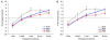 | Figure 1Consecutive changes of postoperative deviation at distant (A) and near (B) fixations according to the operative methods for intermittent exotropia. Plication revealed similar surgical outcomes with resection procedure (A, B). BLR = bilateral rectus muscle recession group; R&P = unilateral lateral rectus muscle recession with medial rectus muscle plication group; R&R = unilateral lateral rectus muscle recession and medial rectus muscle resection group; Mon = month(s). *p < 0.05, **p < 0.01 between BLR and R&P groups by Kruskall-W allis test with post-hoc analysis.
|
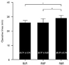 | Figure 2Operative times in three groups according to operative method. Plication consumed significantly shorter duration required for the surgery compared to resection procedure. BLR = bilateral rectus muscle recession group; R&P = unilateral lateral rectus muscle recession with medial rectus muscle plication group; R&R = unilateral lateral rectus muscle recession and medial rectus muscle resection group; min = minutes. *p < 0.001 by Kruskall-W allis test with post-hoc analysis.
|
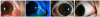 | Figure 3Postoperative status of anterior segment appearance in the right eye of 9-year-old male patient. Dellen was identified at 1 week after lateral rectus muscle recession with medial rectus muscle plication (A, B). Consequently, it was improved by the topical agents after 1 month of the surgery (C, D).
|
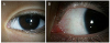 | Figure 4Anterior segment appearance (A) with magnification (B) in the left eye of 4-year-old female patient at a couple of months after medial rectus (MR) muscle plication. Elevation over the plicated site of MR muscle was not grossly apparent.
|
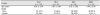
Table 2
Consecutive changes of postoperative deviations at distant fixation according to the operative method analyzed by linear mixed model
Values are presented as mean ± standard deviation unless otherwise indicated.
BLR = bilateral rectus muscle recession group; R&P = unilateral lateral rectus muscle recession and medial rectus muscle plication group; R&R = unilateral lateral rectus muscle recession and medial rectus muscle resection group; SE = standard error; PD = prism diopters; CI = confidence interval.
*Kruskal-Wallis test.
![]()
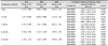
Table 3
Consecutive changes of postoperative deviations at near fixation according to the operative method analyzed by linear mixed model
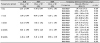
Values are presented as mean ± standard deviation unless otherwise indicated.
BLR = bilateral rectus muscle recession group; R&P = unilateral lateral rectus muscle recession and medial rectus muscle plication group; R&R = unilateral lateral rectus muscle recession and medial rectus muscle resection group; SE = standard error; PD = prism diopters; CI = confidence interval.
*Kruskal-Wallis test.
![]()
 XML Download