Journal List > J Korean Med Sci > v.34(6) > 1116263

Seok, Sohn, and Peck: A Paragonimiasis Mimicking Gastric Submucosal Tumor
jkms-34-e45-g002
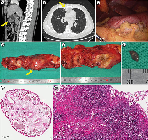
A previously healthy 49-year-old man was referred to the hospital for a symptomatic gastric submucosal tumor that was detected in the computed tomography (CT) during a health screening in July 2017 (Fig. 1A). There was incidental bronchopneumonia in the anterior segment of the right upper lobe on the chest CT (Fig. 1B). The submucosal septate cystic mass was removed by laparoscopic gastric wedge resection. During the operation, multiple tiny nodules were found in the entire peritoneum, omentum, transverse colon, and the liver surface (Fig. 1C). When the mass was dissected, brownish discharge poured out and an oval-shaped worm was found (Fig. 1D-F). The trematode was 5–6 mm long and 4–5 mm wide and was characterized by an oral sucker, a ventral sucker, a multilobed ovary, testes in the posterior region of the body, teguments covered with spines, and well-developed vitellaria (Fig. 1G). Multiple eggs (size, 50–60 µm) were observed in the lymph node which showed granulomatous inflammation with eosinophil infiltration (Fig. 1H). Additional history taking was taken. He is an office worker living in Seoul, and has eaten freshwater crabs several times. Considering the clinical and pathologic findings, praziquantel (25 mg/kg three times a day for two days) was prescribed after the diagnosis of paragonimiasis. Pulmonary paragonimiasis is sometimes mistaken for pulmonary tumors. However, in the case of extrapulmonary paragonimiasis, to suspect parasite infection is not easy before pathologic confirmation. Prior studies have reported some extrapulmonary paragonimiasis identified in the abdominal cavity.123 Here we report the first case of paragonimiasis mimicking gastric submucosal tumor.
This manuscript was written after receiving the patient's agreement of the publication including private clinical records and images.

Figures and Tables

Fig. 1

A case of intra-abdominal paragonimiasis. (A) A gastric submucosal mass is shown in the abdominopelvic CT (yellow arrow). (B) A incidental lung nodule facing pleura are seen in the chest CT. (C) Laparoscopic finding shows multiple tiny nodules on the liver surface and omentum. (D) Gross finding of the gastric submucosal mass (yellow arrow) and lymph nodes. (E) Cross section of the mass. (F) Gross finding of the parasite. (G) Microscopic finding of the parasite (H & E, × 40). (H) Chronic granulomatous inflammation with eosinophil infltration and parasite eggs (H & E, × 200).

CT = computed tomography, H & E = hematoxylin and eosin.
jkms-34-e45-g001

Notes

Disclosure The authors have no potential conflicts of interest to disclose.

Author Contributions

  • Conceptualization: Seok H, Sohn TS, Peck KR.

  • Data curation: Seok H.

  • Investigation: Seok H.

  • Methodology: Seok H.

  • Supervision: Sohn TS, Peck KR.

  • Validation: Peck KR.

  • Writing - original draft: Seok H.

  • Writing - review & editing: Seok H, Sohn TS, Peck KR.

References

1. Oh CR, Kim MJ, Lee KH. A case of intra-abdominal paragonimiasis mimicking metastasis of lung cancer diagnosed by endoscopic ultrasound-guided fine needle aspiration. Korean J Gastroenterol. 2015; 66(1):41–45.
crossref pmid
2. Kim MJ, Kim SH, Lee SO, Choi SH, Kim YS, Woo JH, et al. A case of ectopic peritoneal paragonimiasis mimicking diverticulitis or abdominal abscess. Korean J Parasitol. 2017; 55(3):313–317.
crossref pmid pmc
3. Kim AY. Heterotopic paragonimiasis presented by intra-abdominal masses. Korean J Gastroenterol. 2013; 61(6):351–353.
crossref pmid
TOOLS
ORCID iDs

Hyeri Seok
https://orcid.org/0000-0002-2032-9538

Tae Sung Sohn
https://orcid.org/0000-0002-9553-3234

Kyong Ran Peck
https://orcid.org/0000-0002-7464-9780

Similar articles