

 PDF
PDF ePub
ePub Citation
 Print
Print


Dear Editor,
The mesenchyme of the conjunctiva is a delicate tissue which contains blood vessels, nerves and lymphatics, but its proliferation has only recently been discovered [1]. In 2012, Herwig et al. [2] described a new classification of disease that encompasses the previously unclassified mesenchymal tumors: the conjunctival stromal tumor (COST). The disease presents as a wide spectrum of clinical characteristics, but histopathologic studies have determined that its common feature is typical benign proliferation of spindle cells in the conjunctival stroma. Until now, every reported COST case described tumors located on the bulbar conjunctiva. We present the rare case of a COST located on the palpebral conjunctiva.
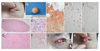
A 43-year-old man with a chief complaint of foreign body sensation visited our clinic with a palpable mass in the left upper eyelid which had been present for one month. No pain or tenderness was noted initially. His past medical history included rheumatoid arthritis for which he was receiving treatment. On examination, his uncorrected visual acuity was 20 / 20 in the right eye and 20 / 40 in the left eye and left corneal erosion was noted. The pupils were round and reactive with no afferent papillary defect. An 8-mm-sized, slight reddish, firm mass was found at the left upper palpebral conjunctiva (Fig. 1A), and a shave biopsy was performed under local anesthesia (Fig. 1B).
Histopathologic sections revealed a nodular mass with squamo-columnar epithelium (Fig. 1C). The mass was composed of interlacing spindle cells with scanty myxoid stroma (Fig. 1D), and the tumor cells disclosed mild to moderate cellularity and mild nuclear pleomorphism (Fig. 1E). Alcian blue special staining revealed a scanty amount of stromal mucin (Fig. 1F). The immunohistochemical staining for tumor cells was positive for vimentin and revealed diffuse, strong positivity for CD34 (Fig. 1G). The stains were negative for smooth muscle actin (Fig. 1H), CD68, and S-100. Only c-kit immunohistochemical staining was reactive in mast cells (Fig. 1I).
After the initial shave biopsy, a 5-mm-sized remnant mass was noticeable (Fig. 1J). During regular follow-up, proliferation of the remnant mass was noted at 2 months after the shave biopsy, resulting in a 1-mm increase in the diameter (Fig. 1K). An excisional biopsy was performed in the operating room, and the histopathologic results were same as the previous shave biopsy. An additional immunohistochemical stain, Ki-67, was performed and shown to be less than 1%. Thereafter, the patient has been disease free for 9 months, and is currently receiving regular follow-up. No other adverse events have been noticed during follow-up.
COST is a very rare disease first described by Herwig et al. [2]. In every case previously described, the tumor was located at the bulbar conjunctiva with mesenchyme proliferation as its unique characteristic, but the disease could not be categorized elsewhere. Herwig et al. [2] proposed a benign mesenchymal proliferation with pseudonuclear inclusions and multinucleated giant cells which showed positive results for immunohistochemistry stains of CD34, vimentin, CD68, and named it COST.
The most likely disease in the differential diagnosis of our case was dermatofibrosarcoma protuberans (DFSP) because the two diseases are clinically and pathologically similar. On microscopic examination, they present as cellular neoplasms composed of fibroblasts in a storiform pattern. DFSP usually stains positively for CD34, hyaluronate, and vimentin, and it stains negatively for factor XIIIa [34]. However, no case originating in the conjunctiva has been reported. Our case was different from DFPS because the mass was small in size compared to a typical DFSP, and mitosis was not noticeable. Other differential diagnoses are myxoma and nodular fasciitis. The former can be excluded by negative stain for alcian blue and the latter by smooth muscle actin negativity.
One notable feature in our case was the difference in pathologic and immunohistochemical features compared to those reported by Herwig et al. [2]. We reported positive immunohistochemical stains for CD34 and vimentin but not for CD68. CD68 is the most widely used marker for identifying macrophages [5]. Because there were no signs of inflammation in our case, it is likely that the expression of macrophages was not identified, and pseudonuclear inclusions and multinucleated giant cell could not be found either. Thus, we suggest that the definition of COST proposed by Herwig et al. [2] needs to be modified.
 XML Download