Abstract
Purpose
Several studies have described age-associated brow drooping in Westerners. However, there are few studies that address brow drooping in the Asian population, and especially in the Korean population. Therefore, we studied brow position changes with age in Korean individuals.
Methods
A total of 300 adults older than 18 years were enrolled. The ImageJ program was used to analyze digital photos of the patients by measuring the following parameters: marginal reflex distance-1, brow-to-pupil distance, nasal ala-lateral brow distance, lateral brow plumb line, and the angle formed by the line from the mid pupil to the midline of the brow and a line from the midline of the brow to the lateral brow. We divided the patients into three groups (18 to 40, 41 to 60, older than 61) and compared them using the ANOVA test.
Results
Group A included 100 patients between 18 and 40 years of age. Group B included 100 patients between 41 and 60 years of age. Group C included 100 patients older than 61 years. There were significant differences between groups A and C and between groups B and C with regard to marginal reflex distance-1, brow-to-pupil distance and the angle. Lateral brow plumb line showed significant difference only between groups A and B. Nasal ala-lateral brow distance was not significantly different across the three groups.
Conclusions
We sought to describe the physiologic facial changes that occur in Korean individuals. We also hoped to establish guidelines for ptosis corrective surgery. We used various parameters to characterize the aging process in Asians. Our data demonstrated that, like Westerners, Koreans experience lateral brow drooping with age; however, this change was only significant in the group aged >61 years.
Changes in brow position influence one's expression of certain negative emotions (such as decreased vitality, tiredness, or grief), as well as facial beauty. Therefore, there is interest in understanding the changes that typically occur with age and in identifying the ideal brow position [1]. The ideal brow position changes; modern Korean women prefer an arch-shaped brow from the perpendicular line passing from the ipsilateral nostril to the line passing through the lateral canthus, ala of nose, and the center of pupil [23]. The aging process typically starts in the upper face before the lower face. Ptosis of the lateral brow is one of the earliest changes with time [4]. In addition, the eyelid skin thins and the eyelid fat shrinks as one ages.
Previous studies have found that the lateral brow descends with age in Western populations, but there are few similar studies in Asian populations [5]. The findings from Western studies cannot necessarily be generalized to Asian populations [6]. Therefore, we investigated brow position in Koreans and the changes associated with age.
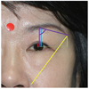
A total of 300 patients over 17 years old who had undergone facial photographs at our clinic between August 2011 and February 2013 were included. This study was approved by the institutional review board of Dong-A University Hospital (DAUHIRB-17-067). Informed consent was obtained from each patient. Patients were excluded if they had a history of facial trauma, strabismus, congenital anomalies, cosmetic surgery, or thick brow tattoos. Patients were also excluded if they had health factors that potentially affect the ocular position, such as hyperthyroidism. Facial photographs were captured using an EOS500 digital camera (Canon, Tokyo, Japan). A 10-mm-diameter red circle was attached to the patients' forehead for these photographs. The following five parameters were obtained using the ImageJ ver. 1.50i (National Institutes of Health, Bethesda, MD, USA): marginal reflex distance-1 (MRD1); brow to pupil distance (BPD); nasal ala-lateral brow distance (NALB); the vertical line extending from the tail of the brow and a horizontal line extending from the lateral canthus (lateral brow plumb line, LBPL); and the angle between the BPD and the line from the upper end point of the BPD to the lateral end of the brow (Angle). These parameters were measured retrospectively on the basis of the red circle (Fig. 1).
The patients were classified into groups A (18 to 40 years old), B (41 to 60 years old), and C (over 60 years old) according to age. The mean ages of patients by group are as follows: 46 for men and 54 for women in group A, 56 for men and 44 for women in group B, and 60 for men and 40 for women in group C. There were no significant differences in sexual distribution across the three groups. The measured parameters were analyzed using ANOVA and the Scheffe test. Statistical analysis was performed using SPSS ver. 12.0 (SPSS Inc., Chicago, IL, USA).
The average MRD1 was 2.99 ± 1.52 mm in group A, 3.15 ± 1.53 mm in group B, and 2.14 ± 1.31 mm in group C. There were significant differences in MRD1 between groups A and C (p < 0.001) and between groups B and C (p < 0.001). In contrast, there was no significant difference between groups A and B (p = 0.756).
The average BPD was 23.11 ± 3.28 mm in group A, 23.26 ± 3.69 mm in group B, and 26.67 ± 9.87 mm in group C. There were significant differences in BPD between groups A and C (p = 0.001) and between groups B and C (p = 0.001). In contrast, there was no difference between groups A and B (p = 0.987).
The average NALB was 79.33 ± 6.96 mm in group A, 78.81 ± 7.63 mm in group B, and 78.67 ± 8.52 mm in group C. There was no significant difference in NALB among the three groups: groups A and B (p = 0.893), groups A and C (p = 0.833), and groups B and C (p = 0.992).
The average LBPL was 18.06 ± 3.12 mm in group A, 20.76 ± 10.31 mm in group B, and 19.35 ± 5.64 mm in group C. There was a significant difference in LBPL only between groups A and B (p = 0.027). In contrast, groups C and A and groups C and B had no significant differences in LBPL (p = 0.437 and p = 0.362, respectively).
The average of Angle was 75.69 ± 10.97° in group A, 74.79 ± 5.83° in group B, and 68.27 ± 8.09° in group C. Groups C and A (p < 0.001) and groups C and B (p < 0.001) had significant differences in Angle. However, there was no significant difference between groups A and B (p = 0.760). There was no gender disparity in any of the groups.
The accurate evaluation of brow position and age-related changes is important for choosing the proper surgical methods [7]. We analyzed various parameters in Asians to reaffirm the characteristics of the aging process in Western people. Sclafani and Jung [8] reported that the brow tail was higher in women than in men; however, the disparity between genders was not found to be statistically significant in our study. The MRD1 and Angle were also not statistically different. However, group C demonstrated a significant decrease compared to those of groups A and B. Two factors that contribute to a descending brow position are a rapid decrease in tissue elasticity and changes in the bony tissue of the superior orbit rim [9]. These changes, which may accelerate after 60 years of age, can explain the significant decreases in MRD1 and Angle in group C. Park et al. [10] reported that MRD1 is highest between ages 30 and 34 and lowest over 60. The group also found that MRD1 decreased with age, with greater changes in men than in women. Lee et al. [11] analyzed the MRD1 of 432 participants and found that there was a significant drop after the age of 60. This result is comparable to our findings. Seo and Ahn [12] also depicted several meaningful changes that occur after the seventh decade, including the increase of upper eyelid tissue, brow ptosis, and an increase in lateral hood width of the eyelid. Although BPD tended to increase with age, there was no significant difference between groups A and B. However, group C had significantly different BPD changes compared to those of groups A and B. Moon et al. [13] found that BPD decreased at the third and fourth decades and increased after the fifth decade. van den Bosch et al. [14] explained that these changes may be caused by age-related interruption of the levator muscle aponeurosis and to involutional atrophy of the orbital fat. It may also be that it occurs as the eyebrows shift upward. Although Glass et al. [4] reported that NALB, LBPL and Angle decreased with age in Western participants, NALB showed no significant difference among the groups, and LBPL was only significantly different between groups A and B. These findings suggest that Asian individuals may have less elastic tissue than Western people, resulting in a less dramatic lateral brow droop with age. In addition, while the angle decreased significantly, there were no significant differences in NALB across the groups. These results suggest that the baseline amount of NALB was too large to reflect the minute amount of brow ptosis. The distance between the lateral canthus and the lateral brow might be a better parameter than NALB. The LBPL also showed no significant differences across groups, except between groups A and B. We suppose that drooping of the lateral canthus may increase the LBPL between groups A and B. In addition, we suspect that acceleration of lateral brow ptosis with age will decrease the LBPL. Angle may reflect not only lateral brow ptosis, but also brow shape. These differences in brow shape vary with racial differences and may explain the discrepancy of results between LBPL and Angle. In addition, because the BPD of group C was significantly higher than that of group A or B, Angle demonstrates significant differences among groups, unlike LBPL. A previous study of Western people revealed an annual decrease of 0.01 mm in MRD1, 0.03 mm of BPD, and 0.22° of Angle [4]. In contrast, we did not find gradually decreases in these parameters; in contrast, there was an increase in the BPD with age. One must also consider the possibility of an upward brow shift, which can create lateral brow drooping, although unrelated to increasing BPD or decreasing Angle.
This study has several limitations. This was a cross-sectional study between age-related groups. We did not analyze the brow position changes according to age in each participant. Therefore, a long-term prospective analysis of brow position changes with age in each participant is needed for more meaningful results. We randomly selected 100 patients for each group to reduce potential errors. In addition, a larger study with more participants and greater age subdivisions may allow for a more detailed analysis.
In conclusion, the Korean brow position changes with age, but these changes are different from those of Westerners. The BPD increased, while Angle tended to decrease with age in Korean individuals. In contrast, NALB did not change significantly with age. We studied facial aging based on multiple parameters. Our data are important with regard to improvements in eyelid surgery.
Figures and Tables
References
1. Hassanpour SE, Khajouei Kermani H. Brow ptosis after upper blepharoplasty: findings in 70 patients. World J Plast Surg. 2016; 5:58–61.


2. Hwang SJ, Kim H, Hwang K, et al. Ideal or young-looking brow height and arch shape preferred by Koreans. J Craniofac Surg. 2015; 26:e412–e416.

3. Kim SK, Cha SH, Hwang K, et al. Brow archetype preferred by Korean women. J Craniofac Surg. 2014; 25:1207–1211.


4. Glass LR, Lira J, Enkhbold E, et al. The lateral brow: position in relation to age, gender, and ethnicity. Ophthalmic Plast Reconstr Surg. 2014; 30:295–300.


5. Kim JS, Ahn HB. Surgical outcomes of levator resection in ptosis patients with deep superior sulcus. J Korean Ophthalmol Soc. 2014; 55:1734–1738.

6. Kim IS, Choi JB, Rah SH, Lee SY. Classification of ptosis in Korea. J Korean Ophthalmol Soc. 2005; 46:1262–1269.
7. Kim HM, Lee TS. Clinical observation and their surgical results of 127 cases of blepharoptosis. J Korean Ophthalmol Soc. 1985; 26:15–22.
8. Sclafani AP, Jung M. Desired position, shape, and dynamic range of the normal adult eyebrow. Arch Facial Plast Surg. 2010; 12:123–127.


9. Knize DM. An anatomically based study of the mechanism of eyebrow ptosis. Plast Reconstr Surg. 1996; 97:1321–1333.


10. Park DM, Song JW, Han KH, Kang JS. Anthropometry of normal Korean eyelids. J Korean Soc Plast Reconstr Surg. 1990; 17:822–834.
11. Lee JS, On KK, Kim JD. Superior visual field on MRD1 and aging changes of MRD1. J Korean Ophthalmol Soc. 1994; 35:884–888.
12. Seo HR, Ahn HB. Morphological changes of the eyelid according to age. J Korean Ophthalmol Soc. 2009; 50:1461–1467.

13. Moon CS, Moon SH, Jang JW. Topographic anatomic difference of the eyelid according to age in Korean. J Korean Ophthalmol Soc. 2003; 44:1865–1871.


 PDF
PDF ePub
ePub Citation
 Print
Print


 XML Download