Journal List > Clin Exp Vaccine Res > v.8(1) > 1115923

Foroutan, Ghaffarifar, Sharifi, Dalimi, and Jorjani: Rhoptry antigens as Toxoplasma gondii vaccine target

Abstract

Toxoplasmosis is a cosmopolitan zoonotic infection, caused by a unicellular protozoan parasite known as Toxoplasma gondii that belongs to the phylum Apicomplexa. It is estimated that over one-third of the world's population has been exposed and are latently infected with the parasite. In humans, toxoplasmosis is predominantly asymptomatic in immunocompetent persons, while among immunocompromised individuals may be cause severe and progressive complications with poor prognosis. Moreover, seronegative pregnant mothers are other risk groups for acquiring the infection. The life cycle of T. gondii is very complex, indicating the presence of a plurality of antigenic epitopes. Despite of great advances, recognize and construct novel vaccines for prevent and control of toxoplasmosis in both humans and animals is still remains a great challenge for researchers to select potential protein sequences as the ideal antigens. Notably, in several past years, constant efforts of researchers have made considerable advances to elucidate the different aspects of the cell and molecular biology of T. gondii mainly on microneme antigens, dense granule antigens, surface antigens, and rhoptry proteins (ROP). These attempts thereby provided great impetus to the present focus on vaccine development, according to the defined subcellular components of the parasite. Although, currently there is no commercial vaccine for use in humans. Among the main identified T. gondii antigens, ROPs appear as a putative vaccine candidate that are vital for invasion procedure as well as survival within host cells. Overall, it is estimated that they occupy about 1%–30% of the total parasite cell volume. In this review, we have summarized the recent progress of ROP-based vaccine development through various strategies from DNA vaccines, epitope or multi epitope-based vaccines, recombinant protein vaccines to vaccines based on live-attenuated vectors and prime-boost strategies in different mouse models.

Introduction

Toxoplasmosis is a cosmopolitan zoonotic infection, caused by a unicellular protozoan parasite known as Toxoplasma gondii that belongs to the phylum Apicomplexa [12]. This obligate intracellular parasite can infect a wide spectrum of warm-blooded vertebrate species such as men, birds, livestocks, marine mammals, etc. [13]. Recently, it has been shown that this parasite can infect snakes [4]. Role of rodents and birds in the spread of infection usually ignored [15]. Probably the global warming dilemma would play a substantial role in the epidemiological distribution of disease [5]. The infection is predominantly transmitted via consumption of raw/undercooked meats contaminated with tissue cysts, drinking water or ingestion of raw/unwashed vegetables contaminated by mature oocysts, and vertically from mother to the fetus [136789]. However, transmission via blood transfusion and organ transplantation occurs rarely [10111213]. The presence of Toxoplasma DNA in meat and meat products indicates the potential risk of food-transmitted toxoplasmosis [1415].
Upon maternal infection, fetus is probably to be exposed with transplacental transmission. Toxoplasmosis may cause the miscarriage in those pregnant mothers that acquired the infection during pregnancy. Based on the gestational age, the consequent complications will be different including deafness, impaired mental development, retardation, microcephaly, hydrocephalus, brain focal lesions, etc. [16]. The infection may result in fetal death, neonatal loss and abortion in some animals, particularly amongst goats and sheep resulting severe economic losses in the industry of veterinary medicine and animal husbandry. Besides, they serve as a source of transmission to people [3].

Need for Vaccine

The present common primary control measures for men and animals T. gondii infection depends on chemotherapy. Since antiparasitic drugs are unable to prevent from the T. gondii infection in both men and animals and also have no effect on the encysted parasites within infected hosts [17], accordingly, discover and development of an effective vaccine urgently needed to prevent and control toxoplasmosis.
Now, the live-attenuated tachyzoites of T. gondii S48 strain, Toxovax is the only commercially licensed vaccine for use in the veterinary industry in some countries that decrease the incidence of abortion in sheep from congenital toxoplasmosis, however, it is inadequate and expensive [18]. Since this vaccine contains the live-attenuated tachyzoites, it is not appropriate for human use, especially in immunocompromised individuals. Besides, there is ascending concerns of safety for use in food-producing animals as well. On the other hands, T. gondii S48 strain may revert to the wild-type virulence, as previously the accidental infections have been reported in farmers [192021].
As we all know, in regard to the major transmission routes of parasite and toxoplasmosis manifestations in the high risk groups, the main targets for vaccination strategy would cover [22].
  • - Vaccination to prevent acute parasitemia and protect against congenital toxoplasmosis

  • - Prevent or reduce tissue cysts in food animals to interrupt the transmission route to humans

  • - Prevention or reduction of oocyst shedding in cats to confine environmental contamination as well as minimize the risk of toxoplasmosis for all intermediate hosts

Rhoptry Proteins

Toxoplasma contains three apical secretory organelles known as the rhoptries, dense granules, and micronemes. Among them the rhoptries are club-shaped and has two distinct portions, including the anterior duct (neck) and the posterior bulb. This unique apical secretory organelle is shared merely in the all apicomplexan parasites [2324]. Rhoptries are contributes in the active penetration of the parasite into the host cell and involved in the biogenesis of parasitophorous vacuole as a peculiar intracellular compartment, in which the parasite multiplies vigorously within it and avoid the intracellular elimination [24]. Rhoptry proteins (ROPs) are contributed in the multiple stages of the invasion of parasites and are also critical for survival within host cells [23242526]. In general, the rhoptries constitutes approximately 1%–30% of the total Toxoplasma cell volume [27]. The researchers were employed the proteomic and genomic approaches to recognize the contents of the rhoptries in T. gondii and other apicomplexan parasites [282930]. They identified 38 ROPs, in which twenty of them were localized to the rhoptry organelle (eleven and nine for the rhoptry bulb and the rhoptry neck, respectively) [293031]. Rhoptry family genes encode many substantial and pivotal proteins that contribute in the virulence and pathogenicity of T. gondii. Due to the key biological role of these proteins, ROPs have lately become popular and focused as hoping vaccine candidates against some parasitic disease such as toxoplasmosis [242526323334]. These proteins are released by rhoptries which are the apical secretory organelles of parasite and mainly involved in the T. gondii invasion into the cytoplasm of host cells [25]. Some of these ROPs (such as ROP5, ROP16, and ROP18) so-called ROP kinases, act as serine-threonine kinases and play a pivotal role in the virulence and pathogenicity of parasite as well as host cell modulation [252635].
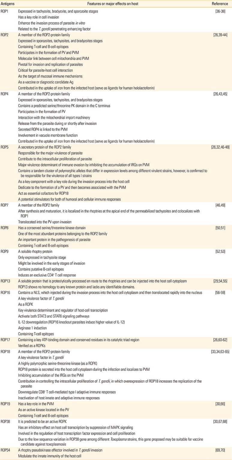
In this review, we have summarized the recent developments via various strategies for ROP-based vaccines in different mouse models. The specific features and main functions of some ROPs have been summarized in Table 1.
In this field, great advances in the development of T. gondii vaccination has been done such as the following:
  • - Several vaccine candidates have been evaluated from ROPs, particularly ROP1, ROP2, ROP5, ROP16, and ROP18.

  • - Novel genetic adjuvants and traditional adjuvants have been surveyed to induce a stronger cellular immunity.

  • - An increasing number of papers have examined the nanoparticles and microparticles as delivery system approaches to enhance a long-lasting protective immunity.

Mouse Models

Mice are being used frequently in experimental vaccine studies against toxoplasmosis as the main biological models to evaluate the outcome of acute and chronic infection before and post challenge with T. gondii strains. These little animals are also used for acute parasitemia and congenital toxoplasmosis or acquired infection. The reason is that toxoplasmosis in mice is histologically alike to that of men. On the other hand, ease of manipulating and cheap maintenance of them compared with larger animals such as livestock and domestic animals are other reasons for widely use of different mouse models [2171]. Furthermore, the immunology of mice is well characterized [72]. It has been shown nearly 99% homology between mouse genome and the human genome [73]. However, some differences exist between the immune systems and immune responses of men and mice [7274].
Type I strains of T. gondii is highly virulent in mouse models, while types II and III are extremely less virulent. The experimental mouse models have different major histocompatibility complex (MHC) haplotypes and different susceptibility to the parasite. For example, BALB/c (H-2d) mice are defined genetically with a low susceptibility to toxoplasmosis [75]. Also, C3H/HeJ (H-2k) and C57BL/6 (H-2b) mouse models with different genetic backgrounds are considered as intermediate and high susceptibility to T. gondii infection, respectively [7677].

DNA Vaccines

The DNA vaccination is a novel method that recruits the plasmid vector in order to transfer and expression of the target gene [7879]. These vaccines can be delivered through various routes, including intramuscular, subcutaneous, or mucosal. It is well known that DNA vaccination is a powerful strategy to provoke and elicit both specific cellular and humoral immune responses [80]. DNA vaccination as a robust method have become a major focus and they have many advantages in comparison to traditional vaccines in several parameters as follows [228081]:
  • - Design (more rapid design as well as can be rapidly isolated and cloned).

  • - Versatility (ease in adapting or improving plasmid sequence, capability to deliver multi-antigen vaccines into a host only with a single dose, ease in formulation with different adjuvants).

  • - Production (cost effective, ease of production, capable of large-scale production, appropriate protein folding for correct epitope expression).

  • - Transport (stability at room temperature and no need to cold chain).

  • - Safety (cannot revert to the pathogenic form, safer than live or attenuated vaccines).

  • - Immune responses (able to induce a long-lasting immunity, elicit efficient and specific humoral and cellular immune responses).

Following the injection, the naked DNA plasmid enter to the cell cytoplasm and express encoded proteins within the host cells [19808283]. Production of specific-IgG antibodies as one of the robust protective immune responses can prevent and inhibit from the attachment of T. gondii to its host cell receptors. Besides, it helps macrophages (MQs) to kill and eliminate the parasite and preventing reactivation [84]. Specific and strong cellular and humoral immune responses are enhanced during the course of T. gondii infection. Generally, the secretion of interferon γ (IFN-γ) from T cells as the adaptive cellular immunity has a crucial role in the controlling and restricting growth of the parasite in both acute and chronic infection stages. Also, this important cytokine, inhibit the reactivation of bradyzoites within dormant tissue cysts [8586]. It is well established that protection against toxoplasmosis generally is developed through both types of CD4+ and CD8+ T cells as cellular immunity arms. However, the role of CD8+ T cells and IFN-γ are evident to be more substantial to limit infection [1982]. It has been reported that DNA vaccination with ROP5, ROP9, ROP16, ROP17, ROP18, and ROP38 strongly increased the percentages of CD4+ T and CD8+ cells in immunized mice [475262678788].
DNA vaccination against T. gondii infection would induce a strong Th1 type immune response (predominance of IgG2a over IgG1) with increased secretion of IFN-γ and interleukin (IL)-2 inflammatory cytokines to confine toxoplasmosis [8586]. Noteworthy, previous studies have shown that DNA vaccination against toxoplasmosis with Th1-type immune response and significant values of IL-2 and IFN-γ (compared with controls), does not guarantee the desirable outcome in mice. It can be concluded that the immunogenicity can not necessarily predictive of brain cyst load and survival rates in mice post challenge [1946528283899091]. However, it should be mentioned DNA vaccinations generally trigger the activation and proliferation of both CD4+ and CD8+ T lymphocytes, along with increasing in essential specific antibodies for limit the infection [1936476782838788]. Chen et al. (2014) [52] showed that pVAX-ROP9 induce a mixed Th1/Th2 response with the predominance of IgG2a levels (as an indicator of Th1-type response) than IgG1 (as an indicator of Th2-type response). In this regard, in the mice immunized with pVAX-ROP9 the production of IFN-γ, IL-2, IL-4, and IL10 were significantly increased, compared with those groups that injected pVAX1 or phosphate buffered saline (p<0.05). Moreover, pVAX-ROP9 considerably enhanced the percentages of CD4+ and CD8+ T cells and prolonged the survival time in Kunming mice post intraperitoneally challenge with 1×10³ tachyzoites of the highly virulent RH strain (12.9±2.9 days, p<0.05). The authors noted that TgROP9 plasmid could be considered as a potent promising vaccine candidate for acute toxoplasmosis [52]. ROP18 as a main virulence factor of T. gondii, involved in the controlling of intracellular proliferation of parasite [3465]. For the first time, this protein has been shown to be a promising vaccine candidate by Yuan et al. (2011) [88]. Briefly, intramuscular vaccination of Kunming mice with the plasmid construct pVAX-ROP18 enhanced both humoral and cellular Th1-biased (predominance of IgG2a levels over IgG1) immune responses and increased activation of CD4+ and CD8+ T cells. Also, pVAX-ROP18 vaccination lead to higher survival time (27.9±15.1 days, p<0.05) than those mice in control groups when challenged with 1×10³ tachyzoites of RH strain [88]. More examples of immunization experiments with DNA vaccines (single antigens) against T. gondii in different mouse models has been listed in Supplementary Table 1.
Accumulating evidence has been shown that multiantigenic DNA vaccinations could overcome the single antigen limitation and enhance the protective immunity against toxoplasmosis either survival duration time and/or brain cyst load [3646648790929394]. More details can be found in Supplementary Table 2. For example, Chen et al. (2015) [47] evaluated the ROP5 and GRA15 antigens alone or in combination. The Kunming mice vaccinated intramuscularly with pVAX-ROP5 or pVAX-GRA15 and then two weeks after the third inoculation, were challenged with 1×10³ tachyzoites of RH strain (type I) intraperitoneally for acute infection and 10 tissue cysts PRU strain (type II) orally for chronic infection. The immunized mice with alone antigen showed dramatically IgG2a titers in sera, high production of IL-12 p40, IL-12 p70, IFN-γ, and IL-2 production as Th1-type immune responses, augmentation of cell-mediated cytotoxic activity with high frequencies of IFN-γ secreting CD8+ T cells, increased survival time (19.4±4.9 and 17.8±3.8 days for pVAX-ROP5 and pVAX-GRA15, respectively) and reduction of brain cysts (57.4% and 65.9% for pVAX-ROP5 and pVAX-GRA15, respectively), compared with control groups. As it was predictable, co-administration of these antigens, boosted the protective efficiency and elicited the cellular and humoral immune responses, compared to single antigen vaccines. Furthermore, significantly prolonged the survival time (22.7±7.2 days) and reduced the number of brain cyst load (79%) [47].
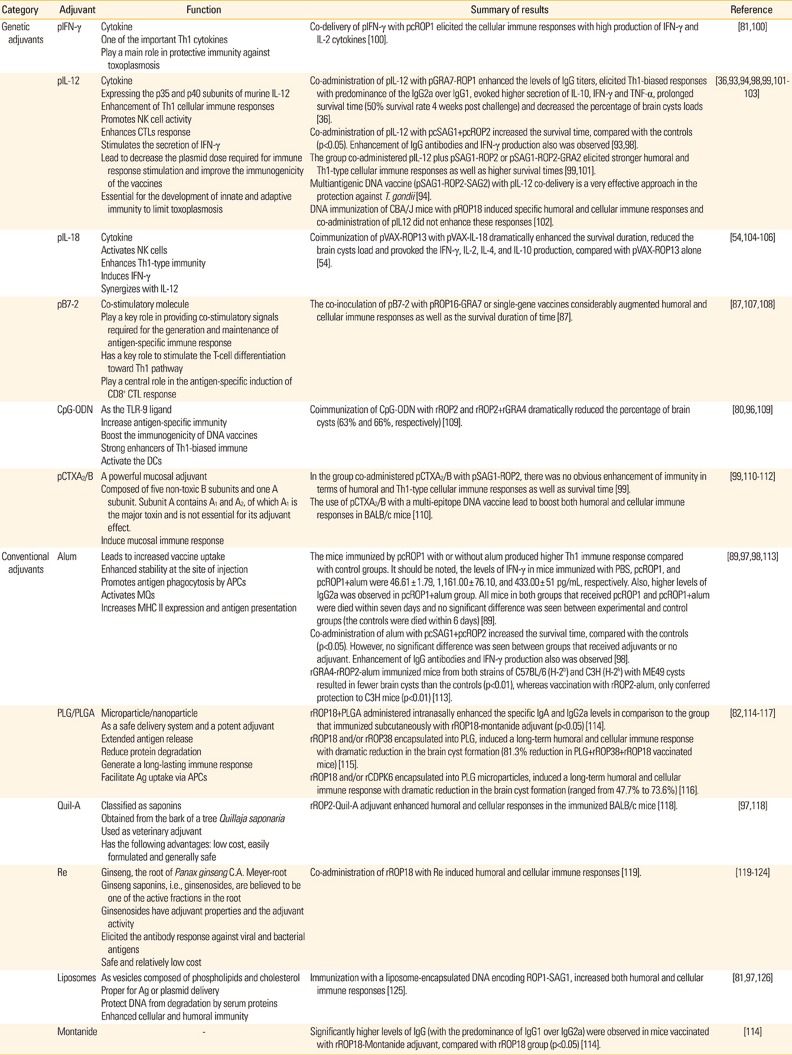
In regard to some delivery problems for DNA vaccines, their immunogenicity is occasionally confined. Several factors are affecting in the immune efficacy of DNA vaccines such as dosage and delivery routes. Albeit, 10 to 100 µg are routinely inoculated intramuscularly in mice [5780]. For instance, the intramuscular injection of DNA vaccines is poorly distributed, inefficiently expressed and quickly degraded, thereby evoke relatively moderate humoral and cell mediated immune responses [9596]. Besides, the degradation of plasmid DNA by lysosomes and DNAses may reduce the expression of plasmid DNA in small experimental animals as well as may affect DNA expression in men [95]. There are several strategies for adjuvanting plasmid DNA to augment their immunogenicity. An adjuvant could boost DNA delivery as well as enhance either the magnitude or time of DNA expression [9697]. Moreover, by recruiting the immune cells to the site of injection, increases the immunostimulatory properties of plasmid. It is speculated, those adjuvants that enhance the magnitude/duration of plasmid DNA expression might help the uptake of DNA into host cells or increase taken up by professional antigen-presenting cells (APCs) as well as protect plasmid against degradation by DNAses [80819697]. Since the immunogenicity of DNA vaccines to stimulate the specific immune responses are often weak, recruiting cytokines and costimulatory molecules as genetic adjuvants or alum as non-genetic adjuvants as well as liposomes as vehicle adjuvants would be enhanced and modulated these immune responses [367880818793979899]. Meanwhile, during recent years to enhance the immunogenicity of DNA vaccines, various strategies have been employed to conquer the present drawbacks and obstacles such as progress and improvement in plasmid design, antigen codon optimization to increase the expression of proteins, utilization of molecular and/or traditional adjuvants, co-expression of molecular adjuvants, electroporation, and prime-boost or combination immunization strategies [8081]. Some examples of adjuvants in vaccination experiments against T. gondii infection in mouse models have been summarized in Table 2 [100101102103104105106107108109110111112113114115116117118119120121122123124125126].
Liposomes as carriers are composed of a diverse cholesterol and phospholipids, can encapsulate or bind plasmid DNA as well as they act as a suitable vehicle adjuvant [9697]. Liposomes also help the entrace of DNA into cells through interaction with the lipid bilayer of the cell membrane and protect DNA from extracellular degradation by serum proteins [126]. Notably, following the formulation of DNA vaccine into liposomes, specific humoral and cellular immune responses considerably could elicit [8197126]. In this context, Chen et al. [125] highlighted that the approach of vaccination with a liposome-encapsulated DNA encoding ROP1-SAG1 of T. gondii, has potential capability to augment the humoral and cell mediated immune responses in BALB/c mice.
Genetic adjuvants have become recently as attractive tools to augment the protective efficacy of DNA vaccine [78]. In general, they are expression plasmid vectors that encoded biologically active molecules such as cytokines (IFN-γ, IL-12, IL18, etc.), chemokines (regulated upon activation normal T-cell expressed and secreted [RANTES]), co-stimulatory molecules (B7-1, B7-2, etc.), etc. (A2/B subunits of cholera toxin [CTXA2/B]) [197882]. It should be noted, genetic adjuvants can be either encoded on a separate vector, or expressed on the same vector as the antigen and thereafter co-administrated with the vaccine [7880819799]. As noted, one of the best approaches to augment the immunogenicity of DNA vaccines is the co-inoculation of plasmids encoding cytokines that has some advantages such as simplicity, ease of cloning procedure, and cost benefits. Since the cytokine is expressed and acts at the site of antigen expression, thus the risk of toxicity of systemically administered cytokines will be minimized [81]. In 2001, Guo et al. [100] evaluated the efficacy of pcIFN-γ as genetic adjuvant in the outcome of a DNA vaccine encoding ROP1 with or without adjuvant. They reported that the proliferation response of spleen T cells, natural killer cells (NK cells) killing activity, the serum concentrations of IFN-γ, IL-2, and nitric oxide in BALB/c mice vaccinated with pcROP1+pcIFN-γ regimen were markedly higher than in those immunized with pcROP1 alone, however, no obvious difference was shown in terms of IgG antibody values between the two groups. The authors concluded that pcIFN-γ is able to considerably elicit the cellular immune responses [100]. The pivotal role of IL-2 for the growth, multiplication, and differentiation of T lymphocytes is well known, as well as contributed in the prevention of parasitic invasion process combined with IFN-γ [85115].
IL-12 is a pivotal proinflammatory cytokine for control and restriction of acute and chronic toxoplasmosis [127]. This critical cytokine is secreted by MQs and dendritic cells (DCs) during antigen stimulation and has a crucial role in the activation of NK cells and development of a Th1-biased immune responses and IFN-γ production which is indispensable for limit to T. gondii [859398127]. IL-12 has been frequently investigated as a proper genetic adjuvant against toxoplasmosis [3693949899101102]. For instance, Quan et al. (2012) [36] reported that co-administration of IL-12 plus pGRA7-ROP1 in BALB/c vaccinated mice leads to increase survival time, in which 28 days post challenge with the lethal dose, 50% of them were survived. Moreover, the number of tissue cysts in the brain significantly reduced [36]. At another study, Khosroshahi et al. (2012) [98] compared the effect of IL-12 and alum as genetic and non-genetic adjuvants, receptively, on the efficiency of cocktail DNA vaccine that encoded psSAG1+psROP2. They found that co-administration of IL-12 and alum with psSAG1+psROP2 enhanced the survival time of experimental groups than controls (p<0.05). Moreover, increased levels of IgG antibody, high levels of IFN-γ and low levels of IL-4 were also observed, compared with control groups. Noteworthy, no significant difference was found between IL-12 and alum adjuvants to induce immune responses [98]. The same results also was found by Zhang et al. (2007) [93] on a multi-antigenic DNA vaccine encoding pSAG1-ROP2 combined with pIL-12.
IL-18 is an important cytokine with diverse functional roles. This cytokine potentially enhances the NK cell activity and also synergize with IL-12 to stimulate NK cell production of IFN-γ by T cells [104105]. Alike to IL-12 cytokine, the predominant role of IL-18 is the boost of immune response toward Th1-type responses. For the mentioned reason, it can confer as a candidate adjuvant to be involved in the Toxoplasma resistance [104105106]. For this purpose, Wang et al. (2012) [54] designed an investigation to evaluate the immunogenicity and immunoprotection of TgROP13 by constructing pVAX-ROP13 alone or with IL-18 as genetic adjuvant. They reported that co-immunization of pVAX-ROP13 with pIL-18 in Kunming mice dramatically (p<0.05) enhanced the survival duration, compared with pVAX-ROP13 alone (32.3±2.7 and 24.9±2.3 days, respectively) against highly virulent RH strain (type I). The rate of reduction of brain cyst load in the mice immunized with pVAX-ROP13 and pVAX-ROP13+pVAX-IL-18 was 39.82% and 66.03%, respectively (p<0.05), after challenge with PRU strain (type II). Besides, co-delivery of pVAX-IL-18 plus pVAX-ROP13 considerably provoked the secretion of IFN-γ, IL-2, IL-4, and IL-10 cytokines, compared with the group that injected pVAX-ROP13 alone. The above-mentioned valuable findings suggest that ROP13 triggered strong humoral and cellular responses against the parasite and can be considered as a potentially efficient vaccine candidate. Also, the use of pVAX-IL-18 as a genetic adjuvant successfully boosted the protective immunity in terms of humoral and cellular responses, prolonged the survival time as well as with fewer brain cysts [54]. Thus, it seems that IL-18 cytokine could be extensively effective as Th1 adjuvant for future studies on other ROPs.
As noted, careful selection of a suitable genetic adjuvant would enhance the protective immunity of DNA vaccines and perhaps is a pivotal strategy to induce an appropriate response [78808197]. B7 costimulatory molecules such as B7-1 (CD80) and B7-2 (CD86) have an important role in providing costimulatory signals which are extremely necessary for the generation and maintenance of antigen-specific immune response. Hence, if a plasmid which expressing B7-1 or B7-2 as costimulatory molecules co-injected with a plasmid DNA vaccine, it is anticipated that can highly stimulate vaccine-elicited specific antibodies and CD8+ responses [87107108]. It has been shown that co-administration of plasmid encoding B7-2 was more efficient than that encoding B7-1 for the generation of antigen-specific cytotoxic T lymphocytes (CTLs) response [128129]. In this regard, Liu et al. (2014) [87] constructed a multiantigenic DNA vaccine expressing the ROP16 and GRA7 (pROP16, pGRA7, and pROP16-GRA7) antigens and a plasmid encoding murine costimulatory molecule B7-2 (pB7-2) as a genetic adjuvant and evaluated the protective efficiency of these antigens with or without pB7-2 in Kunming mice against acute toxoplasmosis. They showed immunization with pROP16-GRA7 produced higher levels of IgG titers (predominance of IgG2a over IgG1), increased the secretion of IFN-γ, enhanced the percentage of CD8+ T cells and median survival times, compared with those of mice received pROP16 or pGRA7 and those in control groups after lethal challenge with 1×10³ tachyzoites of RH strain. Noteworthy, the formulation of pB7-2 with multiantigenic DNA vaccine (pROP16-GRA7) or single-gene vaccine (pROP16 or pGRA7), significantly boosted humoral and cellular immune responses as well as the survival duration of time. The authors proposed that B7-2 would be a feasible and promising genetic adjuvant to increase protective immunity, however, further studies are required in future [87].

Recombinant Protein Vaccines

In several past years, constant efforts of investigators have made considerable advances to elucidate the different aspects of the cell and molecular biology of T. gondii [25262932334549555670130]. These attempts thereby provided great impetus to the present focus on vaccine development, according to the defined subcellular components of the parasite. Among the main identified T. gondii antigens, the ROPs appear as putative vaccine candidates that are vital for invasion procedure as well as survival within host cells [2482]. Recombinant subunit vaccines are one of the current approaches that may offer an alternative way for the development of vaccine candidates against toxoplasmosis in humans and animals. These vaccines are able to elicit systemic humoral and cell mediated responses as well as being apt for large-scale production [82].
ROP5 is responsible for the major virulence of parasite and involved in the intracellular proliferation. This protein significantly decreases the accumulation of immunity-related GTPases in parasitophorous vacuole membrane (PVM), thereby maintains the PVM integrity [2632]. To evaluate the protective efficacy of recombinant form of ROP5 (rROP5), Zheng et al. (2013) [48] performed a study to understand this issue. They vaccinated the BALB/c mice subcutaneously with 100 µg protein+Freund's complete adjuvant in first immunization and 100 µg protein+Freund's incomplete adjuvant (FIA) in second and third immunization. Enhanced IgG titers (p<0.01), mixed Th1/Th2 responses with the predominance of IgG2a over IgG1 (p<0.05), high production of IFN-γ, IL-2, IL-4, and IL-10 cytokines (p<0.05) as well as prolonged survival time (p<0.05) was observed as the outcome of vaccination, compared with control groups. Besides, co-injection of rSAG1 with rROP5 boosted the protective efficiency. As it is clear, immunization with compound polyvalent vaccine has better efficacy than a single antigen. The authors proposed evaluation of the brain cyst burden in both immunized and control groups using low virulence strains of parasites can helpful in future studies [48]. Noteworthy, IL-2 in combination with IFN-γ play a key role in preventing parasitic invasion [85116].
The use of traditional and molecular adjuvants has become attractive recently because of their potential ability in eliciting specific and long-lasting protective immunity [97]. Also, admirable attempts have been made to introduce novel delivery systems to boost protective efficiency [8297]. Aluminum salt-based adjuvants (alum) have been utilized as vaccine adjuvant since 1926 and is the most common used vaccine adjuvant in men. They are widely used in various vaccine formulations with some advantages such as enhance stability and immunogenicity of antigen following the adsorption to the alum particles [97]. It is well known that alum increases the expression of MHC II and adhesion or costimulatory molecules, including intercellular adhesion molecule 1, lymphocyte function-associated antigen 3, and CD40. Also, it has been reported that alum absorption enhances antigen uptake at the site of injection and augment the antigen phagocytosis by professional APCs such as DCs, MQs, and B cells as well as induce the production of some chemokines including the chemokine (C-C motif) ligand 2, the chemokine (C-X-C motif) ligand 1 and CCL11 in mice [97131]. Noteworthy, alum is unable to induce a strong cell mediated response (Th1 or CTL) that are critical to limit and elimination of intracellular parasites [97]. In 2004, a paper by Martin et al. [113] was published that were evaluated the efficacy of alum adjuvant on the immunogenicity of rROP2 and rGRA4 in C57BL/6 (H-2b) and C3H (H-2k) mouse models. These experimental models have different MHC haplotypes and different susceptibility to the parasite. Vaccination with rROP2-rGRA4-alum regimen resulted significantly reduction (p<0.01) in brain cyst load after oral challenge with 20 (sublethal dose) ME49 tissue cysts. While, immunization with rROP2-alum, only was considerably reduced the number of brain cysts in C3H (H-2k) mice (p<0.01). They concluded that use of alum adjuvant could be used in vaccination against T. gondii infection [113].
Oligodeoxynucleotides contained CG motifs (CpG-ODN) were illustrated that could increase antigen-specific immunity to protein vaccines in a variety of hosts ranging from mouse models, humans and various veterinary species [132]. These adjuvants are strong enhancers of Th1-biased immune responses via activation of TLR-9 dependent cascades [80]. Nevertheless, the formulation of CpG-ODN adjuvant as a vaccination approach against T. gondii has been rarely investigated [109110133134135136] and investigations, according to CpG-ODN+ROPs of T. gondii has very limited explored [109]. Lately, were shown co-inoculation of rROP2+CpG-ODN elicit a strong humoral and Th1-biased immune responses with the predominance of IgG2a over IgG1 and enhanced the production of IFN-γ and IL-10 and negligible values of IL-4 cytokines. The brain cyst burden significantly reduced in the C3H/HeN (H-2k) mice vaccinated with rROP2+CpG-ODN (63%, p<0.001) and rROP2+rGRA4+CpG-ODN (66%, p<0.001) following a non-lethal challenge with 20 tissue cysts of Me49 (type II) strain orally. The authors have declared CpG-ODN is a potential adjuvant which can induce strong Th1-type immune responses, however, they proposed more studies are needed because of the different pathogenicity of T. gondii strains [109].
Due to easily proteolytically degradation of recombinant proteins that entails, the more frequent immunizations, a favorable delivery system to protect from degradation would be indispensable [137]. More recently, some studies have focused on polylactide-co-glycolide (PLG), a biodegradable and biocompatible polymer as a safe delivery system for antigens. PLG can extend the protein releasing period to induce a long-lasting immune response and reduce the protein degradation, thereby, increase the uptake of antigen and its presentation by APCs [115117]. PLG also considered as a potent vaccine adjuvant as well as encapsulate the recombinant subunit vaccines and can maintain their antigenicity to elicit an efficient protection [82114115116117]. More recently, rROP18 and rROP38 were encapsulated into PLG microparticles to prolong the antigenicity. Vaccination of Kunming mice with rROP18 and rROP38 entrapped into PLG enhanced significantly humoral and cellular immune responses in terms of total IgG titers (p<0.01), IgG2a subclass (p<0.01), IFN-γ cytokine (p<0.01), and mixed Th1/Th2 immunity responses (but bias to Th1) as well as reduction of brain tissue cysts (p<0.01). The use of mixed antigen (rROP38+rROP18+PLG) boosted the protective immunity. For example, the percentage of CD4+ and CD8+ T cells were enhanced with the brain tissue cyst reduction of 81.3%. The authors have declared that PLG polymer microparticles with preserving the protein immunogenicity for extended duration, could be a promising novel adjuvant, however, further studies are required [115].
As we all know, T. gondii infects a wide spectrum of hosts, mainly via the mucosal surfaces of the digestive tracts. Also, this opportunistic agent can attack all nucleus cells and easily spread throughout the body. Hence, the development of potential vaccine candidate that capable to augment systemic and mucosal immunity has of great priority [138139]. The nasal route of vaccination as a non-invasive and needle-free strategy has high priority in immunization investigations [140]. The i.n route is superior than the oral route because of requires fewer antigen and less proteolytic activity in the nasal cavity. Besides, i.n route could induce both systemic and mucosal protective immunity to recombinant protein antigen [112141]. Increased stimulation of IgG and IgA antibodies have been frequently demonstrated in papers following nasal administration [102112118138140]. The specific IgG antibodies have a key role in restriction of T. gondii infection and involved in the activation and promotion of the classical complement pathway, MQs phagocytosis and block invasion, other vital roles against intracellular parasites [84]. Many evidence showed specific secretory immunoglobulin A (SIgA) has a key role in mucosal surfaces and acts as the first line defense against several infectious agents that colonize mucosal tissues such as Toxoplasma [138139]. In this context, previously, rROP2+Quil-A adjuvant and rROP2-GRA5-GRA7+cholera toxin (CT) adjuvant have been tested as immunogens to vaccinate BALB/c mice intranasally and shown acceptable responses including increased IgG and IgA titers. Moreover, rROP2+Quil-A elicited a significant lymphocyte proliferation response and rROP2-GRA5-GRA7+CT lead to 58.3% protection against brain cyst formation (after oral challenge with 50 cysts from VEG strain), compared with the control group (p<0.05) [112118]. More recently, Wang et al. (2014) [138] were reported that nasal immunization of BALB/c mice, elicited IgG antibody production (p<0.01), promoted mixed Th1/Th2 immune response (p<0.05) with the predominance of IgG2a over IgG1, increased production of IFN-γ (p<0.01), IL-2 (p<0.01), and IL-4 cytokines (p<0.05), enhanced SIgA antibody titers in the nasal, vaginal and intestinal washes of rROP17-vaccinated mice (p<0.01), increased survival rate (75% protection 30 days post challenge with 4×10⁴ tachyzoites of RH strain orally, p<0.01) and reduced the liver and brain parasite burden (p<0.05), compared with the control group. The authors remarked that intranasal vaccination of rROP17 can strongly provoke both systemic and mucosal immunity and would be considered as a potential vaccine candidate against T. gondii infection [138]. The examples of vaccination with protein vaccines against T. gondii in different mouse models have been summarized in Supplementary Tables 3 and 4.

Epitope Mapping and Epitope-Based Vaccines

Recognize and construct novel vaccines for prevent and control of toxoplasmosis in both humans and animals is still remains a great challenge for researchers to select highly potential protein sequences as the ideal antigens [1982]. Bioinformatics is an interdisciplinary science that analyzes the biological data by using defined technologies and algorithms from mathematics, statistics, computer sciences, physics, medicine and biology [142]. Bioinformatics had many advantages than the traditional methods including: affordable and required low-cost, effective, satisfactory precision and accuracy and required lower times [142143]. Recently, this novel science became popular and widely employed for various purposes such as predict protein structures, functions, biological characteristics, and epitopes as well as the design of new vaccines [51130142143]. Particularly, prediction of epitopes is an indispensable tool in the immunogenicity design and reverse Vaccinology [66143]. Several papers were found through database searching about ROP-based vaccines that analyzed the potential B and T cells epitopes using bioinformatics online servers to introduce novel vaccine candidates [2739515966110144145146147]. However, for some of them there is a lack of confirmation of the protective efficacy in mouse models. Since the immunogenicity of the predicted sequences should be approved in suitable animal models, therefore, both in silico and in vivo approaches are required to evaluate the potency of protein as vaccine candidates [82130].
The life cycle of T. gondii is very complex, indicating the presence of a plurality of antigenic epitopes. It has been proven that vaccination with stage-specific antigens only lead to stage-limited protection [130148]. Accordingly, immune responses against T. gondii antigens that are express in various stages of parasite life cycles, are presumably more efficient and such vaccines likely confer increased survival time and lower brain cyst load than control groups. Thus, vaccination with compound polyvalent vaccines probably to be more efficient over a single antigen. Bioinformatics method helps researchers to predict the highly potential B and T cell epitopes [5982110145147]. Recently, the use of multi-epitope vaccines has become popular as a novel strategy in vaccine design against the opportunistic agent of toxoplasmosis, T. gondii. An ideal epitope-based vaccine should contain both B and T-cell epitopes that are vital for eliciting antibody responses and induce CTL responses, respectively [130]. In this context, for the first time, in 2008 Cong et al. [110], constructed a DNA vaccine encoding multi-epitope gene (MEG) including several putative immunodominant T-cell and B-cell epitopes of T. gondii SAG1 (59–67), SAG1 (246–256), GRA1 (176–186), ROP2 (199–216), and GRA4 (235–245) and CpG motif, with or without CTXA2/B as a genetic adjuvant and then tested in BALB/c mice. After immunization, increased levels of IgG antibody in the mice immunized with pVAX1-MEG (p=0.009) and pVAX1-MEG-CTXA2/B group (p=0.006) were recorded, compared than negative controls. Furthermore, in subsets of IgG, the predominance of IgG2a over IgG1 (especially in mice immunized with pVAX1-MEG-CTXA2/B) was observed. In addition, IgG2a levels in the group vaccinated with pVAX1-MEG-CTXA2/B were markedly higher compared with a pVAX1-MEG immunization regimen (p<0.001), whereas similar concentrations of IgG1 titers existed between these groups (p=0.834). CTL activity was enhanced, mainly in mice immunized with pVAX1-MEG-CTXA2/B. After cytokine assay, the results showed pVAX1-MEG-CTXA2/B immunized mice had higher amounts of IFN-γ and IL-2 than pVAX1-MEG group (p=0.009). Eventually, these results lead to prolonged survival time (p<0.05 and p<0.001 in mice immunized with pVAX1-MEG and pVAX1-MEG-CTXA2/B, respectively) following the challenge of mice with 1×10³ tachyzoites of highly virulent RH strain, compared with three control groups. pVAX1-MEG-CTXA2/B immunization regimen resulted 20% survival rate in this group, while all mice in other groups succumbed. As evident, all of the above-mentioned findings suggests that the formulation of CpG motif and CTXA2/B as adjuvants in combination of this DNA vaccine, considerably boosted the protective efficacy against acute infection of T. gondii [110]. Until now, toll-like receptor (TLR-1–TLR-13) genes have been discovered in men that have critical roles in the innate immune system [80149]. Oligodeoxynucleotides contained CG motifs (CpG ODN) as the TLR-9 ligand, was shown to be effective to boost the immunogenicity of DNA vaccines [8096]. CpG motifs are also able to activate the DCs and stimulate the production of some cytokines from them such as type I IFN from CD11c+B220+plasmacytoid DCs or IFN-γ and IL-12p70 from CD11c+CD8+B220DCs [96]. Examples of vaccination experiments with epitope-based vaccines against T. gondii in mouse models have been embedded in Supplementary Table 5.
Previously, the antigenic characteristics of ROP19 and SAG1 were analyzed and compared together using bioinformatics databases. For this purpose, the Immune Epitope Database (IEDB) online service was employed to predict the T-cell epitopes and linear B-cell epitopes of the antigens. The DNASTAR software showed that ROP19 is superior to SAG1 in terms of antigenic index and surface probability. The authors claimed that ROP19 had good linear B-cell epitopes compared to SAG1. Additionally the 50% inhibitory concentration (IC50) values of peptides binding to the MHC class II molecules of ROP19 were also predicted and lower IC50 values (low percentile rank=high level binding) were estimated for ROP19 indicating that ROP19 has viable T-cell epitopes [66]. These researchers, were performed a similar study with same design on ROP54 and SAG1. The linear-B cell epitopes analysis showed the superiority of ROP54 than SAG1 in terms of antigenic index and significant surface probability. Besides, Th-cell epitopes on ROP54 also were analyzed by the bioinformatics methods to predict the capability of binding to MHC class II molecules. Briefly, the minimum percentile ranks for 4 different MHC II alleles were chosen and listed on SAG1 and ROP54. Overall, the IC50 values for ROP54 were estimated lower than SAG1 (low percentile=high binding) which indicates better Th-cell epitopes. The authors concluded that the bioinformatics prediction of ROP54 sequence on linear-B cell epitopes and Th-cell epitopes revealed positive results with high potentiality to become an excellent vaccine candidate for toxoplasmosis [144]. ROP54 as a novel rhoptry protein pseudokinase is associated with the PVM after being injected into the host cell [70144].
In 2016 Zhou et al. [27], for the first time performed a new survey on ROP48 with multiple bioinformatics approaches to predict some characteristics of the protein sequence in terms of physical and chemical features, epitope, and topological structure. They demonstrated that ROP48 was mainly located in the membrane. Moreover, several positive B- and T-cell epitopes with favorable flexibility and surface probability also were identified, which indicated positive antigenicity, suggesting this protein could be a potential DNA vaccine candidate against toxoplasmosis for future studies [27]. Camejo and colleagues reported that deletion of ROP48 in a type II strain (Δrop48 parasites) did not show significant affect on the in vitro growth or virulence in female C57BL/6 J mice [23].

Vaccines Based on Live-Attenuated Vectors

Since Toxoplasma is an obligatory intracellular protozoa, the use of live, attenuated vectors (bacteria or viruses) as vehicles to deliver and express the parasite antigen, can mimic the intracellular niche of T. gondii [1983]. It has been shown that this immunization strategy against T. gondii infection capable to provoke a strong humoral and cell mediated immune responses lead to high protection or complete protection in some studies, because of its intrinsic adjuvant properties and/or mimicry of a natural infection [147150151152153154]. These vaccine types can also be administered intramuscularly, intraoral, intranasal, subcutaneous, and intravenous and induce effective immune responses and protection in terms of enhancing the survival time and/or reduce the brain cyst burden [146147].
Bacille Calmette-Guerin (BCG), an attenuated strain derived from Mycobacterium bovis, has been employed widely during recent decades as a live vaccine against tuberculosis and has peculiar intrinsic adjuvant properties thereby develop the cellular type responses within host [155156]. Moreover, some advantages are cost effectiveness, ease of production, relatively thermostable as well as is unable to revert toward virulent phenotype [153157]. Both M. bovis BCG and T. gondii are obligatory intracellular microbes, thus, the use of recombinant BCG as a foreign antigen delivery system would be very suitable for the vaccine development against toxoplasmosis [153156]. Immunization with recombinant M. bovis BCG expressing TgROP2 (BCG/pMV262-ROP2) was found to elicit both humoral and cellular immune responses and increase survival rate post challenge intraperitoneally with 5×10² tachyzoites of RH strain in BALB/c mice (p<0.05). The authors concluded that M. bovis BCG is an adequate vector to express TgROP2 antigen. Nevertheless, they proposed further studies are required to evaluate the protective efficiency of BCG/pMV262-ROP2 in other mouse models such as C57BL/6 and C3H mice [153].
Vaccinia virus as a prominent vehicle increase the antigen presentation to the immune system of the host and is a powerful immunostimulant against those antigens that normally not identified by the immune system [154158]. A study showed that a new recombinant modified vaccinia virus Ankara (MVA) expressing TgROP2 (MVA ROP2) induced mixed Th1/Th2 immune response (predominance of IgG2a over IgG1) and prolonged the survival duration (11 days vs. 8–9 days, p=0.04) post challenge with 300 tachyzoites of RH strain in female Swiss mice. However, the brain cyst load do not differ between experimental and control mice. Interestingly, all mice vaccinated with ts-4 strain of T. gondii survived following challenge. The authors remarked that MVA ROP2 generated an effective immune response which lead to delaying the mortality time [154].
It is well known that the use of recombinant viral vectors has the excellent ability to elicit effectual expression of the foreign antigens, thereby, help the presentation and stimulation of humoral and cellular immune responses [159]. For example, it has been reported that adenoviral vectors are safe and efficient for transgene expression in vivo as well as having intrinsic adjuvant properties which lead to activation the innate immune response through TLRs and nod-like receptors [160161]. Adenoviruses (Ad) are considered as popular vaccine vectors and have been used extensively, because of their powerful capability to provokation the T-cell mediated immunity [151152]. Since, there is frequently pre-existing immunity against the classically human adenovirus type 5 (AdHu5), alternatively canine adenovirus type 2 (CAV-2) has been suggested as vectors for human gene transfer [162]. Besides, the use of CAV-2 has the following advantages: well-characterized biology of CAV-2, ease of genetic manipulation, suitable for gene transfer into the central nervous system, able to induce strong protective immunity of humoral and cellular immune responses [152163]. Thus, currently CAV-2 is appointed as one of the most applicable non-human adenoviruses for vaccine vector purposes [151152]. In this case, Li et al. (2016) [151] constructed a novel recombinant CAV-2 carrying TgROP16 (CAV-2-ROP16) and then evaluated the immune response and survival status of BALB/c mice. CAV-2-ROP16 was able to elicit significantly both humoral and cell mediated responses (mixed IgG1 and IgG2a levels with the predominance of IgG2a titers, p<0.05) and increased production of IFN-γ, IL-2, and IL-4 (p<0.05). Furthermore, the enhanced survival rate (25% protection 80 days after challenging with 1×10³ tachyzoites of RH strain) was observed, compared with control mice that died within 7 days (p<0.05). They showed this system could markedly enhance the protection with eliciting humoral and cell-type immune responses [151]. The same authors were demonstrated that CAV-2-ROP18 has also been potentially capable to induce the same immune response with CAV-2-ROP16. Briefly, CAV-2-ROP18 immunization elicited a strong IgG antibody response (p<0.05), increased levels of a mixed IgG1 and IgG2a (p<0.05) with the predominance of IgG2a production, enhanced splenocyte proliferation (about 21-fold than control groups, p<0.05), enhanced production of IFN-γ, IL-2, and IL-4 cytokines (p<0.05), increased CTL activity in Kunming mice (p<0.05), and elevated numbers of CD4+ T and CD8+ T cells (p<0.05). The immunized mice with CAV-2-ROP18 showed 40% protection (60-day post infection intraperitoneally with 1×10³ tachyzoites of RH strain), while all control mice died within seven days (p<0.05). Besides, 57.3% reduction of brain tissue cyst burden was recorded following challenge intragastrically via oral gavage with 5 cysts of PRU strain (genotype II) (p<0.05). The authors declared that the potential utilization of a CAV vector carrying the TgROP18 gene in the development of a useful vaccine against chronic and acute toxoplasmosis in future investigations [152]. It should be noted in addition to cell-type immune responses, humoral immunity with high production of antigen-specific IgG antibodies are vital to limit the T. gondii invasion by preventing the attachment of parasites to host cell receptors [84]. Furthermore, CD8+ T cells, especially in synergy with CD4+ T cells are very critical for the control of toxoplasmosis [152164]. Noteworthy, the safety concerns and hazards was outlined regarding the use of live or attenuated vectors and need the more consideration during their development in the future that must be further investigated [165]. Supplementary Table 6 listed the examples of immunization with live-attenuated vectors expressing T. gondii antigens in mouse models.

Prime-Boost Strategies

The prime-boost approaches such as DNA prime/viral vector boost (i.e. using Adenovirus, fowlpox, vaccinia virus, etc.), DNA prime/protein boost, and protein prime/DNA boost have been shown extensively to be an efficient strategy to induce both cellular and humoral immune responses against some micro-organisms like human immunodeficiency virus (HIV), Plasmodium, Leishmania, T. gondii, etc., which would further provide a foundation for the development of appropriate vaccine candidates [81145166167168169170]. Homologous prime-boost approach involves the similar formulation employed in both the prime and boost regimens, while heterologous prime-boost strategies contains different formulations used in more than one immunization [81169]. Notably, the interval between prime and boost is extremely important for vaccine response and excellent efficacy. In addition, the arrangement of vaccination may influence the outcome of prime-boost strategies [80171]. Some papers suggested that heterologous prime-boost regimens are more effectual than the homologous prime-boost [169172]. Heterologous prime-boost strategy predominantly uses a DNA or a viral vector for priming and a protein-based vaccine for boosting [172].
It is well established that different vaccination strategies lead to different immune response. For instance, as noted, heterologous prime-boost regimens strongly elicit both of humoral and cell-mediated immunity against an antigen using each delivery system individually. In the other hand, subunit vaccines often provoke a predominant humoral immune response, whereas DNA vaccines or recombinant live vector-based vaccines mainly elicit an efficient cellular immunity [169]. Li et al. (2011) [168] reported that a DNA prime/protein boost immunization based on ROP2 and SAG1 (pcROP2-SAG1/rROP2-SAG1+FIA), pcROP2-SAG1 and rROP2-SAG1 formulations could elicit similar humoral and cellular immunity against toxoplasmosis. However, the BALB/c mice immunized with the rROP2-SAG1 enhanced humoral response (IgG specific antibodies with the predominance of IgG1 over IgG2a), slightly increased IFN-γ production and more vigorous specific lymphoproliferative responses, compared with other antigen formulations [168]. In another study, Yin et al. (2015) [145] designed an excellent and comprehensive investigation to evaluate the efficacy of a T. gondii vaccine encoding all stages of the parasite antigens. At first, main antigens present in sporozoite, tachyzoites, and bradyzoite stages were predicted for CD8+ T cell epitopes conserved regions based on their binding affinity to human leukocyte antigen (HLA-A*02, HLA-A*03, and HLA-B*07) and H2 (H2-Ld, H2-Dd, and H2-Kd) supertype molecules using bioinformatic algorithms from IEDB online service. Then protein fragments of SAG3101–144, ROP18347–396, MIC6288–347, GRA7182–224, MAG158–125, BAG1156–211, and SPA142–200 were selected and ubiquitin-conjugated multistage antigen segments (UMAS) plasmid DNA were constructed. They reported that among different formulations of prime/boost regimens (DNA/DNA, Ad/Ad, DNA/Ad, and Ad/DNA), priming with DNA and boosting with Ad-UMAS elicited higher values of specific IgG (predominance of IgG2a over IgG1) and higher production of IFN-γ (1,691±35.18 pg/mL) and IL-2 (561±19.68 pg/mL) cytokines were achieved, compared with p-UMAS or Ad-UMAS injection alone (p<0.05). Also p-UMAS prime/Ad-UMAS boost regimen significantly increased survival rate (67%, 28 days post challenge intraperitoneally with 1×10³ tachyzoites of RH strain) compared than controls which died within 8–10 days and reduced the brain cyst load (p<0.01) [145]. Ubiquitin is a 76-amino-acid peptide which documented increase DNA vaccine responses against targeted antigen in the adjuvant setting [145]. Conjugating ubiquitin to a DNA construct was determined to increase the proteasome dependent degradation of endogenously synthesized antigens, thereby elicit cellular immune responses toward the conjugated antigen in animal models [145146173174]. Heterologous vaccination oftentimes shows a powerful synergistic effect in comparison to homologous regimens [80]. Some studies on vaccines against hepatitis B virus [175], hepatitis C virus [176], HIV [177], and T. gondii [145] have been emphasized that the best effective prime-boost approaches recruit priming with a DNA vaccine and then followed by recombinant adenovirus vaccine as boosting. The examples of heterologous prime-boost vaccination against T. gondii in mouse models have been inserted in Supplementary Table 7.

Conclusion

T. gondii can infect a wide spectrum of warm-blooded vertebrate species. Toxoplasmosis is almost asymptomatic in immunocompetent individuals, however, in immunocompromised persons may be cause severe complications or even may result in death if not treated. Since, current common drugs are unable to prevent from T. gondii infection in both humans and animals and also have no effect on the encysted parasites within infected hosts, thus, the development of an effective vaccine urgently needed to prevent and control toxoplasmosis. During the two past decades, the different vaccine types with various strategies have been tested experimentally worldwide. However, currently there is a lack of a licensed commercial vaccine for human applications. The vaccination with stage-specific antigens only lead to stage-limited protection. Accordingly, recently the use of epitope mapping for design of multi-epitope vaccines has become popular as a novel approach against toxoplasmosis. Moreover, these vaccine types remove undesirable factors which often lead to improve the highly specific responses and better protection. The use of live-attenuated vectors as vehicles to deliver and express the antigen are another strategy for vaccine development that demonstrated excellent protection up to 60% for ROP-based antigens. Also, heterologous prime-boost regimens appear very effective that showed up to 67% survival rate. Notably, frequently was shown that the use of traditional and molecular adjuvants as well as delivery systems has become attractive recently because of their potential ability in eliciting specific and long-lasting protective immunity. Collectively, the results are widely diverse, but extremely valuable findings have been obtained, so that they gave promising perspectives for future investigations. It should be mentioned that several limitations might influence the outcomes of experimental vaccine studies because of the following reasons: unsuitable immunization protocol, inadequate evaluation criterion, the vaccine construct, the strain of T. gondii, dosage of inoculum, the delivery route, the various mouse models, etc. The future investigations should be addressed all these facets to minimize the faults. Also optimize immunization protocol and use of different types of delivery systems, genetic and/or non-genetic adjuvants surely would affect the findings.

Notes

No potential conflict of interest relevant to this article was reported.

The authors would like to thank all staff of Department of Parasitology of Tarbiat Modares University, Iran.

References

1. Dubey JP. The history of Toxoplasma gondii: the first 100 years. J Eukaryot Microbiol. 2008; 55:467–475. PMID: 19120791.
2. Rostami A, Riahi SM, Fakhri Y, et al. The global seroprevalence of Toxoplasma gondii among wild boars: a systematic review and meta-analysis. Vet Parasitol. 2017; 244:12–20. PMID: 28917302.
crossref
3. Foroutan M, Dalvand S, Daryani A, et al. Rolling up the pieces of a puzzle: a systematic review and meta-analysis of the prevalence of toxoplasmosis in Iran. Alex J Med. 2018; 54:189–196.
crossref
4. Nasiri V, Teymurzadeh S, Karimi G, Nasiri M. Molecular detection of Toxoplasma gondii in snakes. Exp Parasitol. 2016; 169:102–106. PMID: 27522027.
crossref
5. Khademvatan S, Foroutan M, Hazrati-Tappeh K, et al. Toxoplasmosis in rodents: a systematic review and meta-analysis in Iran. J Infect Public Health. 2017; 10:487–493. PMID: 28237696.
crossref
6. Saki J, Shafieenia S, Foroutan-Rad M. Seroprevalence of toxoplasmosis in diabetic pregnant women in southwestern of Iran. J Parasit Dis. 2016; 40:1586–1589. PMID: 27876989.
crossref
7. Foroutan-Rad M, Khademvatan S, Majidiani H, Aryamand S, Rahim F, Malehi AS. Seroprevalence of Toxoplasma gondii in the Iranian pregnant women: a systematic review and meta-analysis. Acta Trop. 2016; 158:160–169. PMID: 26952970.
8. Foroutan M, Rostami A, Majidiani H, et al. A systematic review and meta-analysis of the prevalence of toxoplasmosis in hemodialysis patients in Iran. Epidemiol Health. 2018; 40:e2018016. PMID: 29748456.
crossref
9. Majidiani H, Dalvand S, Daryani A, Galvan-Ramirez ML, Foroutan-Rad M. Is chronic toxoplasmosis a risk factor for diabetes mellitus? A systematic review and meta-analysis of case-control studies. Braz J Infect Dis. 2016; 20:605–609. PMID: 27768900.
crossref
10. Yousefi E, Foroutan M, Salehi R, Khademvatan S. Detection of acute and chronic toxoplasmosis amongst multi-transfused thalassemia patients in southwest of Iran. J Acute Dis. 2017; 6:120–125.
crossref
11. Foroutan-Rad M, Majidiani H, Dalvand S, et al. Toxoplasmosis in blood donors: a systematic review and meta-analysis. Transfus Med Rev. 2016; 30:116–122. PMID: 27145927.
crossref
12. Wang ZD, Liu HH, Ma ZX, et al. Toxoplasma gondii infection in immunocompromised patients: a systematic review and meta-analysis. Front Microbiol. 2017; 8:389. PMID: 28337191.
crossref
13. Foroutan M, Majidiani H. Toxoplasma gondii: are there any implications for routine blood screening? Int J Infect. 2018; 5:e62886.
crossref
14. Belluco S, Mancin M, Conficoni D, Simonato G, Pietrobelli M, Ricci A. Investigating the determinants of Toxoplasma gondii prevalence in meat: a systematic review and meta-regression. PLoS One. 2016; 11:e0153856. PMID: 27082633.
crossref
15. Kijlstra A, Jongert E. Toxoplasma-safe meat: close to reality? Trends Parasitol. 2009; 25:18–22. PMID: 18951847.
crossref
16. Fallahi S, Rostami A, Nourollahpour Shiadeh M, Behniafar H, Paktinat S. An updated literature review on maternal-fetal and reproductive disorders of Toxoplasma gondii infection. J Gynecol Obstet Hum Reprod. 2018; 47:133–140. PMID: 29229361.
crossref
17. Antczak M, Dzitko K, Dlugonska H. Human toxoplasmosis-Searching for novel chemotherapeutics. Biomed Pharmacother. 2016; 82:677–684. PMID: 27470411.
crossref
18. Buxton D. Toxoplasmosis: the first commercial vaccine. Parasitol Today. 1993; 9:335–337. PMID: 15463799.
crossref
19. Zhang NZ, Chen J, Wang M, Petersen E, Zhu XQ. Vaccines against Toxoplasma gondii: new developments and perspectives. Expert Rev Vaccines. 2013; 12:1287–1299. PMID: 24093877.
20. Hiszczynska-Sawicka E, Gatkowska JM, Grzybowski MM, Dlugonska H. Veterinary vaccines against toxoplasmosis. Parasitology. 2014; 141:1365–1378. PMID: 24805159.
crossref
21. Garcia JL. Vaccination concepts against Toxoplasma gondii. Expert Rev Vaccines. 2009; 8:215–225. PMID: 19196201.
22. Foroutan M, Ghaffarifar F. Calcium-dependent protein kinases are potential targets for Toxoplasma gondii vaccine. Clin Exp Vaccine Res. 2018; 7:24–36. PMID: 29399577.
23. Camejo A, Gold DA, Lu D, et al. Identification of three novel Toxoplasma gondii rhoptry proteins. Int J Parasitol. 2014; 44:147–160. PMID: 24070999.
crossref
24. Dlugonska H. Toxoplasma rhoptries: unique secretory organelles and source of promising vaccine proteins for immunoprevention of toxoplasmosis. J Biomed Biotechnol. 2008; 2008:632424. PMID: 18670609.
25. Bradley PJ, Sibley LD. Rhoptries: an arsenal of secreted virulence factors. Curr Opin Microbiol. 2007; 10:582–587. PMID: 17997128.
crossref
26. El Hajj H, Demey E, Poncet J, et al. The ROP2 family of Toxoplasma gondii rhoptry proteins: proteomic and genomic characterization and molecular modeling. Proteomics. 2006; 6:5773–5784. PMID: 17022100.
27. Zhou J, Wang L, Zhou A, et al. Bioinformatics analysis and expression of a novel protein ROP48 in Toxoplasma gondii. Acta Parasitol. 2016; 61:319–328. PMID: 27078655.
crossref
28. Reid AJ, Vermont SJ, Cotton JA, et al. Comparative genomics of the apicomplexan parasites Toxoplasma gondii and Neospora caninum: Coccidia differing in host range and transmission strategy. PLoS Pathog. 2012; 8:e1002567. PMID: 22457617.
crossref
29. Bradley PJ, Ward C, Cheng SJ, et al. Proteomic analysis of rhoptry organelles reveals many novel constituents for host-parasite interactions in Toxoplasma gondii. J Biol Chem. 2005; 280:34245–34258. PMID: 16002398.
crossref
30. Peixoto L, Chen F, Harb OS, et al. Integrative genomic approaches highlight a family of parasite-specific kinases that regulate host responses. Cell Host Microbe. 2010; 8:208–218. PMID: 20709297.
crossref
31. Lamarque MH, Papoin J, Finizio AL, et al. Identification of a new rhoptry neck complex RON9/RON10 in the Apicomplexa parasite Toxoplasma gondii. PLoS One. 2012; 7:e32457. PMID: 22427839.
crossref
32. El Hajj H, Lebrun M, Fourmaux MN, Vial H, Dubremetz JF. Inverted topology of the Toxoplasma gondii ROP5 rhoptry protein provides new insights into the association of the ROP2 protein family with the parasitophorous vacuole membrane. Cell Microbiol. 2007; 9:54–64. PMID: 16879455.
crossref
33. El Hajj H, Lebrun M, Arold ST, Vial H, Labesse G, Dubremetz JF. ROP18 is a rhoptry kinase controlling the intracellular proliferation of Toxoplasma gondii. PLoS Pathog. 2007; 3:e14. PMID: 17305424.
crossref
34. Saeij JP, Boyle JP, Coller S, et al. Polymorphic secreted kinases are key virulence factors in toxoplasmosis. Science. 2006; 314:1780–1783. PMID: 17170306.
crossref
35. Wei F, Wang W, Liu Q. Protein kinases of Toxoplasma gondii: functions and drug targets. Parasitol Res. 2013; 112:2121–2129. PMID: 23681193.
crossref
36. Quan JH, Chu JQ, Ismail HA, et al. Induction of protective immune responses by a multiantigenic DNA vaccine encoding GRA7 and ROP1 of Toxoplasma gondii. Clin Vaccine Immunol. 2012; 19:666–674. PMID: 22419676.
crossref
37. Bradley PJ, Hsieh CL, Boothroyd JC. Unprocessed Toxoplasma ROP1 is effectively targeted and secreted into the nascent parasitophorous vacuole. Mol Biochem Parasitol. 2002; 125:189–193. PMID: 12467986.
crossref
38. Eslamirad Z, Ghaffarifar F, Shojapour M, Khansarinejad B, Sadraei J. A preliminary study: expression of rhoptry protein 1 (ROP1) Toxoplasma gondii in prokaryote system. Jundishapur J Microbiol. 2013; 6:e10089.
crossref
39. Saavedra R, Becerril MA, Dubeaux C, et al. Epitopes recognized by human T lymphocytes in the ROP2 protein antigen of Toxoplasma gondii. Infect Immun. 1996; 64:3858–3862. PMID: 8751939.
crossref
40. Sinai AP, Joiner KA. The Toxoplasma gondii protein ROP2 mediates host organelle association with the parasitophorous vacuole membrane. J Cell Biol. 2001; 154:95–108. PMID: 11448993.
crossref
41. Saavedra R, de Meuter F, Decourt JL, Herion P. Human T cell clone identifies a potentially protective 54-kDa protein antigen of Toxoplasma gondii cloned and expressed in Escherichia coli. J Immunol. 1991; 147:1975–1982. PMID: 1716289.
42. Liu L, Liu T, Yu L, et al. rROP2(186-533): a novel peptide antigen for detection of IgM antibodies against Toxoplasma gondii. Foodborne Pathog Dis. 2012; 9:7–12. PMID: 22085219.
43. Dziadek B, Dziadek J, Dlugonska H. Identification of Toxoplasma gondii proteins binding human lactoferrin: a new aspect of rhoptry proteins function. Exp Parasitol. 2007; 115:277–282. PMID: 17069806.
crossref
44. Khosroshahi KH, Ghaffarifar F, Sharifi Z, Dalimi A. Expression of complete rhoptry protein 2 (ROP2) gene of Toxoplasma gondii in eukaryotic cell. Afr J Biotechnol. 2008; 7:4432–4436.
45. Carey KL, Jongco AM, Kim K, Ward GE. The Toxoplasma gondii rhoptry protein ROP4 is secreted into the parasitophorous vacuole and becomes phosphorylated in infected cells. Eukaryot Cell. 2004; 3:1320–1330. PMID: 15470260.
46. Wang L, Lu G, Zhou A, et al. Evaluation of immune responses induced by rhoptry protein 5 and rhoptry protein 7 DNA vaccines against Toxoplasma gondii. Parasite Immunol. 2016; 38:209–217. PMID: 26802673.
47. Chen J, Li ZY, Petersen E, Huang SY, Zhou DH, Zhu XQ. DNA vaccination with genes encoding Toxoplasma gondii antigens ROP5 and GRA15 induces protective immunity against toxoplasmosis in Kunming mice. Expert Rev Vaccines. 2015; 14:617–624. PMID: 25749394.
48. Zheng B, Lu S, Tong Q, Kong Q, Lou D. The virulence-related rhoptry protein 5 (ROP5) of Toxoplasma gondii is a novel vaccine candidate against toxoplasmosis in mice. Vaccine. 2013; 31:4578–4584. PMID: 23928460.
crossref
49. Hajj HE, Lebrun M, Fourmaux MN, Vial H, Dubremetz JF. Characterization, biosynthesis and fate of ROP7, a ROP2 related rhoptry protein of Toxoplasma gondii. Mol Biochem Parasitol. 2006; 146:98–100. PMID: 16330111.
crossref
50. Parthasarathy S, Fong MY, Ramaswamy K, Lau YL. Protective immune response in BALB/c mice induced by DNA vaccine of the ROP8 gene of Toxoplasma gondii. Am J Trop Med Hyg. 2013; 88:883–887. PMID: 23509124.
crossref
51. Foroutan M, Ghaffarifar F, Sharifi Z, Dalimi A, Pirestani M. Bioinformatics analysis of ROP8 protein to improve vaccine design against Toxoplasma gondii. Infect Genet Evol. 2018; 62:193–204. PMID: 29705360.
crossref
52. Chen J, Zhou DH, Li ZY, et al. Toxoplasma gondii: protective immunity induced by rhoptry protein 9 (TgROP9) against acute toxoplasmosis. Exp Parasitol. 2014; 139:42–48. PMID: 24602875.
crossref
53. Reichmann G, Dlugonska H, Fischer HG. Characterization of TgROP9 (p36), a novel rhoptry protein of Toxoplasma gondii tachyzoites identified by T cell clone. Mol Biochem Parasitol. 2002; 119:43–54. PMID: 11755185.
crossref
54. Wang PY, Yuan ZG, Petersen E, et al. Protective efficacy of a Toxoplasma gondii rhoptry protein 13 plasmid DNA vaccine in mice. Clin Vaccine Immunol. 2012; 19:1916–1920. PMID: 23015648.
crossref
55. Turetzky JM, Chu DK, Hajagos BE, Bradley PJ. Processing and secretion of ROP13: A unique Toxoplasma effector protein. Int J Parasitol. 2010; 40:1037–1044. PMID: 20359481.
crossref
56. Butcher BA, Fox BA, Rommereim LM, et al. Toxoplasma gondii rhoptry kinase ROP16 activates STAT3 and STAT6 resulting in cytokine inhibition and arginase-1-dependent growth control. PLoS Pathog. 2011; 7:e1002236. PMID: 21931552.
crossref
57. Yuan ZG, Zhang XX, He XH, et al. Protective immunity induced by Toxoplasma gondii rhoptry protein 16 against toxoplasmosis in mice. Clin Vaccine Immunol. 2011; 18:119–124. PMID: 21106780.
58. Saeij JP, Coller S, Boyle JP, Jerome ME, White MW, Boothroyd JC. Toxoplasma co-opts host gene expression by injection of a polymorphic kinase homologue. Nature. 2007; 445:324–327. PMID: 17183270.
crossref
59. Cao A, Liu Y, Wang J, et al. Toxoplasma gondii: vaccination with a DNA vaccine encoding T- and B-cell epitopes of SAG1, GRA2, GRA7 and ROP16 elicits protection against acute toxoplasmosis in mice. Vaccine. 2015; 33:6757–6762. PMID: 26518401.
crossref
60. Wang HL, Yin LT, Zhang TE, et al. Construction, expression and kinase function analysis of an eukaryocyte vector of rhoptry protein 17 in Toxoplasma gondii. Zhongguo Ji Sheng Chong Xue Yu Ji Sheng Chong Bing Za Zhi. 2014; 32:29–33. PMID: 24822361.
61. Qiu W, Wernimont A, Tang K, et al. Novel structural and regulatory features of rhoptry secretory kinases in Toxoplasma gondii. EMBO J. 2009; 28:969–979. PMID: 19197235.
crossref
62. Wang HL, Wang YJ, Pei YJ, et al. DNA vaccination with a gene encoding Toxoplasma gondii Rhoptry Protein 17 induces partial protective immunity against lethal challenge in mice. Parasite. 2016; 23:4. PMID: 26842927.
63. Taylor S, Barragan A, Su C, et al. A secreted serine-threonine kinase determines virulence in the eukaryotic pathogen Toxoplasma gondii. Science. 2006; 314:1776–1780. PMID: 17170305.
64. Qu D, Han J, Du A. Evaluation of protective effect of multiantigenic DNA vaccine encoding MIC3 and ROP18 antigen segments of Toxoplasma gondii in mice. Parasitol Res. 2013; 112:2593–2599. PMID: 23591483.
crossref
65. Fentress SJ, Steinfeldt T, Howard JC, Sibley LD. The arginine-rich N-terminal domain of ROP18 is necessary for vacuole targeting and virulence of Toxoplasma gondii. Cell Microbiol. 2012; 14:1921–1933. PMID: 22906355.
66. Zhou J, Wang L, Lu G, et al. Epitope analysis and protection by a ROP19 DNA vaccine against Toxoplasma gondii. Parasite. 2016; 23:17. PMID: 27055564.
67. Xu Y, Zhang NZ, Tan QD, et al. Evaluation of immuno-efficacy of a novel DNA vaccine encoding Toxoplasma gondii rhoptry protein 38 (TgROP38) against chronic toxoplasmosis in a murine model. BMC Infect Dis. 2014; 14:525. PMID: 25267356.
crossref
68. Xu Y, Zhang NZ, Chen J, et al. Toxoplasma gondii rhoptry protein 38 gene: sequence variation among isolates from different hosts and geographical locations. Genet Mol Res. 2014; 13:4839–4844. PMID: 24446336.
crossref
69. Yang WB, Zhou DH, Zou Y, et al. Vaccination with a DNA vaccine encoding Toxoplasma gondii ROP54 induces protective immunity against toxoplasmosis in mice. Acta Trop. 2017; 176:427–432. PMID: 28935555.
crossref
70. Kim EW, Nadipuram SM, Tetlow AL, et al. The rhoptry pseudokinase ROP54 modulates Toxoplasma gondii virulence and host GBP2 loading. mSphere. 2016; 1:e00045-16.
crossref
71. Garcia JL, Innes EA, Katzer F. Current progress toward vaccines against Toxoplasma gondii. Vaccine Devel Ther. 2014; 4:23–37.
crossref
72. Pifer R, Yarovinsky F. Innate responses to Toxoplasma gondii in mice and humans. Trends Parasitol. 2011; 27:388–393. PMID: 21550851.
crossref
73. Mouse Genome Sequencing Consortium. Waterston RH, Lindblad-Toh K, et al. Initial sequencing and comparative analysis of the mouse genome. Nature. 2002; 420:520–562. PMID: 12466850.
crossref
74. Mestas J, Hughes CC. Of mice and not men: differences between mouse and human immunology. J Immunol. 2004; 172:2731–2738. PMID: 14978070.
crossref
75. Dziadek B, Gatkowska J, Grzybowski M, Dziadek J, Dzitko K, Dlugonska H. Toxoplasma gondii: the vaccine potential of three trivalent antigen-cocktails composed of recombinant ROP2, ROP4, GRA4 and SAG1 proteins against chronic toxoplasmosis in BALB/c mice. Exp Parasitol. 2012; 131:133–138. PMID: 22445587.
crossref
76. Dziadek B, Gatkowska J, Brzostek A, et al. Evaluation of three recombinant multi-antigenic vaccines composed of surface and secretory antigens of Toxoplasma gondii in murine models of experimental toxoplasmosis. Vaccine. 2011; 29:821–830. PMID: 21087690.
crossref
77. Echeverria PC, de Miguel N, Costas M, Angel SO. Potent antigen-specific immunity to Toxoplasma gondii in adjuvant-free vaccination system using Rop2-Leishmania infantum Hsp83 fusion protein. Vaccine. 2006; 24:4102–4110. PMID: 16545504.
crossref
78. Ghaffarifar F. Strategies of DNA vaccines against toxoplasmosis. Rev Med Microbiol. 2015; 26:88–90.
crossref
79. Ghaffarifar F. Plasmid DNA vaccines: where are we now? Drugs Today (Barc). 2018; 54:315–333. PMID: 29911696.
crossref
80. Li L, Petrovsky N. Molecular mechanisms for enhanced DNA vaccine immunogenicity. Expert Rev Vaccines. 2016; 15:313–329. PMID: 26707950.
crossref
81. Saade F, Petrovsky N. Technologies for enhanced efficacy of DNA vaccines. Expert Rev Vaccines. 2012; 11:189–209. PMID: 22309668.
crossref
82. Zhang NZ, Wang M, Xu Y, Petersen E, Zhu XQ. Recent advances in developing vaccines against Toxoplasma gondii: an update. Expert Rev Vaccines. 2015; 14:1609–1621. PMID: 26467840.
83. Lim SS, Othman RY. Recent advances in Toxoplasma gondii immunotherapeutics. Korean J Parasitol. 2014; 52:581–593. PMID: 25548409.
crossref
84. Sayles PC, Gibson GW, Johnson LL. B cells are essential for vaccination-induced resistance to virulent Toxoplasma gondii. Infect Immun. 2000; 68:1026–1033. PMID: 10678903.
85. Denkers EY, Gazzinelli RT. Regulation and function of T-cell-mediated immunity during Toxoplasma gondii infection. Clin Microbiol Rev. 1998; 11:569–588. PMID: 9767056.
86. Suzuki Y, Orellana MA, Schreiber RD, Remington JS. Interferon-gamma: the major mediator of resistance against Toxoplasma gondii. Science. 1988; 240:516–518. PMID: 3128869.
87. Liu Q, Wang F, Wang G, et al. Toxoplasma gondii: immune response and protective efficacy induced by ROP16/GRA7 multicomponent DNA vaccine with a genetic adjuvant B7-2. Hum Vaccin Immunother. 2014; 10:184–191. PMID: 24096573.
88. Yuan ZG, Zhang XX, Lin RQ, et al. Protective effect against toxoplasmosis in mice induced by DNA immunization with gene encoding Toxoplasma gondii ROP18. Vaccine. 2011; 29:6614–6619. PMID: 21762755.
crossref
89. Eslamirad Z, Dalimi A, Ghaffarifar F, Sharifi Z, Hosseini AZ. Induction of protective immunity against toxoplasmosis in mice by immunization with a plasmid encoding Toxoplama gondii ROP1 gene. Afr J Biotechnol. 2012; 11:8735–8741.
crossref
90. Hoseinian Khosroshahi K, Ghaffarifar F, D'Souza S, Sharifi Z, Dalimi A. Evaluation of the immune response induced by DNA vaccine cocktail expressing complete SAG1 and ROP2 genes against toxoplasmosis. Vaccine. 2011; 29:778–783. PMID: 21095254.
crossref
91. Leyva R, Herion P, Saavedra R. Genetic immunization with plasmid DNA coding for the ROP2 protein of Toxoplasma gondii. Parasitol Res. 2001; 87:70–79. PMID: 11199854.
crossref
92. Naserifar R, Ghaffarifar F, Dalimi A, Sharifi Z, Solhjoo K, Hosseinian Khosroshahi K. Evaluation of immunogenicity of cocktail DNA vaccine containing plasmids encoding complete GRA5, SAG1, and ROP2 antigens of Toxoplasma gondii in BALB/C mice. Iran J Parasitol. 2015; 10:590–598. PMID: 26811726.
93. Zhang J, He S, Jiang H, et al. Evaluation of the immune response induced by multiantigenic DNA vaccine encoding SAG1 and ROP2 of Toxoplasma gondii and the adjuvant properties of murine interleukin-12 plasmid in BALB/c mice. Parasitol Res. 2007; 101:331–338. PMID: 17265053.
crossref
94. Cui YL, He SY, Xue MF, Zhang J, Wang HX, Yao Y. Protective effect of a multiantigenic DNA vaccine against Toxoplasma gondii with co-delivery of IL-12 in mice. Parasite Immunol. 2008; 30:309–313. PMID: 18331395.
crossref
95. Bureau MF, Naimi S, Torero Ibad R, et al. Intramuscular plasmid DNA electrotransfer: biodistribution and degradation. Biochim Biophys Acta. 2004; 1676:138–148. PMID: 14746908.
96. Greenland JR, Letvin NL. Chemical adjuvants for plasmid DNA vaccines. Vaccine. 2007; 25:3731–3741. PMID: 17350735.
crossref
97. Montomoli E, Piccirella S, Khadang B, Mennitto E, Camerini R, De Rosa A. Current adjuvants and new perspectives in vaccine formulation. Expert Rev Vaccines. 2011; 10:1053–1061. PMID: 21806399.
crossref
98. Khosroshahi KH, Ghaffarifar F, Sharifi Z, et al. Comparing the effect of IL-12 genetic adjuvant and alum non-genetic adjuvant on the efficiency of the cocktail DNA vaccine containing plasmids encoding SAG-1 and ROP-2 of Toxoplasma gondii. Parasitol Res. 2012; 111:403–411. PMID: 22350714.
crossref
99. Xue M, He S, Zhang J, Cui Y, Yao Y, Wang H. Comparison of cholera toxin A2/B and murine interleukin-12 as adjuvants of Toxoplasma multi-antigenic SAG1-ROP2 DNA vaccine. Exp Parasitol. 2008; 119:352–357. PMID: 18442818.
crossref
100. Guo H, Chen G, Lu F, Chen H, Zheng H. Immunity induced by DNA vaccine of plasmid encoding the rhoptry protein 1 gene combined with the genetic adjuvant of pcIFN-gamma against Toxoplasma gondii in mice. Chin Med J (Engl). 2001; 114:317–320. PMID: 11780322.
101. Xue M, He S, Cui Y, Yao Y, Wang H. Evaluation of the immune response elicited by multi-antigenic DNA vaccine expressing SAG1, ROP2 and GRA2 against Toxoplasma gondii. Parasitol Int. 2008; 57:424–429. PMID: 18562245.
crossref
102. Rashid I, Moire N, Heraut B, Dimier-Poisson I, Mevelec MN. Enhancement of the protective efficacy of a ROP18 vaccine against chronic toxoplasmosis by nasal route. Med Microbiol Immunol. 2017; 206:53–62. PMID: 27757545.
crossref
103. Gazzinelli RT, Wysocka M, Hayashi S, et al. Parasite-induced IL-12 stimulates early IFN-gamma synthesis and resistance during acute infection with Toxoplasma gondii. J Immunol. 1994; 153:2533–2543. PMID: 7915739.
104. Hyodo Y, Matsui K, Hayashi N, et al. IL-18 up-regulates perforin-mediated NK activity without increasing perforin messenger RNA expression by binding to constitutively expressed IL-18 receptor. J Immunol. 1999; 162:1662–1668. PMID: 9973427.
105. Zhang T, Kawakami K, Qureshi MH, Okamura H, Kurimoto M, Saito A. Interleukin-12 (IL-12) and IL-18 synergistically induce the fungicidal activity of murine peritoneal exudate cells against Cryptococcus neoformans through production of gamma interferon by natural killer cells. Infect Immun. 1997; 65:3594–3599. PMID: 9284124.
crossref
106. Marshall DJ, Rudnick KA, McCarthy SG, et al. Interleukin-18 enhances Th1 immunity and tumor protection of a DNA vaccine. Vaccine. 2006; 24:244–253. PMID: 16135392.
crossref
107. Maue AC, Waters WR, Palmer MV, et al. CD80 and CD86, but not CD154, augment DNA vaccine-induced protection in experimental bovine tuberculosis. Vaccine. 2004; 23:769–779. PMID: 15542201.
crossref
108. Santra S, Barouch DH, Jackson SS, et al. Functional equivalency of B7-1 and B7-2 for costimulating plasmid DNA vaccine-elicited CTL responses. J Immunol. 2000; 165:6791–6795. PMID: 11120800.
crossref
109. Sanchez VR, Pitkowski MN, Fernandez Cuppari AV, et al. Combination of CpG-oligodeoxynucleotides with recombinant ROP2 or GRA4 proteins induces protective immunity against Toxoplasma gondii infection. Exp Parasitol. 2011; 128:448–453. PMID: 21554876.
110. Cong H, Gu QM, Yin HE, et al. Multi-epitope DNA vaccine linked to the A2/B subunit of cholera toxin protect mice against Toxoplasma gondii. Vaccine. 2008; 26:3913–3921. PMID: 18555564.
crossref
111. Hajishengallis G, Hollingshead SK, Koga T, Russell MW. Mucosal immunization with a bacterial protein antigen genetically coupled to cholera toxin A2/B subunits. J Immunol. 1995; 154:4322–4332. PMID: 7722290.
112. Igarashi M, Kano F, Tamekuni K, et al. Toxoplasma gondii: evaluation of an intranasal vaccine using recombinant proteins against brain cyst formation in BALB/c mice. Exp Parasitol. 2008; 118:386–392. PMID: 18154953.
crossref
113. Martin V, Supanitsky A, Echeverria PC, et al. Recombinant GRA4 or ROP2 protein combined with alum or the gra4 gene provides partial protection in chronic murine models of toxoplasmosis. Clin Diagn Lab Immunol. 2004; 11:704–710. PMID: 15242945.
114. Nabi H, Rashid I, Ahmad N, et al. Induction of specific humoral immune response in mice immunized with ROP18 nanospheres from Toxoplasma gondii. Parasitol Res. 2017; 116:359–370. PMID: 27785602.
crossref
115. Xu Y, Zhang NZ, Wang M, et al. A long-lasting protective immunity against chronic toxoplasmosis in mice induced by recombinant rhoptry proteins encapsulated in poly (lactide-co-glycolide) microparticles. Parasitol Res. 2015; 114:4195–4203. PMID: 26243574.
crossref
116. Zhang NZ, Xu Y, Wang M, et al. Vaccination with Toxoplasma gondii calcium-dependent protein kinase 6 and rhoptry protein 18 encapsulated in poly(lactide-co-glycolide) microspheres induces long-term protective immunity in mice. BMC Infect Dis. 2016; 16:168. PMID: 27090890.
crossref
117. Jain S, O'Hagan DT, Singh M. The long-term potential of biodegradable poly(lactide-co-glycolide) microparticles as the next-generation vaccine adjuvant. Expert Rev Vaccines. 2011; 10:1731–1742. PMID: 22085176.
crossref
118. Igarashi M, Zulpo DL, Cunha IA, et al. Toxoplasma gondii: humoral and cellular immune response of BALB/c mice immunized via intranasal route with rTgROP2. Rev Bras Parasitol Vet. 2010; 19:210–216. PMID: 21184696.
crossref
119. Qu D, Han J, Du A. Enhancement of protective immune response to recombinant Toxoplasma gondii ROP18 antigen by ginsenoside Re. Exp Parasitol. 2013; 135:234–239. PMID: 23896123.
crossref
120. Li Y, Xie F, Chen J, Fan Q, Zhai L, Hu S. Increased humoral immune responses of pigs to foot-and-mouth disease vaccine supplemented with ginseng stem and leaf saponins. Chem Biodivers. 2012; 9:2225–2235. PMID: 23081923.
crossref
121. Yang ZG, Ye YP, Sun HX. Immunological adjuvant effect of ginsenoside Rh4 from the roots of Panax notoginseng on specific antibody and cellular response to ovalbumin in mice. Chem Biodivers. 2007; 4:232–240. PMID: 17311234.
122. Sun J, Hu S, Song X. Adjuvant effects of protopanaxadiol and protopanaxatriol saponins from ginseng roots on the immune responses to ovalbumin in mice. Vaccine. 2007; 25:1114–1120. PMID: 17069940.
crossref
123. Rivera E, Hu S, Concha C. Ginseng and aluminium hydroxide act synergistically as vaccine adjuvants. Vaccine. 2003; 21:1149–1157. PMID: 12559792.
crossref
124. Hu S, Concha C, Lin F, Persson Waller K. Adjuvant effect of ginseng extracts on the immune responses to immunisation against Staphylococcus aureus in dairy cattle. Vet Immunol Immunopathol. 2003; 91:29–37. PMID: 12507847.
crossref
125. Chen H, Chen G, Zheng H, Guo H. Induction of immune responses in mice by vaccination with Liposome-entrapped DNA complexes encoding Toxoplasma gondii SAG1 and ROP1 genes. Chin Med J (Engl). 2003; 116:1561–1566. PMID: 14570624.
126. Jorritsma SHT, Gowans EJ, Grubor-Bauk B, Wijesundara DK. Delivery methods to increase cellular uptake and immunogenicity of DNA vaccines. Vaccine. 2016; 34:5488–5494. PMID: 27742218.
crossref
127. Yap G, Pesin M, Sher A. Cutting edge: IL-12 is required for the maintenance of IFN-gamma production in T cells mediating chronic resistance to the intracellular pathogen, Toxoplasma gondii. J Immunol. 2000; 165:628–631. PMID: 10878333.
128. Agadjanyan MG, Kim JJ, Trivedi N, et al. CD86 (B7-2) can function to drive MHC-restricted antigen-specific CTL responses in vivo. J Immunol. 1999; 162:3417–3427. PMID: 10092797.
129. Iwasaki A, Stiernholm BJ, Chan AK, Berinstein NL, Barber BH. Enhanced CTL responses mediated by plasmid DNA immunogens encoding costimulatory molecules and cytokines. J Immunol. 1997; 158:4591–4601. PMID: 9144471.
130. Wang Y, Wang G, Cai J, Yin H. Review on the identification and role of Toxoplasma gondii antigenic epitopes. Parasitol Res. 2016; 115:459–468. PMID: 26581372.
crossref
131. Mbow ML, De Gregorio E, Ulmer JB. Alum's adjuvant action: grease is the word. Nat Med. 2011; 17:415–416. PMID: 21475229.
crossref
132. Bode C, Zhao G, Steinhagen F, Kinjo T, Klinman DM. CpG DNA as a vaccine adjuvant. Expert Rev Vaccines. 2011; 10:499–511. PMID: 21506647.
crossref
133. Liu S, Shi L, Cheng YB, Fan GX, Ren HX, Yuan YK. Evaluation of protective effect of multi-epitope DNA vaccine encoding six antigen segments of Toxoplasma gondii in mice. Parasitol Res. 2009; 105:267–274. PMID: 19288132.
crossref
134. El-Malky M, Shaohong L, Kumagai T, et al. Protective effect of vaccination with Toxoplasma lysate antigen and CpG as an adjuvant against Toxoplasma gondii in susceptible C57BL/6 mice. Microbiol Immunol. 2005; 49:639–646. PMID: 16034207.
135. Spencer JA, Smith BF, Guarino AJ, Blagburn BL, Baker HJ. The use of CpG as an adjuvant to Toxoplasma gondii vaccination. Parasitol Res. 2004; 92:313–316. PMID: 14727185.
crossref
136. Saavedra R, Leyva R, Tenorio EP, et al. CpG-containing ODN has a limited role in the protection against Toxoplasma gondii. Parasite Immunol. 2004; 26:67–73. PMID: 15225293.
crossref
137. Sinha VR, Trehan A. Biodegradable microspheres for protein delivery. J Control Release. 2003; 90:261–280. PMID: 12880694.
crossref
138. Wang HL, Zhang TE, Yin LT, et al. Partial protective effect of intranasal immunization with recombinant Toxoplasma gondii rhoptry protein 17 against toxoplasmosis in mice. PLoS One. 2014; 9:e108377. PMID: 25255141.
crossref
139. Velge-Roussel F, Marcelo P, Lepage AC, Buzoni-Gatel D, Bout DT. Intranasal immunization with Toxoplasma gondii SAG1 induces protective cells into both NALT and GALT compartments. Infect Immun. 2000; 68:969–972. PMID: 10639474.
140. Lycke N. Recent progress in mucosal vaccine development: potential and limitations. Nat Rev Immunol. 2012; 12:592–605. PMID: 22828912.
crossref
141. Wu HY, Russell MW. Induction of mucosal and systemic immune responses by intranasal immunization using recombinant cholera toxin B subunit as an adjuvant. Vaccine. 1998; 16:286–292. PMID: 9607044.
crossref
142. Romano P, Giugno R, Pulvirenti A. Tools and collaborative environments for bioinformatics research. Brief Bioinform. 2011; 12:549–561. PMID: 21984743.
crossref
143. Flower DR, Macdonald IK, Ramakrishnan K, Davies MN, Doytchinova IA. Computer aided selection of candidate vaccine antigens. Immunome Res. 2010; 6(Suppl 2):S1.
crossref
144. Zhou J, Lu G, Wang L, et al. Structuraland antigenic analysis of a new rhoptry pseudokinase gene (ROP54) in Toxoplasma gondii. Acta Parasitol. 2017; 62:513–519. PMID: 28682759.
crossref
145. Yin H, Zhao L, Wang T, Zhou H, He S, Cong H. A Toxoplasma gondii vaccine encoding multistage antigens in conjunction with ubiquitin confers protective immunity to BALB/c mice against parasite infection. Parasit Vectors. 2015; 8:498. PMID: 26420606.
crossref
146. Wang T, Yin H, Li Y, Zhao L, Sun X, Cong H. Vaccination with recombinant adenovirus expressing multi-stage antigens of Toxoplasma gondii by the mucosal route induces higher systemic cellular and local mucosal immune responses than with other vaccination routes. Parasite. 2017; 24:12. PMID: 28367800.
147. Cong H, Yuan Q, Zhao Q, et al. Comparative efficacy of a multi-epitope DNA vaccine via intranasal, peroral, and intramuscular delivery against lethal Toxoplasma gondii infection in mice. Parasit Vectors. 2014; 7:145. PMID: 24685150.
crossref
148. Alexander J, Jebbari H, Bluethmann H, Satoskar A, Roberts CW. Immunological control of Toxoplasma gondii and appropriate vaccine design. Curr Top Microbiol Immunol. 1996; 219:183–195. PMID: 8791700.
crossref
149. Oldenburg M, Kruger A, Ferstl R, et al. TLR13 recognizes bacterial 23S rRNA devoid of erythromycin resistance-forming modification. Science. 2012; 337:1111–1115. PMID: 22821982.
150. Cong H, Gu QM, Jiang Y, et al. Oral immunization with a live recombinant attenuated Salmonella typhimurium protects mice against Toxoplasma gondii. Parasite Immunol. 2005; 27:29–35. PMID: 15813720.
crossref
151. Li XZ, Lv L, Zhang X, et al. Recombinant canine adenovirus type-2 expressing TgROP16 provides partial protection against acute Toxoplasma gondii infection in mice. Infect Genet Evol. 2016; 45:447–453. PMID: 27742446.
crossref
152. Li XZ, Wang XH, Xia LJ, et al. Protective efficacy of recombinant canine adenovirus type-2 expressing TgROP18 (CAV-2-ROP18) against acute and chronic Toxoplasma gondii infection in mice. BMC Infect Dis. 2015; 15:114. PMID: 25886737.
crossref
153. Wang H, Liu Q, Liu K, et al. Immune response induced by recombinant Mycobacterium bovis BCG expressing ROP2 gene of Toxoplasma gondii. Parasitol Int. 2007; 56:263–268. PMID: 17587637.
crossref
154. Roque-Resendiz JL, Rosales R, Herion P. MVA ROP2 vaccinia virus recombinant as a vaccine candidate for toxoplasmosis. Parasitology. 2004; 128(Pt 4):397–405. PMID: 15151145.
crossref
155. Flynn JL. Recombinant BCG as an antigen delivery system. Cell Mol Biol (Noisy-le-grand). 1994; 40(Suppl 1):31–36. PMID: 7950859.
156. Frommel D, Lagrange PH. BCG: a modifier of immune responses to parasites. Parasitol Today. 1989; 5:188–190. PMID: 15463209.
crossref
157. Shepard CC, Walker LL, van Landingham R. Heat stability of Mycobacterium leprae immunogenicity. Infect Immun. 1978; 22:87–93. PMID: 365752.
crossref
158. Levine AJ. The origins of the small DNA tumor viruses. Adv Cancer Res. 1994; 65:141–168. PMID: 7879664.
crossref
159. Yang TC, Millar JB, Grinshtein N, Bassett J, Finn J, Bramson JL. T-cell immunity generated by recombinant adenovirus vaccines. Expert Rev Vaccines. 2007; 6:347–356. PMID: 17542750.
crossref
160. Appledorn DM, Patial S, Godbehere S, Parameswaran N, Amalfitano A. TRIF, and TRIF-interacting TLRs differentially modulate several adenovirus vector-induced immune responses. J Innate Immun. 2009; 1:376–388. PMID: 20375595.
crossref
161. Appledorn DM, Patial S, McBride A, et al. Adenovirus vector-induced innate inflammatory mediators, MAPK signaling, as well as adaptive immune responses are dependent upon both TLR2 and TLR9 in vivo. J Immunol. 2008; 181:2134–2144. PMID: 18641352.
crossref
162. Zak DE, Andersen-Nissen E, Peterson ER, et al. Merck Ad5/HIV induces broad innate immune activation that predicts CD8(+) T-cell responses but is attenuated by preexisting Ad5 immunity. Proc Natl Acad Sci U S A. 2012; 109:E3503–E3512. PMID: 23151505.
crossref
163. Paillard F. Advantages of non-human adenoviruses versus human adenoviruses. Hum Gene Ther. 1997; 8:2007–2009. PMID: 9414248.
164. Casciotti L, Ely KH, Williams ME, Khan IA. CD8(+)-T-cell immunity against Toxoplasma gondii can be induced but not maintained in mice lacking conventional CD4(+) T cells. Infect Immun. 2002; 70:434–443. PMID: 11796568.
165. Detmer A, Glenting J. Live bacterial vaccines: a review and identification of potential hazards. Microb Cell Fact. 2006; 5:23. PMID: 16796731.
166. Abdian N, Gholami E, Zahedifard F, Safaee N, Rafati S. Evaluation of DNA/DNA and prime-boost vaccination using LPG3 against Leishmania major infection in susceptible BALB/c mice and its antigenic properties in human leishmaniasis. Exp Parasitol. 2011; 127:627–636. PMID: 21187087.
crossref
167. Moore AC, Hill AV. Progress in DNA-based heterologous prime-boost immunization strategies for malaria. Immunol Rev. 2004; 199:126–143. PMID: 15233731.
crossref
168. Li WS, Chen QX, Ye JX, Xie ZX, Chen J, Zhang LF. Comparative evaluation of immunization with recombinant protein and plasmid DNA vaccines of fusion antigen ROP2 and SAG1 from Toxoplasma gondii in mice: cellular and humoral immune responses. Parasitol Res. 2011; 109:637–644. PMID: 21404064.
crossref
169. Kardani K, Bolhassani A, Shahbazi S. Prime-boost vaccine strategy against viral infections: mechanisms and benefits. Vaccine. 2016; 34:413–423. PMID: 26691569.
crossref
170. Chea LS, Amara RR. Immunogenicity and efficacy of DNA/MVA HIV vaccines in rhesus macaque models. Expert Rev Vaccines. 2017; 16:973–985. PMID: 28838267.
crossref
171. Ledgerwood JE, Zephir K, Hu Z, et al. Prime-boost interval matters: a randomized phase 1 study to identify the minimum interval necessary to observe the H5 DNA influenza vaccine priming effect. J Infect Dis. 2013; 208:418–422. PMID: 23633407.
crossref
172. Lu S. Heterologous prime-boost vaccination. Curr Opin Immunol. 2009; 21:346–351. PMID: 19500964.
crossref
173. Chen JH, Yu YS, Liu HH, et al. Ubiquitin conjugation of hepatitis B virus core antigen DNA vaccine leads to enhanced cell-mediated immune response in BALB/c mice. Hepat Mon. 2011; 11:620–628. PMID: 22140385.
crossref
174. Schwartz AL, Ciechanover A. The ubiquitin-proteasome pathway and pathogenesis of human diseases. Annu Rev Med. 1999; 50:57–74. PMID: 10073263.
175. Kosinska AD, Johrden L, Zhang E, et al. DNA prime-adenovirus boost immunization induces a vigorous and multifunctional T-cell response against hepadnaviral proteins in the mouse and woodchuck model. J Virol. 2012; 86:9297–9310. PMID: 22718818.
crossref
176. Rollier C, Verschoor EJ, Paranhos-Baccala G, et al. Modulation of vaccine-induced immune responses to hepatitis C virus in rhesus macaques by altering priming before adenovirus boosting. J Infect Dis. 2005; 192:920–929. PMID: 16088843.
crossref
177. Kibuuka H, Kimutai R, Maboko L, et al. A phase 1/2 study of a multiclade HIV-1 DNA plasmid prime and recombinant adenovirus serotype 5 boost vaccine in HIV-Uninfected East Africans (RV 172). J Infect Dis. 2010; 201:600–607. PMID: 20078213.
crossref

Supplementary Material

Supplementary materials are available at Clinical and Experimental Vaccine Research website (http://www.ecevr.org).

Supplementary Table 1

Baseline characteristics of included studies based on immunization experiments with T. gondii DNA-encoding ROPs in mouse models (single antigens)
cevr-8-4-s001.pdf

Supplementary Table 2

Baseline characteristics of included studies based on immunization experiments with T. gondii DNA-encoding ROPs in mouse models (mixed antigens)
cevr-8-4-s002.pdf

Supplementary Table 3

Baseline characteristics of included studies based on immunization experiments with protein vaccines against T. gondii in mouse models (single antigens)
cevr-8-4-s003.pdf

Supplementary Table 4

Baseline characteristics of included studies based on immunization experiments with protein vaccines against T. gondii in mouse models (mixed antigens)
cevr-8-4-s004.pdf

Supplementary Table 5

Examples of immunization experiments with epitope-based vaccines against T. gondii in mouse models
cevr-8-4-s005.pdf

Supplementary Table 6

Examples of immunization with live-attenuated vectors expressing T. gondii antigens in mouse models
cevr-8-4-s006.pdf

Supplementary Table 7

Examples of heterologous prime-boost immunization against T. gondii in mouse models
cevr-8-4-s007.pdf

References

cevr-8-4-s008.pdf
Table 1

The main features and functions of some ROPs

cevr-8-4-i001
Antigens Features or major effects on host Reference
ROP1 Expressed in tachyzoite, bradyzoite, and sporozoite stages [363838]
Has a key role in cell invasion
Enhance the invasion process of parasite in vitro
Related to the T. gondii penetrating enhancing factor
ROP2 A member of the ROP2-protein family [26394041424344]
Expressed in sporozoites, tachyzoites, and bradyzoites stages
Containing T-cell and B-cell epitopes
Participates in the formation of PV and PVM
Molecular link between cell mitochondria and PVM
Pivotal for invasion and replication of parasites
Critical for parasite-host cell interaction
As the target of mucosal immune mechanisms
As a vaccine or diagnostic candidate Ag
Contributed in the uptake of iron from the infected host (serve as ligands for human hololactoferrin)
ROP4 A member of the ROP2-protein family [264345]
Expressed in sporozoites, tachyzoites, and bradyzoites stages
Contains a predicted serine/threonine PK domain in the C terminus
Participates in the formation of PV
Interaction with the mitochondrial import machinery
Release from the parasite during or shortly after invasion
Secreted ROP4 is linked to the PVM
Involvement in vacuole membrane function
Contributed in the uptake of iron from the infected host (serve as ligands for human hololactoferrin)
ROP5 A secretory protein of the ROP2 family [2632464748]
Responsible for the major virulence of parasite
Contribute to the intracellular proliferation of parasite
Major virulence determinant of immune evasion by inhibiting the accumulation of IRGs on PVM
Contains a tandem cluster of polymorphic alleles that differ in expression levels among different virulent strains, however, is confirmed to be responsible for the virulence of all types I strains
As a key component with a key role during the invasion process into the host cell
Dedicate to the formation of a PV and then becomes associated with the PVM
Act as essential cofactors for ROP18
A potential stimulators for both of humoral and cellular immune responses
ROP7 A member of the ROP2 family [4649]
After synthesis and maturation, it is localized in the rhoptries at the apical end of the permeabilized tachyzoites and colocalizes with ROP1
Translocated into the PV upon invasion
ROP8 Has a conserved serine/threonine kinase domain [5051]
One of the most abundant proteins belonging to the ROP2 family
An important protein in the pathogenesis of parasite
Containing T-cell and B-cell epitopes
ROP9 A soluble rhoptry protein [5253]
Only expressed in tachyzoite stage
Might be involved in the early stages of invasion
Contains putative B-cell epitopes
Induces an exclusive CD4+ T cell response
ROP13 A soluble protein that is proteolytically processed en route to the rhoptries and can be injected into the host cell cytoplasm [295455]
ROP13 shows no homology to any known protein and lacks any identifiable domains.
ROP16 Contains a NLS, which injected during the invasion process into the host cell cytoplasm and then translocated rapidly into the nucleus [56575859]
A key virulence factor of T. gondii
As a ROPK
Key virulence determinant and regulator of host-cell transcription
Activate both STAT3 and STAT6 signaling pathways
IL-12 downregulation (ROP16 knockout parasites induce higher value of IL-12)
Arginase 1 induction
Containing T-cell epitopes
ROP17 Containing a key ATP-binding domain and conserved residues in its catalytic triad region [26606162]
Verified as a ROPKs
ROP18 A member of the ROP2-protein family [3334636465]
A key virulence factor in T. gondii
A highly polymorphic serine-threonine kinase (as a ROPK)
ROP18 protein is secreted into the host cell cytoplasm during the infection and localizes to PVM
Inhibiting accumulation of the IRGs on the PVM
Contribution in controlling the intracellular proliferation of T. gondii, in which overexpression of ROP18 increases the replication of the parasite
Downregulate CD8+ T cell-mediated type I adaptive immune responses
Inactivation of host innate and adaptive immune responses
ROP19 Has a key role in the PVM [3066]
As an active kinase located in the PV
Containing T-cell and B-cell epitopes
ROP38 It is predicted to be an active ROPK [306768]
Has an inhibitory effect on host cell transcription by suppression of MAPK signaling
Involved in the regulation of host transcription factor expression and cell proliferation
Due to the low sequence variation in ROP38 gene among different Toxoplasma strains, this gene proposed may be suitable for vaccine candidate against toxoplasmosis
ROP54 A rhoptry pseudokinase effector involved in T. gondii invasion [6970]
Modulate the innate immunity of the host cell

ROP, rhoptry protein or rhoptry antigens; T. gondii, Toxoplasma gondii; PV, parasitophorous vacuole; PVM, parasitophorous vacuole membrane; Ag, antigen; PK, protein kinase; IRGs, immunity-related GTPases; NLS, nuclear localization sequence; ROPK, rhoptry protein kinase; IL, interleukin; MAPK, mitogen activated protein kinase.

Table 2

Examples of adjuvants in vaccination experiments against Toxoplasma gondii infection in mouse models

cevr-8-4-i002
Category Adjuvant Function Summary of results Reference
Genetic adjuvants pIFN-γ Cytokine Co-delivery of pIFN-γ with pcROP1 elicited the cellular immune responses with high production of IFN-γ and IL-2 cytokines [100]. [81100]
One of the important Th1 cytokines
Play a main role in protective immunity against toxoplasmosis
pIL-12 Cytokine Co-administration of pIL-12 with pGRA7-ROP1 enhanced the levels of IgG titers, elicited Th1-biased responses with predominance of the IgG2a over IgG1, evoked higher secretion of IL-10, IFN-γ and TNF-α, prolonged survival time (50% survival rate 4 weeks post challenge) and decreased the percentage of brain cysts loads [36]. [3693949899101102103]
Expressing the p35 and p40 subunits of murine IL-12 Co-administration of pIL-12 with pcSAG1+pcROP2 increased the survival time, compared with the controls (p<0.05). Enhancement of IgG antibodies and IFN-γ production also was observed [9398].
Enhancement of Th1 cellular immune responses The group co-administered pIL-12 plus pSAG1-ROP2 or pSAG1-ROP2-GRA2 elicited stronger humoral and Th1-type cellular immune responses as well as higher survival times [99101].
Promotes NK cell activity Multiantigenic DNA vaccine (pSAG1-ROP2-SAG2) with pIL-12 co-delivery is a very effective approach in the protection against T. gondii [94].
Enhances CTLs response DNA immunization of CBA/J mice with pROP18 induced specific humoral and cellular immune responses and co-administration of pIL12 did not enhance these responses [102].
Stimulates the secretion of IFN-γ
Lead to decrease the plasmid dose required for immune response stimulation and improve the immunogenicity of the vaccines
Essential for the development of innate and adaptive immunity to limit toxoplasmosis
pIL-18 Cytokine Coimmunization of pVAX-ROP13 with pVAX-IL-18 dramatically enhanced the survival duration, reduced the brain cysts load and provoked the IFN-γ, IL-2, IL-4, and IL-10 production, compared with pVAX-ROP13 alone [54]. [54104105106]
Activates NK cells
Enhances Th1-type immunity
Induces IFN-γ
Synergizes with IL-12
pB7-2 Co-stimulatory molecule The co-inoculation of pB7-2 with pROP16-GRA7 or single-gene vaccines considerably augmented humoral and cellular immune responses as well as the survival duration of time [87]. [87107108]
Play a key role in providing co-stimulatory signals required for the generation and maintenance of antigen-specific immune response
Has a key role to stimulate the T-cell differentiation toward Th1 pathway
Play a central role in the antigen-specific induction of CD8+ CTL response
CpG-ODN As the TLR-9 ligand Coimmunization of CpG-ODN with rROP2 and rROP2+rGRA4 dramatically reduced the percentage of brain cysts (63% and 66%, respectively) [109]. [8096109]
Increase antigen-specific immunity
Boost the immunogenicity of DNA vaccines
Strong enhancers of Th1-biased immune
Activate the DCs
pCTXA2/B A powerful mucosal adjuvant In the group co-administered pCTXA2/B with pSAG1-ROP2, there was no obvious enhancement of immunity in terms of humoral and Th1-type cellular immune responses as well as survival time [99]. [99110111112]
Composed of five non-toxic B subunits and one A subunit. Subunit A contains A1 and A2, of which A1 is the major toxin and is not essential for its adjuvant effect. The use of pCTXA2/B with a multi-epitope DNA vaccine lead to boost both humoral and cellular immune responses in BALB/c mice [110].
Induce mucosal immune response
Conventional adjuvants Alum Leads to increased vaccine uptake The mice immunized by pcROP1 with or without alum produced higher Th1 immune response compared with control groups. It should be noted, the levels of IFN-γ in mice immunized with PBS, pcROP1, and pcROP1+alum were 46.61±1.79, 1,161.00±76.10, and 433.00±51 pg/mL, respectively. Also, higher levels of IgG2a was observed in pcROP1+alum group. All mice in both groups that received pcROP1 and pcROP1+alum were died within seven days and no significant difference was seen between experimental and control groups (the controls were died within 6 days) [89]. [899798113]
Enhanced stability at the site of injection Co-administration of alum with pcSAG1+pcROP2 increased the survival time, compared with the controls (p<0.05). However, no significant difference was seen between groups that received adjuvants or no adjuvant. Enhancement of IgG antibodies and IFN-γ production also was observed [98].
Promotes antigen phagocytosis by APCs rGRA4-rROP2-alum immunized mice from both strains of C57BL/6 (H-2b) and C3H (H-2k) with ME49 cysts resulted in fewer brain cysts than the controls (p<0.01), whereas vaccination with rROP2-alum, only conferred protection to C3H mice (p<0.01) [113].
Activates MQs
Increases MHC II expression and antigen presentation
PLG/PLGA Microparticle/nanoparticle rROP18+PLGA administered intranasally enhanced the specific IgA and IgG2a levels in comparison to the group that immunized subcutaneously with rROP18-montanide adjuvant (p<0.05) [114]. [82114115116117]
As a safe delivery system and a potent adjuvant rROP18 and/or rROP38 encapsulated into PLG, induced a long-term humoral and cellular immune response with dramatic reduction in the brain cyst formation (81.3% reduction in PLG+rROP38+rROP18 vaccinated mice) [115].
Extended antigen release rROP18 and/or rCDPK6 encapsulated into PLG microparticles, induced a long-term humoral and cellular immune response with dramatic reduction in the brain cyst formation (ranged from 47.7% to 73.6%) [116].
Reduce protein degradation
Generate a long-lasting immune response
Facilitate Ag uptake via APCs
Quil-A Classified as saponins rROP2-Quil-A adjuvant enhanced humoral and cellular responses in the immunized BALB/c mice [118]. [97118]
Obtained from the bark of a tree Quillaja saponaria
Used as veterinary adjuvant
Has the following advantages: low cost, easily formulated and generally safe
Re Ginseng, the root of Panax ginseng C.A. Meyer-root Co-administration of rROP18 with Re induced humoral and cellular immune responses [119]. [119120121122123124]
Ginseng saponins, i.e., ginsenosides, are believed to be one of the active fractions in the root
Ginsenosides have adjuvant properties and the adjuvant activity
Elicited the antibody response against viral and bacterial antigens
Safe and relatively low cost
Liposomes As vesicles composed of phospholipids and cholesterol Immunization with a liposome-encapsulated DNA encoding ROP1-SAG1, increased both humoral and cellular immune responses [125]. [8197126]
Proper for Ag or plasmid delivery
Protect DNA from degradation by serum proteins
Enhanced cellular and humoral immunity
Montanide - Significantly higher levels of IgG (with the predominance of IgG1 over IgG2a) were observed in mice vaccinated with rROP18-Montanide adjuvant, compared with rROP18 group (p<0.05) [114]. [114]

IFN-γ, interferon-γ; Th, T helper; ROP, rhoptry protein or rhoptry antigens; IL, interleukin; GRA, dense granule antigens; NK cells, natural killer cells; CTLs, cytotoxic T lymphocytes; TNF-α, tumor necrosis factor α; CpG ODN, oligodeoxynucleotides contained CG motifs; TLR, Toll-like receptor; DCs, dendritic cells; CTXA2/B, A2/B subunits of cholera toxin; APCs, antigen-presenting cells; MQs, macrophages; MHC, major histocompatibility complex; PBS, phosphate-buffered saline; PLGA, polylactide-co-glycolide acid; Ag, antigen; PLG, polylactide-co-glycolide; Re, Ginsenoside Re; SAG, surface antigens.

TOOLS
Similar articles