

 PDF
PDF Citation
 Print
Print


INTRODUCTION
Hypersensitivity reactions can be seen with various common and novel drugs and may be potentially life-threatening. As a result the patient may be relegated to secondary therapeutic options which may be less effective. Drug hypersensitivity reactions are classified as allergic and non-allergic according to the nomenclature of the task force of the European Academy of Allergy and Clinical Immunology and the American Academy of Asthma Allergy and Immunology [1]. Allergic drug hypersensitivity reactions are seen either through IgE-mediated mast cell activation or occur via mast cell activation without demonstrable IgE involvement. These reactions range widely in clinical severity from mild pruritus to anaphylaxis, which is the most serious reaction that occurs through the activation of mast cells [2]. Drug hypersensitivity reactions are also classified as immediate or nonimmediate depending on the time of the onset of symptoms [3]. In immediate reactions either IgE-mediated or a nonspecific histamine release can play a role whereas in some nonimmediate reactions a T cell-mediated mechanism has been shown [34].
Drug desensitization is a therapeutic method that enables patients to take medication that previously caused hypersensitivity reactions. Desensitization to a drug by administering gradually increasing doses to reach the total cumulative therapeutic dose induces a temporary tolerance state to the drug and consequently minimizes or completely inhibits the hypersensitivity reactions [12]. Desensitization is performed both in IgE-mediated reactions and also in those where drug-specific IgE cannot be demonstrated. This transient unresponsiveness yields a hypersensitive state after every re-exposure of the same drug after the drug has been discontinued, such as in treatments like chemotherapy. The tolerance state is lost within a few hours or days, depending on the drug used. Therefore, this procedure must be repeated for every drug exposure after long periods of drug intervals [1]. These longer periods are intervals significantly greater than the half-lives of the drugs [5]. In immediate reactions, rapid drug desensitization is used whereas in nonimmediate reactions slower protocols are performed only in restricted clinical situations such as mild uncomplicated exanthemas and fixed drug eruptions [23].
The mechanism of this procedure is partially understood with mast cells as key effector cells seeming to be the main target especially in rapid drug desensitization. In IgE mediated reactions after successful rapid desensitization to a specific drug, the skin test reactivity becomes negative confirming the inhibition of mast cell activation in this procedure [6]. This unresponsiveness state of mast cells is antigen specific and does not occur with other stimuli [1]. Recently, a reproducible in vitro mouse model of antigen-specific, rapid mast cell/IgE desensitization demonstrated a complete abolition of the acute phase of mast cell activation and a lack of late-phase mediators generation. The model suggested that the main mechanism in rapid drug desensitization is likely to be related to the stabilization of membrane bound IgE receptor carrying the responsible antigen [7].
Although it is assumed that drug tolerance does not indicate a permanent state, it is a pharmacologic tolerance as opposed to an immunologic tolerance, increasing the number of successful desensitizations in a patient markedly reduces the rate of reactions indicating perhaps a true tolerance upon repeated drug allergen exposures similar to allergen immunotherapy [89]. In a few case reports and studies there are indications of the influence of regulatory cells. Therefore, studying the role of regulatory T cells in peripheral blood in desensitized patients may provide insight into the pathophysiology of the desensitized state of this procedure [2]. Our aim was to evaluate the effects of drug desensitization on some cytokine levels in patients desensitized for immediate or nonimmediate drug hypersensitivity reactions.
MATERIALS AND METHODS
Patients with a hypersensitivity reaction to any drug for whom desensitization was planned with the culprit drug were included in the study. Those patients who have contraindications for drug desensitization such as those with uncontrolled asthma or cardiac disease, hemodynamically unstable patients or those who have experienced severe life-threatening immunocytotoxic reactions, vasculitis, or bullous skin diseases like Stevens-Johnson syndrome (SJS)/toxic epidermal necrolysis (TEN), and drug-induced hypersensitivity syndrome/drug reaction with eosinophilia and systemic symptoms (DRESS) were excluded from the study [1]. Patients who could tolerate the same drugs and healthy subjects who were not exposed to these drugs were also enrolled as control subjects.
After written informed consent was obtained from the subjects, the recommended diagnostic tests of drug hypersensitivity to each drug were conducted. The drug hypersensitivity reactions in each patient's medical history were classified according to recent recommendations [10]. Immediate reactions included urticaria, angioedema, rhinitis, conjunctivitis, bronchospasm, gastrointestinal symptoms such as nausea, vomiting, diarrhea, abdominal pain and anaphylaxis that occurred within 1–6 hours after the last drug administration. Nonimmediate reactions consisted of delayed urticaria, maculopapular eruptions, fixed drug eruptions, vasculitis, toxic epidermal necrolysis, SJS, DRESS, acute generalized exanthematous pustulosis and symmetrical drug-related intertriginous and flexural exanthemas, internal organ involvements such as hepatitis, renal failure, pneumonitis, anemia, neutropenia, thrombocytopenia which occur at any time at least 1 hour after the initial drug administration.
Skin tests were conducted with skin prick tests for all drugs and were continued with intradermal tests for parenteral drugs [11]. If a definite diagnosis could not be performed after skin tests and detailed anamnesis and if the initial reaction was different from anaphylaxis and other severe cutaneous drug hypersensitivity reactions such as DRESS, TEN, or SJS, challenge tests with culprit drugs were conducted as recommended [12].
After definite hypersensitivity reactions were diagnosed, serum samples from patients, control patients and healthy subjects were collected and stored in -20°C until further analysis.
A 12-step standard rapid desensitization protocol was used for immediate reactions [2] and prolonged protocols were used for nonimmediate reactions according to the previously described protocols. Tailor made protocols were generated where no related protocols had been published for individual drugs, as shown in Table 1 [13141516171819].
Table 1
Demographic features, results of diagnostic tests, and desensitization protocols with culprit drugs in the patient group

| Patient No. | Age | Sex | Primary disease | Culprit drug (oral/parenteral) | Reaction time | Initial reaction type | Skin test results | DPT result | Published desensitization protocol/duration | Result of desensitization | |
|---|---|---|---|---|---|---|---|---|---|---|---|
| Prick | ID | ||||||||||
| 1 | 67 | F | Multiple myeloma | Lenalidomide (oral) | Immediate | Urticaria | NP | NP | + | Philips et al. [13] | Successful |
| 1 day | |||||||||||
| 2 | 46 | F | Endometrium cancer | Carboplatin (parenteral) | Immediate | Anaphylaxis | − | + | NP | Castells [14] | Successful |
| 1 day | |||||||||||
| 3 | 32 | F | Congenital adrenal hyperplasia | Dexamethasone (oral) | Immediate | Angioedema | − | − | + | No published protocol tailor-made | Successful |
| 4 | 31 | F | Iron deficiency anemia | Iron carboxymaltose (parenteral) | Immediate | Anaphylaxis | − | + | NP | Rodríguez-Jiménez et al. [15] | Successful |
| 1 day | |||||||||||
| 5 | 24 | M | Testicular cancer | Bleomycin (parenteral) | Immediate | Urticaria | − | + | NP | Castells [14] | Successful |
| 1 day | |||||||||||
| 6 | 45 | F | Iron deficiency anemia | Fe III (parenteral) | Immediate | Anaphylaxis | − | + | NP | Rodríguez-Jiménez et al. [15] | Successful |
| 1 day | |||||||||||
| 7 | 60 | F | Ovarian cancer | Carboplatin (parenteral) | Immediate | Anaphylaxis | NP | NP | NP | Castells [14] | Successful |
| 1 day | |||||||||||
| 8 | 61 | M | Lung cancer | Cisplatin (parenteral) | Immediate | Anaphylaxis | − | + | NP | Castells [14] | Successful |
| 1 day | |||||||||||
| 9 | 55 | F | Endometrium cancer | Carboplatin (parenteral) | Immediate | Anaphylaxis | − | + | NP | Castells [14] | Successful |
| 1 day | |||||||||||
| 10 | 51 | F | Multiple sclerosis | Methyl prednisolone (parenteral) | Immediate | Anaphylaxis | − | − | NP | Castells [14] | Successful |
| 1 day | |||||||||||
| 11 | 55 | M | Samter's disease | Aspirin (oral) | Immediate | Respiratory exacerbation | NP | NP | + | Hope et al. [16] | Successful |
| 3 days | |||||||||||
| 12 | 21 | M | Fabry disease | Fabryzyme (parenteral) | Immediate | Anaphylaxis | − | + | NP | Erdoğdu et al. [17] | Successful |
| 1 day | |||||||||||
| 13 | 28 | F | Gaucher | Imıgluseraze (parenteral) | Immediate | Urticaria and angioedema | − | + | NP | Erdoğdu et al. [17] | Successful |
| 1 day | |||||||||||
| 14 | 38 | F | Lymphoma | Rituximab (parenteral) | Immediate | Urticaria and angioedema | − | − | + | Brennan et al. [9] | Successful |
| 1 day | |||||||||||
| 15 | 66 | M | Coronary artery disease | Aspirin (oral) | Immediate | Urticaria | NP | NP | + | Hope et al. [16] | Successful |
| 3 days | |||||||||||
| 16 | 54 | F | Colon cancer | Oxaliplatin (parenteral) | Immediate | Anaphylaxis | − | + | NP | Castells [14] | Successful |
| 1 day | |||||||||||
| 17 | 74 | F | Multiple myeloma | Lenalidomide (oral) | Nonimmediate | Egzematous rash | NP | NP | + | Lee et al. [18] | Successful |
| 6 weeks | |||||||||||
| 18 | 51 | M | Soft tissue sarcoma | Pazopanib (oral) | Nonimmediate | Maculopapular rash | NP | NP | + | Demir et al. [19] | Successful |
| 28 days (modified) | |||||||||||
| 19 | 43 | F | Graves' disease | Thyromazol (oral) | Nonimmediate | Egzematous rash | − | NP | + | No published protocol | Successful |
| Tailor-made | |||||||||||
| 20 | 65 | M | Multiple myeloma | Lenalidomide (oral) | Nonimmediate | Egzematous rash | NP | NP | + | Lee et al. [18] | Successful |
| 6 weeks | |||||||||||
| 21 | 74 | M | Colon cancer | Capacitabine (oral) | Nonimmediate | Maculopapular rash | NP | NP | + | Demir et al. [19] | Successful |
| 16 days | |||||||||||
| 22 | 36 | F | Multiple sclerosis | Methyl prednisolone (parenteral) | Immediate | Urticaria | − | + | NP | Castells [14] | Successful |
| 1 day | |||||||||||
| 23 | 55 | M | Samter's disease | Aspirin (oral) | Immediate | Respiratory exacerbation, urticaria | NP | NP | + | Hope et al. [16] | Unsuccessful |
| 3 days | |||||||||||
| 24 | 35 | F | Samter's disease | Aspirin (oral) | Immediate | Respiratory exacerbation, urticaria | NP | NP | + | Hope et al. [16] | Unsuccessful |
| 3 days | |||||||||||
| 25 | 26 | M | Chronic myeloid leukemia | Nilotinib (oral) | Nonimmediate | Maculopapular rash | NP | NP | + | Demir et al. [19] | Unsuccessful |
| 16 days | |||||||||||
| 26 | 67 | M | Multiple myeloma | Lenalidomide (oral) | Nonimmediate | Urticaria | NP | NP | + | Lee et al. [18] | Unsuccessful |
| 6 weeks | |||||||||||
![]()
Interleukin (IL)-4, IL-5, interferon (IFN)-γ, and IL-10 levels were determined with the bead-based Milliplex MAP multiplex technology (Millipore Corp., Billerica, MA, USA) in the peripheral serum samples of the patients before the desensitization and within 24 hours after the procedure had been completed. The same cytokine levels were also measured in control patient and healthy subjects to present baseline levels. The Ethical Commitee of Istanbul Faculty of Medicine approved the study.
Statistical analysis
The cytokine levels measured in the 3 groups of subjects were nonparametric variables and were therefore analyzed with a Kruskal-Wallis H test. The analysis of cytokine levels before and after desensitization in the patient group was performed with a Wilcoxon Signed Ranks test. A Mann-Whitney U test was used to compare the cytokine levels in chemotherapeutic desensitization with other drug desensitizations. Results were evaluated at a p < 0.05 significance level.
RESULTS
A total of 26 patients (16 female [61.5%]; mean age 48.46 ± 15.97 years old) for whom a desensitization protocol was planned for hypersensitivity reactions to certain drugs were included in the study. Table 1 shows the culprit drug, the primary disease, the type of hypersensitivity reaction in their history, the skin test and the provocation test results, and the outcome of the desensitization for each patient. All of the provocation tests except for 2 were previously published and were cited in Table 1. We used tailor made desensitization protocols for dexamethasone and thyromazol since we could not find a published protocol for either.
During the desensitization procedure, 2 patients experienced dyspepsia and urticaria with acetylsalicylic acid and did not want to continue the procedure. One patient experienced a maculopapular rash with a low dose of nilotinib and could not tolerate the therapeutically effective dose. Another patient who experienced urticaria with lenalidomide during the procedure did not give consent for further desensitization. As a result these four patients were grouped as the ‘unsuccessful desensitization group’ whereas 22 patients successfully completed the procedure and formed the ‘successful desensitization group’.
Additionally, 10 patients (5 female [50%]; mean age 47.4 ± 15.4 years old) who could tolerate the same group of drugs were included. Six of them had malignancy including colon, testicular, breast and lung cancers, lymphoma or multiple myeloma and could well tolerate methylprednisolone, aspirin, capecitabine, bleomycine, carboplatin, cisplatin, lenolidomide or rituximab. One patient with Basedow Graves disease could tolerate thyromazol. Another patient diagnosed with Fabry Disease could use imigluseraze and 1 patient with iron deficiency anemia could tolerate parenteral iron. 5 healthy subjects (3 female [60%]; mean age 34.2 ± 5.6 years old) were also enrolled in the study.
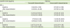
Table 2 demonstrates the baseline cytokine levels in patients, control patients and healthy controls. There was not a statistically significant difference between baseline cytokine levels in three the groups. Additionally, a significant difference in baseline levels was not observed among patients with malignancy and those with other primary diseases.
Table 2
Comparison of serum baseline cytokine levels of the study groups

![]()
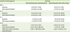
When postdesensitization cytokine levels were measured, IL-10 levels were significantly higher than their initial levels in the successful desensitization group (p = 0.005) whereas the other cytokine levels were not significantly different. In the unsuccessful desensitization group, none of the cytokine levels significantly changed after the attempt of desensitization (Table 3).
Table 3
Comparison of postdesensitization cytokine levels with initial values
Values are presented as median (range).
IFN, interferon; IL, interleukin; NS, not significant.
*p = 0.005.
![]()
The rise in IL-10 levels was greater in chemotherapeutic desensitization than the desensitization with other drugs (p = 0.006) (Table 4). Demographic factors, the primary disease or the reaction type in the history were not related to the rise in IL-10.
Table 4
Comparison of cytokine levels in chemotherapeutic desensitization with desensitizations for other drugs
Values are presented as median (range).
IFN, interferon; IL, interleukin; NS, not significant.
*p = 0.006.
![]()
DISCUSSION
This is the first study evaluating cytokine involvement in desensitization to various drugs in different clinical pictures and presents the absolute rise in IL-10 within 24 hours after successful procedures regardless of the hypersensitivity reaction type or the culprit drug.
Most of our knowledge about the mechanism of desensitization relies on the findings of rapid desensitization. This method was found to be effective both in IgE mediated and in non-IgE mediated immediate reactions due to direct mediator release from mast cells [20]. Therefore, the role of mast cells in immediate reactions and their unresponsive states were examined in previous studies [2122]. Three hypotheses were asserted: depletion of activating signal transduction components, subthreshold depletion of mediators, and internalization of FcεRI through progressive cross-linking at a low antigen concentration [5]. Recently, a mouse model indicated that the main mechanism responsible for the unresponsiveness of mast cells is the impairment of the internalization of IgE and FcεRIα resulting in the bondage of the cross-linked receptors to the cell membrane. This unresponsiveness of mast cells through rapid desensitization was shown to be antigen specific and did not induce anergy [7].
Immediate desensitization is not known to prevent the occurrence of non-IgE-mediated reactions, like maculopapular eruptions or severe cutaneous bullous reactions [1]. An important point to be addressed is that in most nonimmediate reactions sensitization cannot be documented by positive skin tests because of their low sensitivity and specificity and therefore the underlying mechanism in these reactions is largely unknown [2324]. Slow desensitization protocols can be successfully performed in some nonimmediate reactions and although not completely understood, cells other than mast cells are likely to be involved.
T cells and their cytokines were examined in a few studies dealing with specific illnesses or in rare case reports of those who underwent drug desensitization. In a recent study in patients with aspirin-exacerbated respiratory disease (AERD), the effect of aspirin desensitization on T-cell cytokines was examined and revealed no difference in the percentage of CD4+ T lymphocytes expressing IL-2, IL-4, and IFN-γ levels 1 month after desensitization when compared to baseline levels [25]. Although a significant difference could not be found in the first month of tolerance, perhaps long-term effects could have been seen in the following months, which was not mentioned in the study. In another study of patients with AERD who underwent aspirin desensitization, intracellular IL-10 and IFN-γ levels decreased after 1 month of desensitization and came to a level similar to healthy subjects [26]. The authors tried to explain the decrease in intracellular IL-10 as a result of the control of inflammation and suggested that IFN- γ and IL-10 expression in CD4+ T lymphocytes might be related to the pathogenesis of AERD and the mechanism of aspirin desensitization. We can speculate in accordance with this finding, that the secreted levels of these cytokines might have increased in the serum although the authors did not measure the levels. If the cytokines in the patients' sera could have been measured, a finding similar to our study might have been expected.
These studies evaluating T-cell related cytokines in aspirin desensitization provide limited information about the role of T cells, however the low number of patients evaluated in these studies and desensitization only to aspirin for a specific disease does not allow further understanding of the mechanism of all types of drug desensitization.
Teraki and Shiohara [23] showed that in allopurinol induced fixed drug eruptions, the number of CD25+CD4+ T cells in the lesion increased after desensitization whereas the number of CD8+ T cells decreased from 91% of CD3+ cells to 35% during the procedure. They proposed that the peripheral Treg cells migrating into the lesion might have a suppressive effect on the effector function of CD8+ T cells in the lesion. In another case report, an increase of IL-10 and IL-6 levels was observed in the culture of peripheral blood mononuclear cell one month after the tolerance induced by allopurinol desensitization in a patient who had experienced a fixed drug eruption due to allopurinol [27]. These 2 patients as examples of the desensitization of a nonimmediate drug eruption revealed the possible role of Treg cells in the induction of desensitization to such reactions.
Regulatory CD25+CD4+T cells suppress immune responses via cell-to-cell interactions and/or the production of cytokines such as IL-10 and transforming growth factor-β [2829] which is also demonstrated in allergen immunotherapy. Therefore the presence of these cells or their cytokines in the blood samples or skin biopsy specimens of the patients can provide further information about the mechanism of drug desensitization. We observed the cytokines known to be secreted primarily from Th2, Th1, and peripheral Treg cells. Although we know that complex and diverse clinical manifestations caused by the functional heterogeneity of T cells occur in immediate and nonimmediate hypersensitivity reactions due to drugs, it is interesting to observe the increase in IL-10 in different types of reactions. The higher levels of IL-10 observed in chemotherapeutic desensitizations is also of note.
Our study has several limitations. Cytokine levels were measured only after the completion of the procedure whereas subsequent measurements were not taken. Each procedure was the first attempted desensitization for that patient so the effect of multiple desensitization was not observed. It was previously shown that patients receiving multiple desensitizations showed a decrease in the severity and frequency of reactions with subsequent procedures which may better explain the existence of immunologic tolerance in drug desensitizations [8]. Although the diversity of the drugs and the reactions are acceptable in our study, the low number of control subjects affects the significance of our findings.
In conclusion, the successful desensitization independent of the hypersensitivity reaction type seems to be related with the increase of IL-10. In order to further understand the mechanism of successful desensitization, regulatory mechanisms should be examined.
 XML Download