

 PDF
PDF ePub
ePub Citation
 Print
Print


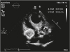
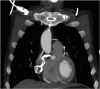
An 80-year-old gentleman with history of coronary artery bypass surgery, repaired abdominal artery aneurysm and repaired bilateral iliac arteries aneurysms was admitted for subacute right lower limb ischemia from partially thrombosed right popliteal artery aneurysm. Before his scheduled surgery an echocardiogram was ordered to complete work-up for possible cardiac cause of thromboembolism. Echocardiogram showed a big rounded atrial mass just proximal to the tricuspid valve at the right atrioventricular junction (Movie 1 and Figure 1). The mass was non-obstructive but appeared to be mobile. This finding triggered a CT angiogram of the thorax. It revealed a large calcified and thrombosed right coronary artery aneurysm compressing the right atrium externally (Movie 2 and Figure 2). He underwent a successful femoral-popliteal bypass surgery and recovered well post-operatively.
Yi Chuan Tham, MD1 , Kok Hooi Yap, MBChB1, Jack Kian Ch'ng, MBBCh2
, Kok Hooi Yap, MBChB1, Jack Kian Ch'ng, MBBCh2

 XML Download