

 PDF
PDF ePub
ePub Citation
 Print
Print


Many pins are used to secure the cutting block to the femur or tibia during total knee arthroplasty (TKA). Sometimes, pins are so deeply inserted or impacted that perforate medial femoral condyle. But, it is difficult to know because the perforated sites of femoral condyle are covered with soft tissue like periosteum or synovium. In this situation, it can be misidentified as the medial femoral condyle fracture in postoperative radiographs. Medial femoral condylar fracture started from the superomedial corner of intercondylar box can be happened in posterior cruciate substituting (PS) type TKA. It is associated with an excessive box cut and additional surgery to fix the medial femoral condyle is needed. However cortical perforation of the medial femoral condyle does not require additional surgery. Therefore, it is important to distinguish cortical perforation of the medial femoral condyle from the medial femoral condyle fracture or intercondylar fracture. To our knowledge, however, there have no reports describing intraoperative cortical perforation and misinterpretation. The authors present two cases of intraoperative cortical perforation, and describe the radiological characteristics. Informed consent was obtained from the patients for publication of this study.
Case Reports
1. Case 1
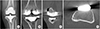
A 66-year-old female patient visited our hospital for left knee joint pain that had developed approximately 3 years prior. In a bone mineral density examination utilizing dual-energy X-ray absorptiometry, her T-score was -0.8 (L1-4) and -0.9 (femur total). Her body mass index (BMI) was 27.3 kg/m2. She was diagnosed with osteoarthritis and did not have any history about osteoporosis, neurologic disorders, chronic steroid use, inflammatory arthropathy and previous surgery. She received TKA with the Vega® knee system (Aesculap, Tuttligen, Germany). The surgery was performed by the single surgeon with routine procedure. We could not recognize any abnormal signs related to a medial condylar fracture during surgery, but a vertical line suggesting a medial condylar fracture was observed in postoperative radiography after TKA. Computed tomography (CT) was performed to evaluate the fracture. A vertical line was identified on some coronal and sagittal cuts. On an axial cut, the line was localized on the posteromedial cortex (Fig. 1). We regarded it as an undisplaced medial condylar fracture and planned to fix the fracture site. However, it was not a medial condylar fracture but a cortical perforation. We found an orifice on medial femoral condyle during revision surgery. It was the orifice formed by the pin insertion too deeply. Therefore, we did not any procedure to fix it.
2. Case 2
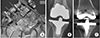
A 59-year-old female patient received TKA with the e.motion® pro knee system (Aesculap). The patient's T-score was -1.3 (L1-4) and 0.1 (femur total). BMI was 28.4 kg/m2. She was diagnosed with osteoarthritis and did not have any other history. The surgery was performed with the same procedure. During surgery, we inspected the medial femoral condyle carefully for demonstrating a cortical perforation. After distal femur cutting, the femoral anteroposterior (A/P) and rotational block was secured with the medial and lateral pins. The sharp tip of medial pin was palpable. The soft tissue covered it was removed and the pin perforation was identified (Fig. 2A). Pin perforation made a hole. We also identified that postoperative radiologic findings on radiograph and CT scan were same to case 1 (Fig. 2B, C).
Discussion
Our major finding was that an intraoperative cortical perforation of the medial femoral condyle could be misidentified as a medial condylar fracture on postoperative radiographs. Intraoperative cortical perforation of the medial femoral condyle could be made during medial pin insertion of the femoral A/P and rotational block. If the medial pin was inserted or impacted deeply, the pin could perforate the medial cortex of the medial femoral condyle.
Cortical perforation of the medial femoral condyle had two features on radiologic evaluation. First, the line was in the vertical orientation. We could find vertical lines on postoperative radiography and coronal CT scan. If the line was traced from proximal to distal on a coronal CT scan, the distal end could be expected to reach the middle of the medial condyle, although we could not exactly observe this due to artifacts caused by the femoral implant. This was the most important difference between cortical perforation and medial condylar fracture. The medial condylar fracture was related to surgical technique, such as an improper bone cutting, an aggressive impaction of the boxed posterior stabilized femoral component, or an angular insertion or removal of the trial component.1) Medial condylar fracture started from the superomedial corner of the intercondylar box. Therefore, fracture line was more oblique than the line of a cortical perforation.
Second, a fine line was localized on the posteromedial cortex area. In axial CT scan, a fine line found on the posteromedial cortex and it was on around orifice thought to be inserted medial pin. Cortical perforation happened on the medial femoral condyle. The reason of cortical perforation of the medial femoral condyle was associated with the anatomy of distal femur. Medial supracondylar ridge is oblique and lateral supracondylar ridge is vertical. Therefore, cortical perforation was mainly happened on medial femoral condyle. Also, we assumed that the posteromedial cortex of the femur is harder than the anteromedial side; therefore, the posteromedial cortex could be perforated as if it were disrupted, and a fine line was formed around the pin orifice that looks like an incomplete fracture.
Intraoperative periprosthetic femoral fractures could be divided into two groups: diaphyseal and metaphyseal fractures. 2) Diaphyseal femoral fractures, such as anterior or anterolateral cortical penetration, usually occurred while the surgeon was inserting an intramedullary guide.3) The metaphyseal femoral fracture was a complete fracture of one or two condyles.4) Lombardi et al.1) reported the complications of intraoperative distal femur intercondylar fractures. There were 40 fractures in 898 PS type TKAs (4.4%). They ascribed this high fracture rate to intercondylar box cut. If box cut placed excessively medial or lateral, it made the respective condyle weakening. Therefore, intraoperative cortical perforation of medial femoral condyle should be distinguished from intraoperative periprosthetic femoral fractures.
Taken together, our findings suggest a surgeon who encounters an unexpected intercondylar fracture line on postoperative radiography should try to distinguish it from cortical perforation with CT scan. If it is cortical perforation, additional surgery is unnecessary. To prevent cortical perforation, attention must be paid when inserting a pin through a femoral A/P and rotational block.
 XML Download