

 PDF
PDF ePub
ePub Citation
 Print
Print


Abstract
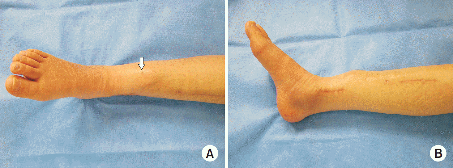
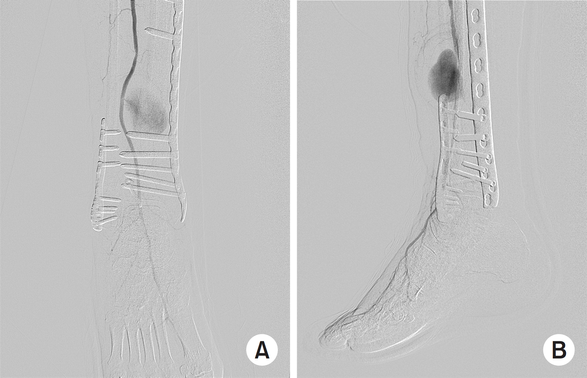
This paper reports a pseudoaneurysm of the anterior tibial artery after reduction with pointed bone reduction forceps on a spiral fracture of the distal tibia. Most reported injuries occurred at the proximal part of anterior tibial artery during drilling of the proximal tibia. To the best of the authors’ knowledge, injury of the distal part of anterior tibial artery has never been reported. This paper describes a 54-year-old woman with a pseudoaneurysm of the anterior tibial artery clinically detected 11 weeks after the index surgery. This report highlights the need for surgeons to be aware of and careful about this complication during and after surgical intervention.
Go to : 
References
1. Gahlot N, Kanojia RK. Anterior tibial artery pseudoaneurysm due to interlocking bolt of tibial nail: a case report and review. Acta Orthop Traumatol Turc. 51:77–83. 2017.

2. Dreyfus U, Fishman J. False aneurysm of the posterior tibial artery complicating fracture of the tibia and fibula. J Trauma. 20:186–187. 1980.
3. Han KJ, Won YY, Khang SY. Pseudoaneurysm after tibial nailing. Clin Orthop Relat Res. 418:209–212. 2004.
4. Sundararajan SR, Rajagopalakrishnan R, Rajasekaran S. Ruptured pseudoaneurysm of the lateral plantar artery after tibiotalocalcaneal fusion with retrograde nail-a rare complication. J Foot Ankle Surg. 57:393–395. 2018.
5. Hussain W, Balach T, Leland JM. Vascular injury involving proximal medial-to-lateral oblique locking screw insertion in tibial intramedullary nailing. Acta Orthop Belg. 77:414–418. 2011.
6. Kim WY, Lee SW, Kim KS, Lee JY. Superior gluteal artery pseudoaneurysm caused by pelvic C-clamp blind application. Hip Pelvis. 29:145–149. 2017.
7. Mróz I, Kielczewski S, Pawlicki D, et al. Blood vessels of the shin – anterior tibial artery – anatomy and embryology – own studies and review of the literature. Folia Med Cracov. 56:33–47. 2016.
Go to :
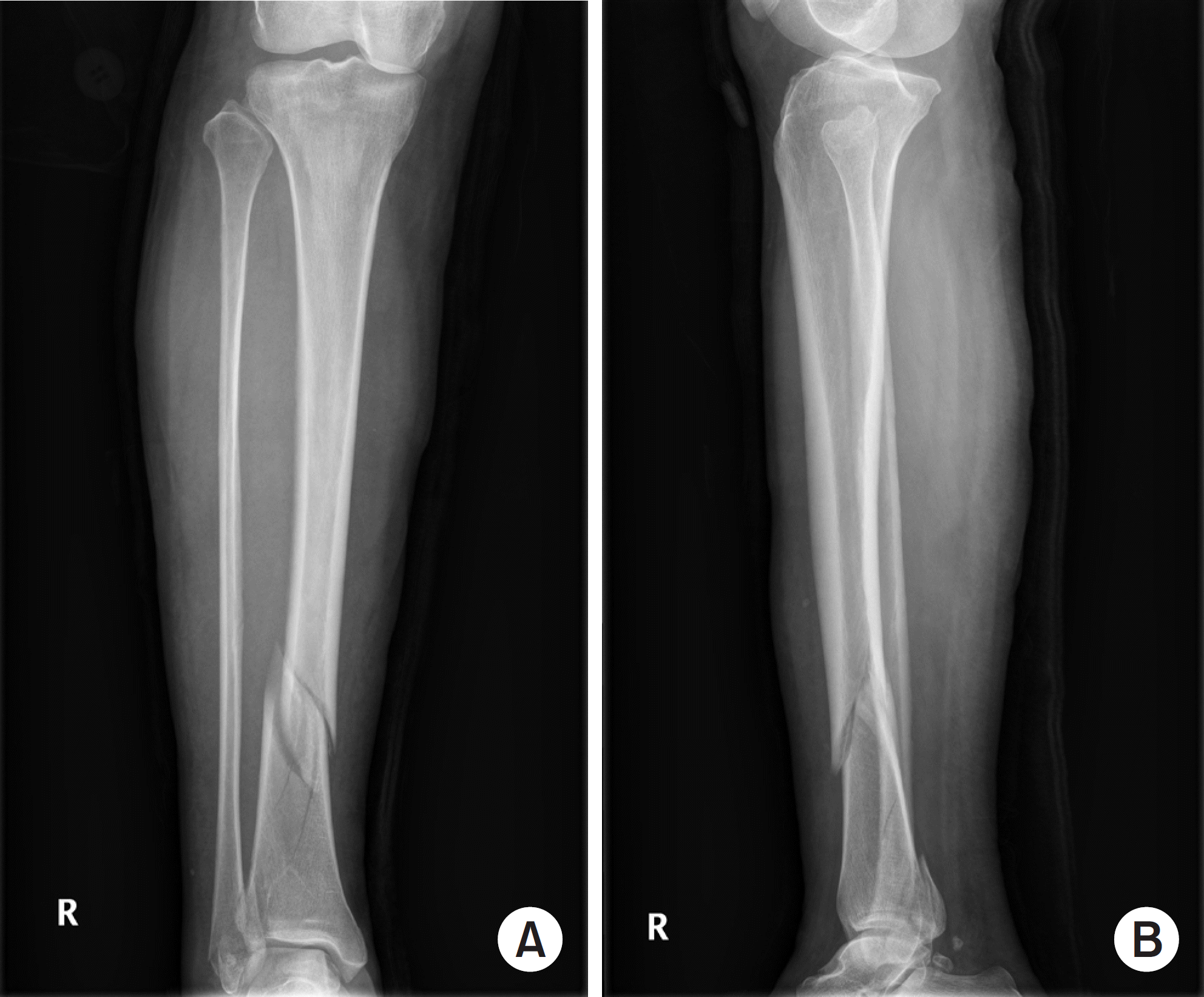 | Fig. 1.Anteroposterior (A) and lateral (B) plain radiographs of the right tibia and fibula show a spiral fracture with a wedge fragment. |
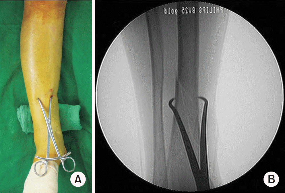 | Fig. 2.Intraoperative photograph (A) and fluoroscopic image (B) applied pointed bone reduction forceps. |
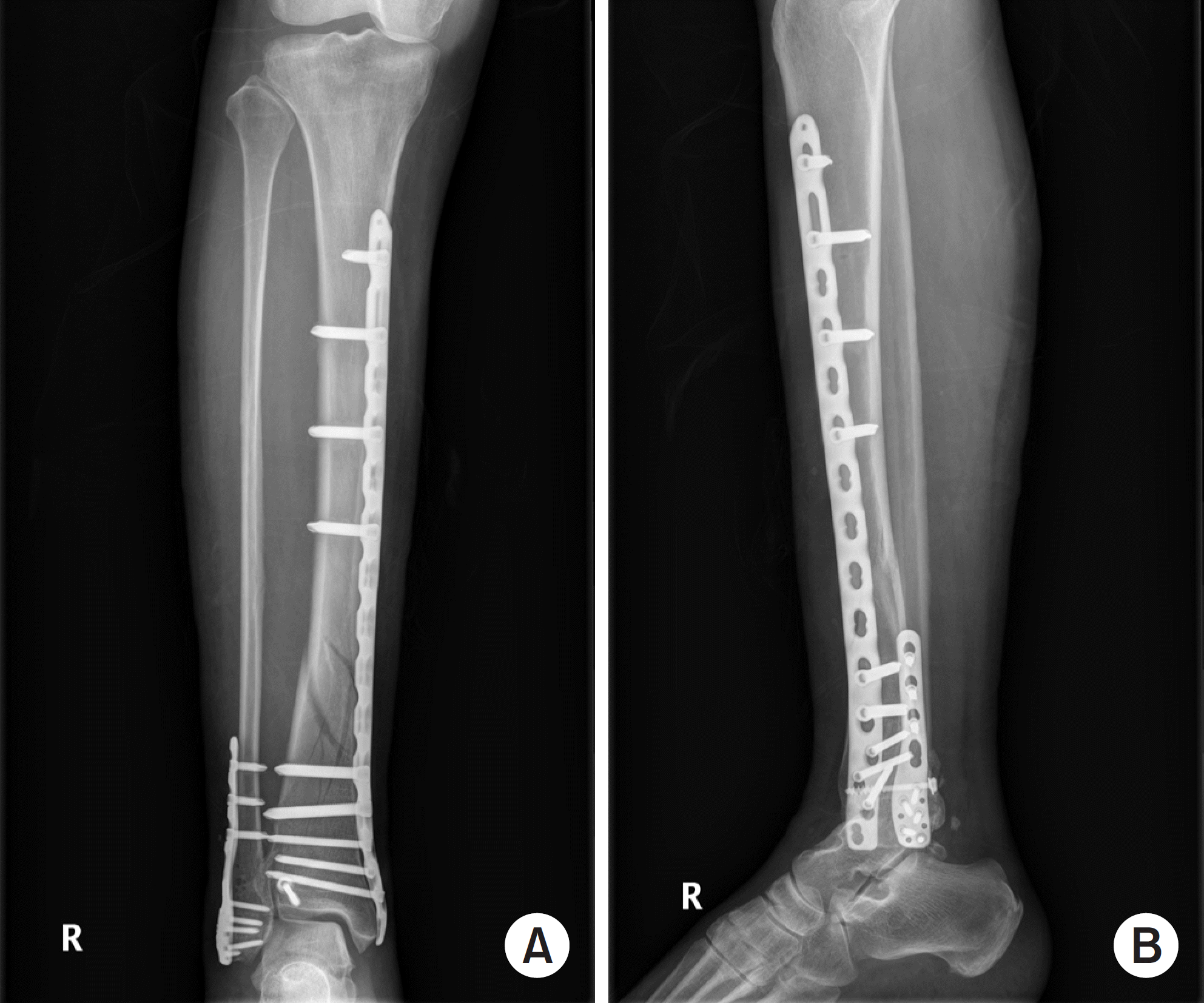 | Fig. 3.Immediate postoperative anteroposterior (A) and lateral (B) radiographs showed acceptable reduction of both the tibia and fibula. |
 XML Download