

 PDF
PDF ePub
ePub Citation
 Print
Print


Abstract
Sclerosing mesenteritis is a rare benign disease with a prevalence of 0.16–3.4% and is characterized by chronic nonspecific inflammation and extensive fibrosis in the adipose tissue of the mesentery although the exact pathogenesis is still elusive. A 65-year-old woman was referred with suspicion of an abdominal mass and biliary stones on abdominal ultrasonography and CT. Bile duct stones were confirmed by endoscopic ultrasonography and successfully treated by endoscopic retrograde cholangiography with stone removal. Furthermore, a 4.7 cm conglomerated mass on small intestinal mesentery was suspected as sclerosing mesenteritis based on the features on abdominal MRI. However, because it could not be differentiated from malignancy without histologic examination, laparoscopic excisional biopsy was performed; it showed only inflammatory cells with extensive fibrosis. Therefore, the abdominal mass was confirmed as sclerosing fibrosis and the patient was followed-up without any treatments because no mass-related symptoms accompanied the findings. Six months later, abdominal CT showed no significant change in the mass. Herein, we report a rare case of incidentally found idiopathic sclerosing mesenteritis.
Go to : 
References
1. Badet N, Sailley N, Briquez C, Paquette B, Vuitton L, Delabrousse É. Mesenteric panniculitis: still an ambiguous condition. Diagn Interv Imaging. 2015; 96:251–257.

2. Min BH, Shinn YC. Idiopathic mesenteric fibrosis. J Korean Surg Soc. 1976; 18:73–76.
3. Chung W, Park KC. A case report of sclerosing mesenteritis. Eulji J Med. 1982; 5:85–86.
4. Cha SJ, Lim HM, Chang ST, Park YW, Yoo JH, Song KY. Primary mesenteritis: a case report. J Korean Surg Soc. 1991; 41:819–829.
5. Jung TY, Lee CW. A case report of the mesenteric panniculitis. J Korean Surg Soc. 1992; 43:916–919.
6. Park KH, Chang HK, Choi SY, et al. Sclerosing mesenteritis associated with skin panniculitis and pleural thickening. Korean J Med. 1999; 57:103–107.
7. Park SD, Bae OS. Cases of postoperative mesenteric panniculitis. J Korean Soc Coloproctol. 2002; 18:128–132.
8. Lee SY, Park DE, Chae KM. Sclerosing mesenteritis. J Korean Surg Soc. 2006; 71:218–221.
9. Li LY, Cho YK, Lee JH, et al. Two cases of mesenteric panniculitis treated by medical management. Korean J Med. 2006; 71:S934–S939.
10. Kim EG, Kang YW, Yoon SG, Kim HD, Kim KY. Four cases of post-operative sclerosing mesenteritis. J Korean Soc Coloproctol. 2007; 23:374–380.
11. Jung SH, Paik CN, Jung JH, Lee KM, Chung WC, Yang JM. Simultaneous colonic obstruction and hydroureteronephrosis due to mesenteric fibromatosis. Gut Liver. 2009; 3:215–217.
12. Bae JH, Kim SH, Ahn SB, et al. A case of idiopathic sclerosing mesenteritis with retroperitoneal fibrosis. Korean J Gastroenterol. 2011; 58:221–225.
13. Lee HJ, Kim JI, Ahn JW, et al. Spontaneous regression of sclerosing mesenteritis presenting as a huge mass. Korean J Gastroenterol. 2012; 59:317–320.
14. Kim EJ, Kim EY, Song JE, et al. A case of IgG4-related sclerosing mesenteritis associated with Crohn's disease. Korean J Gastroenterol. 2014; 63:176–182.
15. Lee SJ, Park CK, Yang WI, Kim SK. IgG4-related sclerosing mesenteritis. J Pathol Transl Med. 2016; 50:309–311.
16. Avincsal MO, Otani K, Kanzawa M, et al. Sclerosing mesenteritis: a real manifestation or histological mimic of IgG4-related disease? Pathol Int. 2016; 66:158–163.
17. Emory TS, Monihan JM, Carr NJ, Sobin LH. Sclerosing mesenteritis, mesenteric panniculitis and mesenteric lipodystrophy: a single entity? Am J Surg Pathol. 1997; 21:392–398.
18. Chen TS, Montgomery EA. Are tumefactive lesions classified as sclerosing mesenteritis a subset of IgG4-related sclerosing disorders? J Clin Pathol. 2008; 61:1093–1097.
19. Deshpande V, Zen Y, Chan JK, et al. Consensus statement on the pathology of IgG4-related disease. Mod Pathol. 2012; 25:1181–1192.
20. Sharma P, Yadav S, Needham CM, Feuerstadt P. Sclerosing mesenteritis: a systematic review of 192 cases. Clin J Gastroenterol. 2017; 10:103–111.
Go to :
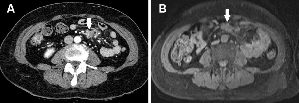 | Fig. 1.Initial abdominal CT (A) and MRI (B) images show a 4.7 cm conglomerated mesenteric mass with enhancement at periumbilical area (arrows). CT, computed tomography; MRI, magnetic resonance imaging. |
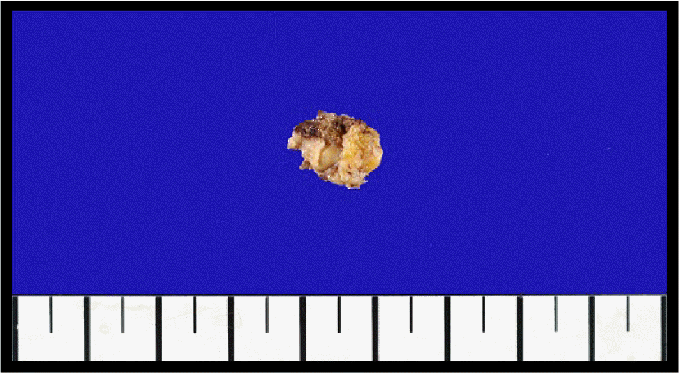 | Fig. 2.Gross finding of laparoscopic excisional biopsy of the mesenteric mass. The submitted specimen was a piece of irregular fibroadipose tissue measuring 1.2×0.9×0.5 cm in size. |
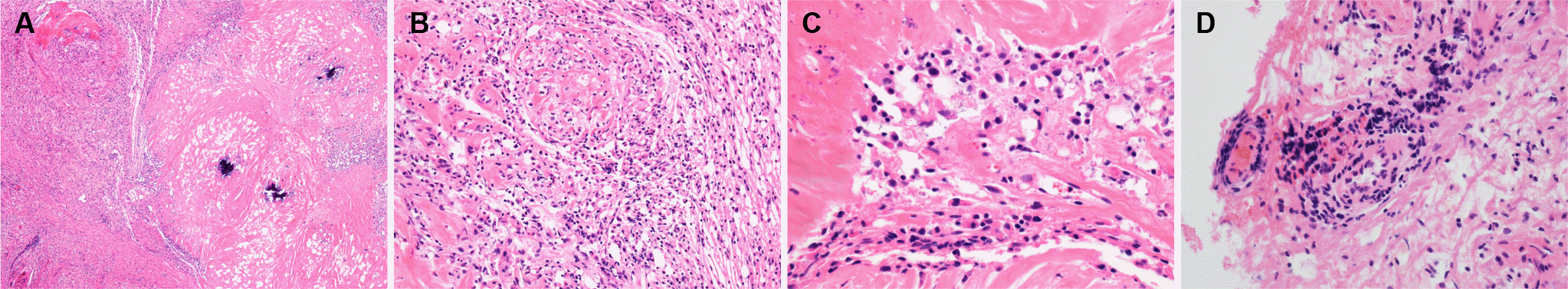 | Fig. 3.Histopathologic findings of sclerosing mesenteritis. (A) Diffuse fibrosclerosis with infiltrate of inflammatory cells and dystrophic calcification (H&E, ×40). (B) A moderate infiltrate of chronic inflammatory cells with collagen deposition (H&E, ×200). (C) The inflammatory infiltrate was abundant plasma cells and lymphocytes in marked fibrosclerosis (left upper corner) (H&E, ×400). (D) Obliterative phlebitis: in high power view of histologic findings, there are foci of a heavy infiltrate of perivenular and intravascular inflammatory cells with obliteration of vascular lumen (H&E, ×400). |
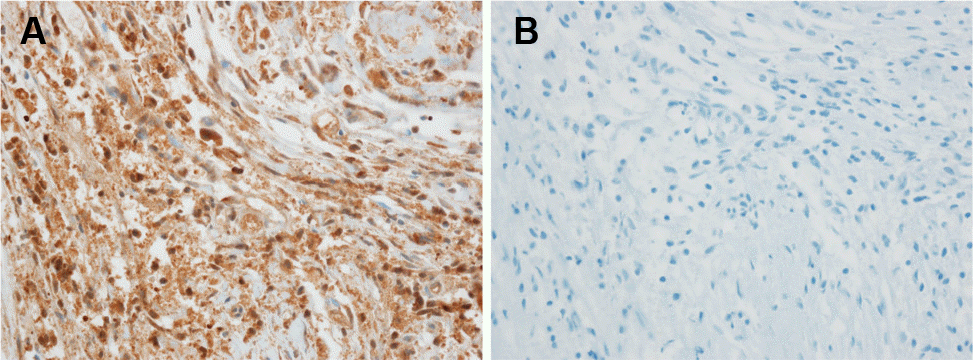 | Fig. 4.Immunohistochemical staining shows markedly increased IgG-positive plasma cells (A, ×400) and complete IgG4 negativity (B,×400). Ig, immunoglobulin. |
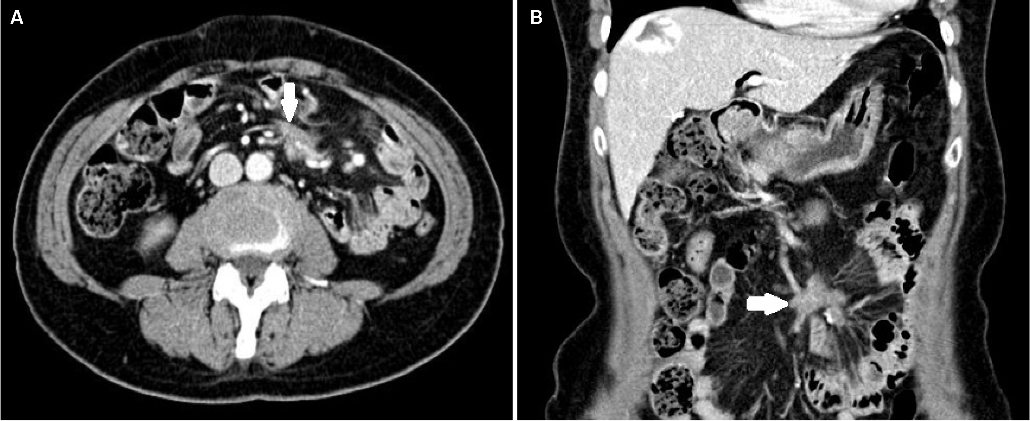 | Fig. 5.Abdominal computed tomography of axial (A) and coronal (B) images after six months of follow-up show no specific change in size or nature of the mass lesion (arrows). |
Table 1.
Reported Cases of Sclerosing Mesenteritis in Korea
 XML Download