Journal List > Chonnam Med J > v.55(1) > 1112109

Gasparri and Roncati: Paired Box Gene 8 (Pax8) Is also an Immunomarker of B-Cell Lineage Which Can Be Source of Diagnostic Pitfalls
Paired box 8 (PAX8) is a transcription factor, which in humans is encoded by the PAX8 gene, located on the chromosome 2, cytoband q14.1.1 It belongs to the paired box protein family, consisting of nine members from PAX1 to PAX9.1 This family of transcription factors is so called because all the members share the same N-terminal paired box domain composed of about 120 amino acids. The PAX proteins are further subdivided into four groups on the basis of the presence (or absence) in their molecular structure of an octopeptide and of a paired-type homeobox at the C-terminal domain: PAX8 is placed in the second group together with PAX5 and PAX2.2
PAX8 is a master regulator in thyroid development; moreover, it plays an important role in the organogenesis of the thymus, kidneys, Müllerian system, eyes, and brain.34567
Several immunohistochemical studies have demonstrated the importance of PAX8 as immunomarker for different epithelial neoplasms arising in the thyroid and parathyroid gland, thymus, female and male genital tract or kidneys, including the Wilms' tumor.8910111213
Some authors have also reported PAX8 expression in pancreatic islet cells and in neuroendocrine tumors of the pancreas, as well as in carcinoids arising in the duodenum, rectum and stomach.1415
Consequently, the pathologists exploit PAX8 immunostaining to differentiate primary and metastatic PAX8-positive tumors from PAX8-negative ones.912
As mentioned above, PAX5 is inserted in the same group as PAX8, however, unlike PAX8, PAX5 is encoded by a gene located on chromosome 9, cytoband p13.2, and it plays a significant role in early B-cell differentiation as well as in neural development and spermatogenesis.31617
PAX5 immunostaining is used by pathologists in the diagnosis of most B-cell non-Hodgkin lymphomas/leukemias, Hodgkin lymphomas, where it faintly depicts Reed-Sternberg and Hodgkin cells, and in the differential diagnosis between plasmacytoma (PAX5-negative) and lymphoplasmacytic lymphoma/marginal zone lymphoma with plasmacytoid differentiation (PAX5-positive).16171819
As it is well known, a secondary follicle, which arises from a primary follicle after an antigenic stimulation of B cells, is provided with a germinal center and a mantle zone. Externally to the mantle zone there is the marginal zone, which is difficult to distinguish, except when it is expanded, as in the course of benignant or lymphomatous proliferations.20212223
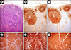
Literature has reported that PAX8 stains B lymphocytes and B-cell lymphomas.242526 To prove the validity of this statement, we have at first immunohistochemically evaluated the PAX5 and PAX8 expression in 50 consecutive normal lymph nodes removed for staging purposes in as many patients affected by epithelial malignancies. In all the tested cases, PAX8 was labeled at the germinal center, the mantle, and the marginal zone, exactly as PAX5 (Fig. 1). Subsequently, we have enrolled 25 cases of germinal center-derived lymphomas, 25 cases of mantle cell lymphoma and 25 cases of marginal zone lymphomas, coming from patients of any age, gender, or ethnicity, all resulted immunohistochemically positive for PAX5 and PAX8, as deriving from mature B cells (Fig. 1).
Since PAX8 is not involved in the process of B-lymphocyte commitment maintaining the same immunohistochemical profile of PAX5, many authors have wondered if that of PAX8 was a real positive. For example, Moretti and colleagues have tested PAX8 and PAX5 in reactive lymph nodes, in diffuse large B-cell lymphomas, and in Hodgkin lymphomas using for PAX5 a monoclonal antibody, and for PAX8 both a N-terminal polyclonal antibody (pPAX8), and a C-terminal monoclonal antibody (mPAX8).27 The results were the same for pPAX8 and PAX5, that were both positive in all lymphomas and reactive lymph nodes, while mPAX8 was negative in all the tested cases.27 In a similar manner, Morgan et al.28 investigated pPAX8, mPAX8 and PAX5 in a cohort of B-cell lymphomas, obtaining the same expression profile for pPAX8 and PAX5. In conclusion, we can state that PAX8 and PAX5 immunohistochemistry are overlapping in the routine diagnostic practice and, therefore, PAX8 positivity in lymphomas should not be surprising. On the other hand, this evidence should be kept in mind by the pathologists in order to avoid diagnostic pitfalls and mistakes.

Figures and Tables

FIG. 1

Reactive lymphadenitis (A, hematoxylin & eosin; 4×): the germinal centers, the mantles and the marginal zone are brown-stained by immunohistochemistry for PAX8 (B, clone MRQ-50, Ventana; 5×) and PAX5 (C, clone SP34, Ventana; 5×) in an overlapping manner involving the B-cell nuclei. Germinal center-derived lymphoma (D, 4×), mantle cell lymphoma (E, 4×) and marginal zone lymphoma (F, 4×) are strongly labeled by PAX8 immunohistochemistry [chromogen: 3,3′ diaminobenzidine tetrahydrochloride hydrate].

cmj-55-70-g001

Notes

CONFLICT OF INTEREST STATEMENT None declared.

References

1. Mansouri A, Hallonet M, Gruss P. Pax genes and their roles in cell differentiation and development. Curr Opin Cell Biol. 1996; 8:851–857.
crossref pmid
2. Treisman J, Harris E, Desplan C. The paired box encodes a second DNA-binding domain in the paired homeo domain protein. Genes Dev. 1991; 5:594–604.
crossref pmid
3. Chi N, Epstein JA. Getting your Pax straight: Pax proteins in development and disease. Trends Genet. 2002; 18:41–47.
crossref pmid
4. Dehbi M, Pelletier J. PAX8-mediated activation of the wt1 tumor suppressor gene. EMBO J. 1996; 15:4297–4306.
crossref pmid pmc
5. Poleev A, Fickenscher H, Mundlos S, Winterpacht A, Zabel B, Fidler A, et al. PAX8, a human paired box gene: isolation and expression in developing thyroid, kidney and Wilms' tumors. Development. 1992; 116:611–623.
crossref pmid
6. Mansouri A, Chowdhury K, Gruss P. Follicular cells of the thyroid gland require Pax8 gene function. Nat Genet. 1998; 19:87–90.
crossref pmid
7. Bouchard M, Souabni A, Mandler M, Neubüser A, Busslinger M. Nephric lineage specification by Pax2 and Pax8. Genes Dev. 2002; 16:2958–2970.
crossref pmid pmc
8. Nonaka D, Tang Y, Chiriboga L, Rivera M, Ghossein R. Diagnostic utility of thyroid transcription factors Pax8 and TTF-2 (FoxE1) in thyroid epithelial neoplasms. Mod Pathol. 2008; 21:192–200.
crossref pmid
9. Laury AR, Perets R, Piao H, Krane JF, Barletta JA, French C, et al. A comprehensive analysis of PAX8 expression in human epithelial tumors. Am J Surg Pathol. 2011; 35:816–826.
crossref pmid
10. Tacha D, Zhou D, Cheng L. Expression of PAX8 in normal and neoplastic tissues: a comprehensive immunohistochemical study. Appl Immunohistochem Mol Morphol. 2011; 19:293–299.
pmid
11. Bowen NJ, Logani S, Dickerson EB, Kapa LB, Akhtar M, Benigno BB, et al. Emerging roles for PAX8 in ovarian cancer and endosalpingeal development. Gynecol Oncol. 2007; 104:331–337.
crossref pmid
12. Ozcan A, Shen SS, Hamilton C, Anjana K, Coffey D, Krishnan B, et al. PAX 8 expression in non-neoplastic tissues, primary tumors, and metastatic tumors: a comprehensive immunohistochemical study. Mod Pathol. 2011; 24:751–764.
crossref pmid
13. Ozcan A, Liles N, Coffey D, Shen SS, Truong LD. PAX2 and PAX8 expression in primary and metastatic müllerian epithelial tumors: a comprehensive comparison. Am J Surg Pathol. 2011; 35:1837–1847.
crossref pmid
14. Long KB, Srivastava A, Hirsch MS, Hornick JL. PAX8 expression in well-differentiated pancreatic endocrine tumors: correlation with clinicopathologic features and comparison with gastrointestinal and pulmonary carcinoid tumors. Am J Surg Pathol. 2010; 34:723–729.
crossref
15. Sangoi AR, Ohgami RS, Pai RK, Beck AH, McKenney JK, Pai RK. PAX8 expression reliably distinguishes pancreatic well-differentiated neuroendocrine tumors from ileal and pulmonary well-differentiated neuroendocrine tumors and pancreatic acinar cell carcinoma. Mod Pathol. 2011; 24:412–424.
crossref pmid
16. Adams B, Dörfler P, Aguzzi A, Kozmik Z, Urbánek P, Maurer-Fogy I, et al. Pax-5 encodes the transcription factor BSAP and is expressed in B lymphocytes, the developing CNS, and adult testis. Genes Dev. 1992; 6:1589–1607.
crossref pmid
17. Torlakovic E, Torlakovic G, Nguyen PL, Brunning RD, Delabie J. The value of anti-pax-5 immunostaining in routinely fixed and paraffin-embedded sections: a novel pan pre-B and B-cell marker. Am J Surg Pathol. 2002; 26:1343–1350.
crossref pmid
18. Desouki MM, Post GR, Cherry D, Lazarchick J. PAX-5: a valuable immunohistochemical marker in the differential diagnosis of lymphoid neoplasms. Clin Med Res. 2010; 8:84–88.
crossref pmid pmc
19. Feldman AL, Law ME, Inwards DJ, Dogan A, McClure RF, Macon WR. PAX5-positive T-cell anaplastic large cell lymphomas associated with extra copies of the PAX5 gene locus. Mod Pathol. 2010; 23:593–602.
crossref pmid pmc
20. Roncati L, Maiorana A. Ectopic extra-nodal in situ follicular neoplasia (ISFN). J Hematopathol. 2016; 9:151–153.
crossref
21. Roncati L, Maiorana A. IgA plasmablastic large B-cell lymphoma. Diagnosis (Berl). 2017; 4:105–107.
crossref pmid
22. Roncati L. De novo CD5-positive primary gastric diffuse large B-cell lymphoma coexpressing MYC and BCL6: towards a proper subset of double-hit, triple-hit and, maybe, quadruple-hit B-cell lymphomas? Chonnam Med J. 2018; 54:80–82.
crossref
23. Roncati L, Barbolini G, Piacentini F, Piscioli F, Pusiol T, Maiorana A. Prognostic factors for breast cancer: an immunomorphological update. Pathol Oncol Res. 2016; 22:449–452.
crossref pmid
24. Parakh R, Cheng L, Tretiakova M. PAX8-positive B-cell lymphoma in adrenal gland masquerading as metastatic renal cell carcinoma. Int J Surg Pathol. 2018; 26:721–724.
crossref pmid
25. Hirsch MS, Nascimento AF. PAX8 distinguishes diffuse large B-cell lymphoma mimicking sarcoma. Case Rep Pathol. 2017; 2017:6714549.
crossref
26. Hubbard EW, Nodit L, Van Meter S. Undifferentiated malignant neoplasm involving parotid and thyroid: sampling and PAX8 cross-reactivity can obscure the diagnosis of lymphoma. Case Rep Pathol. 2016; 2016:3291549.
crossref
27. Moretti L, Medeiros LJ, Kunkalla K, Williams MD, Singh RR, Vega F. N-terminal PAX8 polyclonal antibody shows cross-reactivity with N-terminal region of PAX5 and is responsible for reports of PAX8 positivity in malignant lymphomas. Mod Pathol. 2012; 25:231–236.
crossref pmid
28. Morgan EA, Pozdnyakova O, Nascimento AF, Hirsch MS. PAX8 and PAX5 are differentially expressed in B-cell and T-cell lymphomas. Histopathology. 2013; 62:406–413.
crossref pmid
TOOLS
Similar articles