 PDF
PDF ePub
ePub Citation
 Print
Print


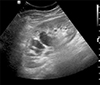
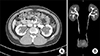
A 36-year-old man visited our hospital with a suspected bilateral hydronephrosis that was detected during a routine check-up. Ultrasonography at the local clinic revealed a wide hypoechoic area in the center of both kidneys instead of a central echogenic complex (Fig. 1). Oliguria or abnormal laboratory values were not observed. On an axial view of the computed tomography with intravenous pyelogram (CT IVP) scan, low attenuated lesions with a cauliflower appearance were observed in the pelvises and calyces of both kidneys (Fig. 2A). No hydronephrosis or obstructive lesions in the urinary tract were observed on reconstructed intravenous pyelogram (IVP) image (Fig. 2B). A parapelvic cyst is a hypoechoic lesion located in the renal pelvis, which is often misdiagnosed as hydronephrosis because of its thin wall.12 In conclusion, CT IVP is a useful test for the differential diagnosis of parapelvic cysts.
- About Synapse
- How to Participate
- KAMJE Databases


- About Synapse
- How to Participate
- KAMJE Databases
Journal List > Chonnam Med J > v.55(1) > 1112106
- TOOLS
 XML Download
- Similar articles