

 PDF
PDF ePub
ePub Citation
 Print
Print


INTRODUCTION
1. History of immuno-inert biomaterials
Biomaterials have been historically developed with the aim to transplant then into the human body as substitutes for damaged tissue or a biological function.12 Biomaterials may be metals, ceramics, polymers, or even living cells and tissue. They can be used as surface coatings, fibers, films, or particles for use in biomedical products such as heart valves, hip joint replacements, dental implants, or drug delivery carriers. The first generation of biomaterials was developed under the concept of immune evasion because the biggest challenge of “foreign objects” has been the action of the immune system. Therefore, it was preferable that they be ‘inert’ and not interact with the biology of the host organism such as proteins, lipids, nucleic acids, sugars, and amino acids. One of the most successful approaches to produce bio-inert materials has been the used of polyethylene glycol (PEG).3 For example, covalent attachment of PEG to proteins or drugs called “PEGylation” has been shown to improve the safety and efficiency, and many PEGylated pharmaceuticals are currently on the market. This technology can be also used as a means to design anti-fouling surfaces. Grafting PEG to solid surfaces significantly reduces protein adsorption and cell adhesion.456 In addition to PEG, zwitterionic polymers have recently been extensively developed for anti-fouling purpose.789 Among them, one of the cell membrane lipids, phosphatidylcoline (PtdCho)-inspired polymers such as 2-methacryloyoxyethyl phosphorylcholine (MPS) have been used in various medical devices.101112 These polymers have extended the applications of biomaterials from traditional implants to biosensing, prodrug carriers, subcellular bioimaging, and cell manipulation.
2. Challenges of immuno-modulating biomaterials
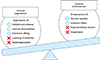
Since studies of bio-inert biomaterials are already well advanced, many researchers have recently switched their focus from bio-inert to bio-modulating materials that promote or inhibit immune responses. However, controlling the immune system with biomaterials is still a challenge due to their methodological complexity. For example, anti-tumor activity is expected when immune systems are activated, while excess activation can cause unexpected symptoms such as allergies (Fig. 1). On the other hand, suppressing the immune system lowers inflammation, but also leads to diminished resistance against infections. Therefore, to obtain immunotherapy with a biomaterial, it is necessary to perfectly understand the biological reactions induced by implanted materials and to adequately design the shape, physical properties, and chemical properties of the material. Fig. 2 shows three types of biomaterials according to their association with the immune system. The first category is called “immuno-inert biomaterials” as described above. The second type is called “immuno-activating biomaterials” designed to exhibit anti-tumor and drug-responsive properties. The third type is known as “immuno-tolerant biomaterials” which suppress and modulate unnecessary uncontrollable inflammation and inhibit rejection responses.
IMMUNE-ACTIVATING BIOMATERIALS
1. Adjuvant materials
As mentioned above, much effort has been previously made to design biomaterials to minimize the host's immune responses against implanted materials. However, biomaterials can also be designed to activate the host's immune responses and/or provide therapeutic effects. The first application of this concept was the use of non-biological adjuvant materials such as γ-polyglutamic acid (γ-PGA),13 poly (lactic-co-glycolic acid) (PLGA),14 or poly (ε-caprolactone) (PCL).15 These biodegradable materials can increase the host's immune response to vaccines. New types of pH-responsive nanoparticles have also been developed because the degradation rate for such biodegradable polymers is not fast enough for effective antigen deliveries.16 The nanoparticles with pH-cleavable crosslinkers are rapidly hydrolyzed under lysosomal acidic conditions (pH 5) and release antigens into dendritic cells (DCs). By contrast, Stayton and co-workers proposed antigen delivery strategies that mimic virus infection mechanisms. They focused on polypropylic acid (PPAA), which has a pKa around pH 6.0–6.5, making it hydrophilic at a biological pH and dramatically hydrophobic at endosomal pH due to the protonation of carboxyl groups along its backbone.17 Due to the polymer phase transition, PPAA disrupted lipid bilayer membranes and antigens tethered to PPAA by disulfide bonds were released under endosomal reductive conditions.
2. Hemagglutinating virus of Japan-envelope (HVJ-E)
Anti-tumor immunity can be also induced by viral envelopes such as Hemagglutinating Virus of Japan-envelope (HVJ-E).18 HVJ-E is a purified product prepared through the complete inactivation of the genome in HVJ by UV irradiation. HVJ-E was originally developed as a novel vector for plasmid DNA, peptides, and drugs because of the fusion proteins of the HVJ such as HN and F are retained after inactivation. Recently, Kaneda and colleagues have reported the tumor-suppressing ability of the inactivated, replication-defective HVJ-E itself.19 HVJ-E alone induced tumor-specific antitumor immunity by eliciting IL-6 production in dendritic cells (DCs) and eradicated 60% to 80% of tumors growing in mice without exogenous gene expression. Thus, HVJ-E has recently attracted a lot of attention as a new type of therapeutic material for cancer immunotherapy. However, one of the greatest challenges for viral-based vectors is that they tend to fuse with virtually all cells and induce hemagglutination in the bloodstream. Therefore, it has been required to increase the tumor-targeting ability as well as reduce nonspecific biding. To achieve this, the chemical modification of the viral vector is one of the most direct approaches. For example, surface-modified adenoviral vectors (Ads) with a multivalent reactive poly [N-(2-hydroxypropyl) methacrylamide] (PHPMA)-based copolymer has been shown successfully to shield from recognition by antibodies.20 The authors have fashioned HVJ-E with hyaluronic acid (HA) to act as a ligand for CD44 to improve its anti-tumor activity.2122 The functionalized HVJ-E is expected to improve the retention time in the blood stream by inhibiting interactions between HVJ-E and red blood cells. Also, the HA layer can be selectively degraded in cancer tissues by hyaluronan-targeting enzymes. A layer-by-layer (LbL) assembly has been employed for the decoration of the HVJ-E. We have succeeded in forming HA/glycol chitosan (GC) multilayers on HVJ-E for up to six assembly cycles. The outermost HA layer was selectively degraded by hyaluronidase. The HA/GC-coated HVJ-E can be thought of as a potential bomb for cancer immunotherapy because of its ability to target CD44 while preventing nonspecific binding in the bloodstream.
IMMUNE-SUPPRESSING BIOMATERIALS
1. Induction of immune tolerance
In addition to immune-activating biomaterials, researchers have been recently developing immuno-tolerant or suppressing materials for the treatment of transplant rejections and autoimmune diseases. In general, the immune system effectively eliminates foreign substances such as pathogenic microorganisms, but does not respond to self-biomolecules, symbiotic microorganisms, or environmental substances. However, in autoimmune diseases, the immune system misidentifies some molecules as foreign and elicits a response against them. For example, chronic inflammatory disorders such as inflammatory bowel disease, allergic reactions, cancer, obesity, diabetes, collagen disease, and Alzheimer's disease are all caused by the impaired maintenance of immune homeostasis. To induce immune tolerance, several strategies have been developed by using various biomaterials for the delivery of adjuvants, antigens, and drugs. For example, PLGA particles that contained costimulatory anti-sense oligonucleotides for DCs and T cells successfully inhibited immune responses.23 In another example, PLGA particles modified with anti-DEC205 antibodies, anti-CD1c antibodies, and P-2 peptides were also developed.24 These examples show the potential application of targeting PLGA particles to maintain immunological tolerance.
However, previous immune-suppressive biomaterials as mentioned above simply operate as delivery carriers. In other words, the materials themselves do not have any anti-inflammatory properties. However, in recent years, material-based immuno-suppressive systems have been developed, and a few specific examples will be introduced here. Stable, organic free radicals such as 2, 2, 6, 6-tetramethylpiperidine 1-oxyl (TEMPO) are often used as catalysts for oxidation reactions in organic synthesis and can act as radical scavengers that detect radical generation reacting systems. Yoshitomi et al.25 focused on the radical scavenging ability of TEMPO for removing reactive oxygen species (ROS) at inflammatory regions. Normally, large amounts of ROS are produced during inflammation, and exposure to this oxidative stress can cause various diseases such as arteriosclerosis. However, reactive oxygen must be selectively removed as it plays an important role during intracellular aerobic respiration in mitochondria. They succeeded in improving the cognitive function of mice that model Alzheimer disease by orally administering TEMPO-functionalized PEG.26
2. Apoptotic cell-inspired biomaterials
Ideally, biomaterials should mimic the living tissue or biomolecules from a mechanical, chemical, biological, and functional point of view. As mentioned in the introduction, one of the most successful biocompatible polymers, MPC polymer, was developed by learning from mammalian cell membrane lipid PtdCho. MPC polymers have been extensively applied to various medical devices, including an artificial joint (Aquala®, Japan Medical Materials), cardiovascular stent (Endeavor®: Medtronic), left ventricular assist device (EVAHEART®, Sun Medical), oxygenator (PrimO2x®: Sorin), and soft contact lenses (Proclear®: Cooper Vision). Therefore, we discuss the potential to create a novel immune-suppressing biomaterial by focusing on the lipid membrane in the last section.
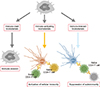
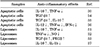
In general, cell membranes have a lipid bilayer constructed by phospholipids such as electrically neutral sphingomyelin (Sph) and PtdCho that occupy the outside membrane and anionically charged phosphatidylethanolamine (PtdEA), PtdSer, and phosphatidylinositol (PtdIno) that are located on the cytosol side.27 Cell membranes keep this heterogeneous structure by active transportation at inner sites by scramblase, and the cytoskeleton is therefore stabilized by anionic phospholipids. However, apoptosis induces the destruction of these highly maintained asymmetrical structures, forcing PtdSer to be exposed on the surface of apoptotic cell membranes, which are then recognized by immune cells (Fig. 3).28 Apoptosis is a genetically programmed cell death mechanism and is necessary to regulate embolic development and maintain homeostasis. Many studies also revealed that PtdSer acts as a trigger molecule for these anti-inflammatory effects.29 Voll et al.30 reported that apoptosis induced leukocytes to downregulate inflammatory cytokine production and upregulate anti-inflammatory cytokines for the production of immune cells. Several therapeutic effects of PtdSer liposomes have been also reported for chronic heart failure,31 Alzheimer's disease32 and osteoporosis.33 Table 1 summarizes the recent progress of anti-inflammatory therapies using apoptotic cells and PtdSer liposomes.3031323334353637
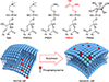
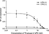
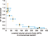
We have recently designed apoptotic cell membrane-inspired polymers which have a phosphoryl serine (PS) group because PtdSer mimetic polymers are very promising materials for anti-inflammatory therapies (Fig. 4).3839 However, the synthesis of PtdSer-mimetic polymers has been difficult as serine has two reactive groups (the amino- and carboxy-groups) that limit the effective and selective modification routes. Therefore, we used a phosphoramidite method which is commonly used in solid phase syntheses of DNA. The advantage of using this reaction is its high selectivity under mild conditions when forming a phosphate ester between two hydroxyl groups.39 First, we synthesized the methacryloyloxyethyl phosphorylserine (MPS) monomer using 2-hydroxyethyl methacrylate, N-α-(t-butoxycarbonyl)-L-serine t-butyl ester, and t-butyl tetraisopropylphosphorodiamidite. Next, poly (MPS) was obtained by free radical polymerization and then deprotection and oxidation of the PtdSer using t-butyl peroxide and trifluoroacetic acid. We applied poly (MPS) to lipopolysaccharide (LPS)-activated RAW 264.7 macrophages to investigate the immune-suppressive effects. Amoeboid cell extensions of macrophages in LPS stimulated conditions were inhibited in the poly (MPS)-treated groups. In addition, the effective suppression of NF-κB in macrophages was observed over 10 mM of PS groups (Fig. 5).38 To investigate how macrophages recognize poly (MPS), the localization of rhodamine-modified poly (MPS) on macrophages was studied. A homogeneous distribution of the poly (MPS) in the cytoplasm was observed. By calculating the up-taken amount of poly (MPS) in the cell, we found that 50 pg or more of poly (MPS) within each macrophage is necessary to inhibit inflammatory behavior (Fig. 6). Furthermore, we investigated the time-dependent immunosuppression induction of poly (MPS). The NF-κB expression was inhibited after 3 h incubating, indicating that a minimum time for immunosuppression is 3 h. The findings in this study are expected to open new doors in developing advanced immuno-suppressive agents and materials for anti-inflammatory therapies such as autoimmune diseases.
CONCLUSION
This review article summarizes recent research examples of immuno-modulating biomaterials and systems. These materials are classified into three categories: (1) immuno-inert, (2) immuno-activating, and (3) immune-tolerant biomaterials. Particularly, we mainly focused on the immuno-tolerant biomaterials in this article. In previous immuno-tolerant biomaterial systems, the materials themselves did not have any anti-inflammatory properties. However, the ability to engineer biological activity into synthetic materials has enabled the design of biomaterials which harness the host's immune responses and provide therapeutic effects. One of the examples is apoptotic cell-membrane mimetic polymers. Advantages of using such polymeric materials for anti-inflammatory therapy is cost-effectiveness as it is inexpensive compared to molecular targeted drugs. However, controlling the immune system is still a challenge due to its methodological complexity. For example, excess activation of the immune system may cause unexpected symptoms such as allergies, while suppressing of the immune system also leads to diminished resistance against infections. Therefore, to obtain immunotherapy with a biomaterial, it is necessary to design the shape, physical properties, and chemical properties of the material adequately. In the future, biomaterials will assume an even greater role in anti-inflammatory therapies through biologically inspired design.

 XML Download